
Abstract
A high prevalence of uterine leiomyoma has been reported in Baltic gray seals aged 15 years and above. Studies on Baltic seals during the 1970s revealed high tissue concentrations of the organochlorines bis(chlorophenyl)-1,1,1-trichloroethane (DDT) and polychlorinated biphenyls (PCBs), lowered reproduction rate, and pathologic changes. In the second half of the 1970s, decreases of PCB and DDT in Baltic biota occurred, and the prevalence of pregnancies in Baltic seals increased. Between 1975 and 1997, 53 Baltic gray seal females of age 15–40 years were found dead and sent to the Swedish Museum of Natural History. Seals were autopsied and 34/53 (64%) had uterine leiomyomas. Samples from 15 were sufficiently well preserved for histologic examination. Uterine leiomyomas were found most commonly in the uterine corpus but also were observed in the uterine horns, cervix, and vagina. Cut surfaces of the leiomyomas appeared as whorled white fibrous tissue. Histologically, spindle cells were arranged in a whorl-like pattern. The nuclei were rod-like and strikingly uniform in shape and size. Mitotic figures were rare. Immunohistochemical staining of the tumors showed a positive reaction to antibodies recognizing smooth muscle actin. Reproductively active gray seals have an ovarian corpus luteum or albicans for most of the year. In 22/34 (65%) gray seals with uterine leiomyomas, ovaries did not contain corpora. In gray seals without macroscopically detected uterine leiomyoma, ovaries from 6/19 (32%) seals had no corpora. It is possible that the development of leiomyoma in the seals is associated with organochlorines and the previous low reproductive activity.
At the end of the 1960s, the Baltic environment was found to be severely polluted by the organochlorines bis(chlorophenyl)-1,1,1-trichloroethane (DDT) and polychlorinated biphenyls (PCBs). 22 , 23 Some PCBs, DDT, and their metabolites have antiestrogenic or estrogenic effects. 15 , 17 , 19 , 28 , 41 Studies on Baltic seals revealed that organochlorines and especially PCB seemed to be responsible for significant reductions in reproduction. 20 , 33 Furthermore, experimental studies in The Netherlands have shown that common seals, feeding on fish from PCB-polluted coastal waters, failed to reproduce. 36 Premature parturition has been observed in California sea lions that had tissue concentrations of PCBs and DDT compounds 2.4 to 8.0 times higher than those of females giving birth to apparently normal pups at full term. 18
In the middle of the 1970s, chemical and pathologic studies on Baltic seals were initiated. Autopsy results showed that a disease complex was common in adult specimens. 7 , 8 Lesions were found in claws, skull, intestine, kidneys, arteries, adrenals, and female reproductive organs, suggesting hormonal imbalance, metabolic disorder, and immune suppression. One of these lesions, uterine leiomyoma, had a high prevalence among gray seals more than 15 years old. 6 In seals, the histologic features of this tumor have not been described. Prevalence in other seal populations outside the Baltic Sea has previously not been reported, although leiomyoma and sqamous cell carcinoma of the uterus of a gray seal from British waters has been described. 29 A time-trend pathologic study showed that 42% of the adult female Baltic gray seals collected between 1977 and 1986 had uterine occlusions and stenosis and 53% had uterine leiomyoma. 6 During the 1970s there was evidence of decreases of DDT and PCB concentrations in Baltic biota. 10 , 11 , 31 , 33 The reductions have continued at a rate of about 5–10% a year. 10 From 1987 to 1996, the prevalence of uterine occlusion and stenosis decreased to 11%, while the prevalence of leiomyoma decreased to 42%. The prevalence of pregnancies increased from 9 to 60% during this period. 6 The aim of this study is to describe uterine leiomyomas in Baltic gray seals and to investigate occurrence of the tumors in relation to reproductive activity, evidenced as the presence of ovarian corpora.
Materials and Methods
Fifty-three Baltic gray seal females of age 15–40 years were found dead and were autopsied between 1975 and 1997. Of these, 34 seals had grossly detected uterine leiomyomas, and 15 of these seals were sufficiently well preserved for histologic examination (Table 1). Of those selected for histologic examination, 14 seals were given complete autopsies, whereas in the 15th seal, only the head, lungs, spleen, intestines, liver, kidneys, ovaries, and uterus were examined. Of the 22 seals excluded from the present histologic study, 15 had complete autopsies, whereas in the remaining ones, only the reproductive organs and various other organs were examined. The seals included in the histologic study were collected and autopsied between 1979 and 1996. The dates when the seal carcasses were found included every month of the year except June and December (Table 1). Samples from one uterine leiomyoma from each seal and one ovarian corpus, when present, were fixed in 10% buffered formalin, dehydrated, embedded in paraffin. and sectioned at 5 μm. The sections were stained with hematoxylin and eosin (HE). Immunohistochemical staining was done on three specimens for detection of smooth muscle actin using a monoclonal antibody, clone 1A4, M851 (DAKO, Denmark). The antibody was diluted 1:200. Slides were processed in an automatic histochemical stainer (Nexes Ventana Medical System 3865, Tucson, USA).
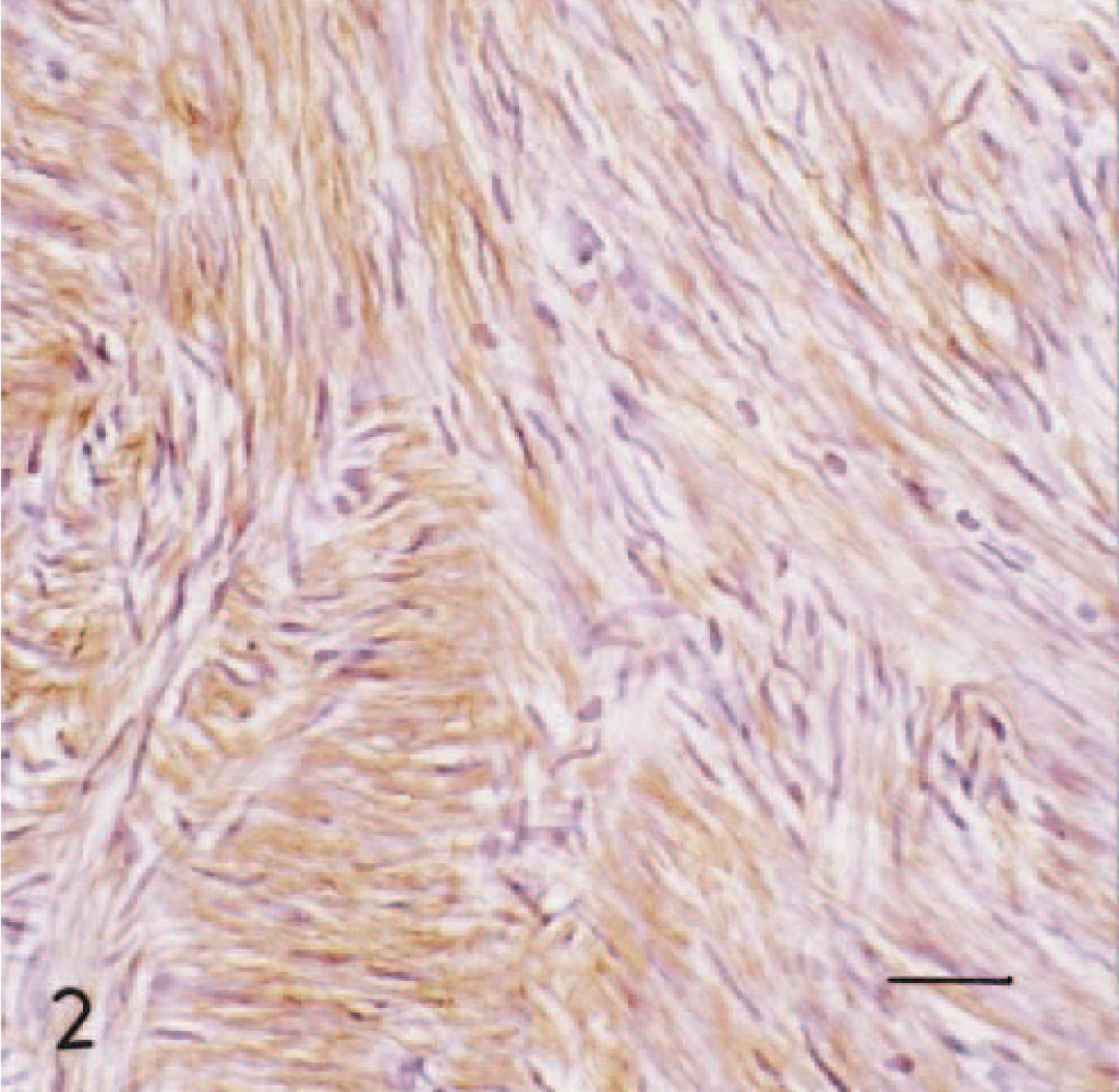
Gray seal females included in the histological study of uterine leiomyomas. The age in years and cause of death; gunshot, drowning caused by entangelment in fishing equipment, disease, or unknown are presented, as well as other macroscopic findings in the uterus. In the ovaries, the presence of corpus luteum and/or albicans is indicated.
∗ CA = corpus albicans; CL = corpus luteum; FE = fishing equipment.
† The age of this seal was estimated at autopsy and was not determined by sectioning of a tooth.
The age of the seals was determined according to the method of Johnston and Watt by examination of the annual growth pattern in cementum zones in decalcified tooth sections. 25
Results
In the gray seals examined, leiomyomas were most commonly found in the wall of the uterine corpus but also were observed in the uterine horns, cervix, and the vagina. They were often multiple and ranged from 10 to 95 mm in diameter. In one seal (#5) there was central necrosis in one leiomyoma. The cut surfaces of the leiomyomas usually appeared whorled and had a white, firm texture, compared with the adjacent myometrium.
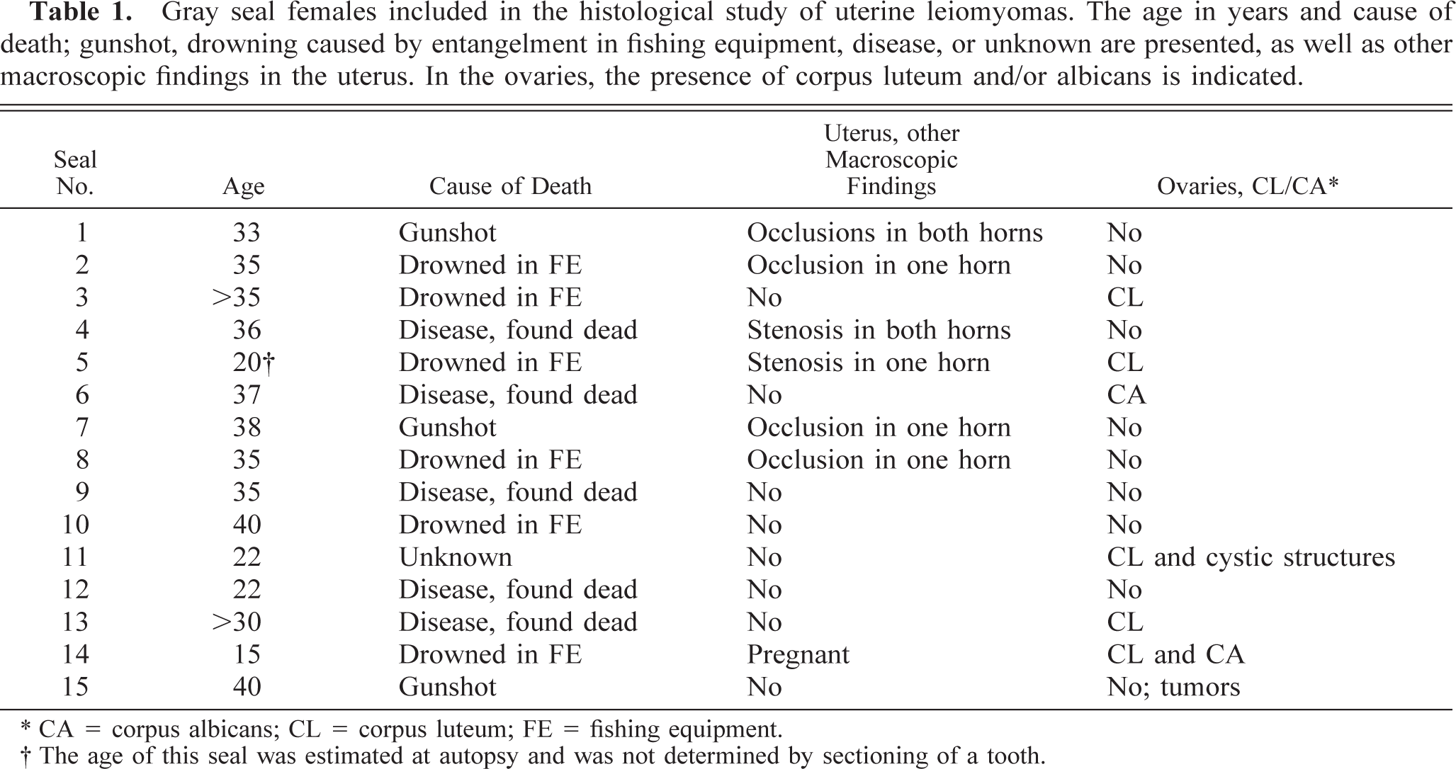
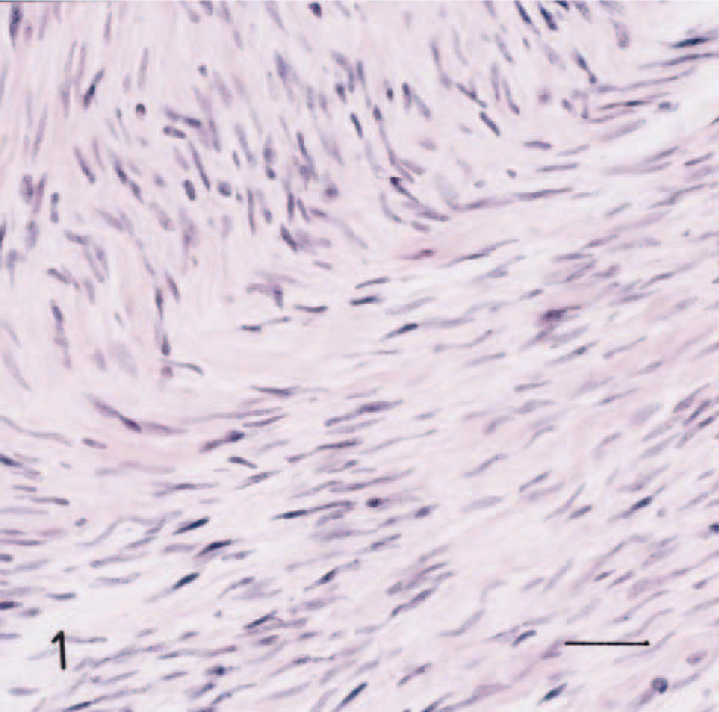
Microscopically, the tumors were formed by interlacing bundles of spindle cells arranged in a whorl-like pattern. The nuclei were rod-like and strikingly uniform in shape and size (Fig. 1). The stroma was well vascularized, and in some areas edema was present. Some areas of hyaline degeneration were also seen (Fig. 1). There was a moderate amount of fibrous connective tissue. Even in areas with high cellularity, atypia was mild or absent, and mitotic figures were rare. Under immunohistochemical analysis, the cytoplasm of the tumor cells showed a positive reaction to antibodies against smooth muscle actin in the three tumors tested. The stroma did not show any reaction (Fig. 2).
Uterine leiomyoma in gray seal no. 5. Section was stained with HE. Hyaline material is seen as pink deposits. Bar = 14 μm.
Uterine leiomyoma in gray seal no. 6. Section was stained with antibodies against smooth muscle actin. The tumor cells showed a cytoplasmic positive reaction, cells were rather uniform with elongated nuclei, and atypia was mild or absent. Paraffin section, avidin–biotin–peroxidase complex method and Mayer's hematoxylin counterstain. Bar = 27μm.
Altogether, 34 seals (64%) had grossly detected uterine leiomyomas. In 22/34 (63%) there were no corpora lutea or albicans in the ovaries as signs of recent reproductive activity. Uterine leiomyomas were not detected macroscopically in 19/53 (36%) seals. Among these seals, 13/19 (68%) had corpora lutea or albicans in the ovaries and 6/19 (32%) did not. In two seals included in the present histologic study, there were pathologic changes in the ovaries. In one seal (#15) there was a tumor-like corpus in each ovary. Histologically, these corpora resembled benign mesenchymal tumors, with closely packed spindle-shaped stromal cells arranged in a storiform pattern. No mitoses were seen. The stroma was hyaline. The microscopic findings are consistent with fibroma. These ovaries were not sufficiently well preserved for further immunohistochemical investigations. In the other seal (#11), several cystic structures were macroscopically noted in one of the ovaries. The ovaries from this seal had not been preserved in formalin. Cystic or tumorlike structures in ovaries were also noted macroscopically in two seals with grossly observed leiomyomas that were excluded from the histologic study.
The causes of death in the gray seals included in the histologic study were drowning, caused by entanglement in fishing equipment (six), gunshot to the head because of disease symptoms (three), disease (five), and unknown (one). The cause of death in one seal was unknown because a complete autopsy was not performed (see Materials and Methods). One of the gray seals (#14) was pregnant, with a fetus of 44.5 cm crown-rump length. In 6 of the 15 histologically examined females, uterine occlusions or stenosis were also present. The stenotic uterine horn formed, in the cranial part, a large dilation, with a narrow center, and in the caudal part, a more or less evident dilation. The occluded horn was sealed by a thin (<1 mm) membrane in the narrow part. There were often accumulations of fluid in the cranial part of the occluded horn.
Altogether, 29 gray seal females were completely autopsied. Macroscopic findings occurring with frequencies of 50% or more in 21 autopsied seals with leiomyoma were cecal ulcer (57%), periodontitis (76%), arteriosclerosis (95%), and adrenocortical hyperplasia (95%). The frequency of these findings in eight seals without leiomyoma were cecal ulcer (62%), periodontitis (25%), arteriosclerosis (62%), and adrenocortical hyperplasia (87%).
Discussion
Uterine leiomyoma is a common tumor of “middle aged” and older dogs but is uncommon in other domestic species. In the dog, these tumors are often associated with ovarian follicular cysts or estrogen-secreting tumors. Cystic or tumorlike structures in ovaries were observed in 4/34 gray seals with leiomyomas. When bitches are castrated early in life, leiomyomas do not develop, and if present in intact bitches, they regress following castration. 26 In humans, uterine leiomyoma is the most common tumor of the female genital tract, occurring in about 30% of women at 35 years of age. 16 Little is known about the etiology and pathogenesis of leiomyoma. However, epidemiologic studies have detected some factors that affect risk: there is increased risk among black women and decreased risk among postmenopausal women, women who have had live births, and women who smoke cigarettes. 42 The occurrence of leiomyomas has also been shown to decrease with increasing number of term pregnancies. 40 It is generally accepted that leiomyomas arise from immature smooth muscle cells in the uterine wall. These tumors appear to be ovarian-steroid dependent, as evidenced by growth during the reproductive years, increase in size during pregnancy or during estrogen therapy, and regression following menopause. 30 , 37 , 39 Histologically, leiomyomas in gray seals resembled those described in humans. 38
As with dogs and humans, uterine leiomyomas in Baltic gray seals seem to be common at middle age. Baltic gray seals at this age often have pathologic changes that have been associated with high tissue concentrations of organochlorines. 6 , 7 , 9 , 20 , 32 Commercial PCB products have experimentally been shown to cause reproductive impairment in several species at certain doses. In mink, PCBs have been reported to induce body weight loss, thymic atrophy, dermatopathy, hepatic lesions, and immunotoxicity. 1 , 2 PCBs also induce fetal death and placental vascular and trophoblastic lesions in the mink. 1 , 3–5 , 24 , 27 , 34 , 35
The breeding season of gray seals in the Baltic starts in the middle of February. Usually these seals give birth to a single pup. Gestation lasts about 350 days, including a period of delayed implantation of about 150 days. 21 Estrus and mating occur 14–18 days after parturition. 12 After ovulation, plasma progesterone concentration increases and remains elevated for most of gestation, including the period of delayed implantation. During the final month of gestation, plasma progesterone increases further and then declines sharply at parturition. 13 , 14 Regression of the corpus luteum and transformation into the corpus albicans starts at parturition, and the corpus has disappeared 1 year after formation, at the latest. 14 Accordingly, reproductively active gray seals have an ovarian corpus and are normally exposed to endogenous progesterone for most of the year. However, 63% of the seals with uterine leiomyoma appeared to be reproductively inactive, as evidenced by absence of ovarian corpora. The reason for the absence of corpora may be interrupted pregnancy, absence of implantation with early involution of the corpus luteum, or absence of ovulation. It is possible that an absence or a shortened exposure-time per year of endogenous progesterone, possibly in combination with exposure to exogenous estrogenic substances, could contribute to development of leiomyoma. In the Baltic gray seal, uterine occlusions and adrenocortical hyperplasia are believed to have contributed in particular to the previously observed decreased reproductive capability. Uterine occlusions may originate from interrupted pregnancies, and adrenocortical hyperplasia may affect both the endocrine and immune systems. Whether uterine leiomyomas in the Baltic gray seals are directly or indirectly associated with organochlorines or with some other factor will be further investigated.
Footnotes
Acknowledgements
We thank Euro Chlor and CEFIC-EMSG in Brussels, Belgium, for financing the investigation.