
Abstract
Rhodococcus equi is an uncommon cause of systemic pyogranulomatous infections in goats with macroscopic similarities to caseous lymphadenitis caused by Corynebacterium pseudotuberculosis. Caprine cases have previously been reported to be caused by avirulent R. equi strains. Six cases of R. equi infection in goats yielding 8 R. equi isolates were identified from 2000 to 2017. Lesions varied from bronchopneumonia, vertebral and humeral osteomyelitis, and subcutaneous abscesses, to disseminated infection involving the lungs, lymph nodes, and multiple visceral organs. Isolates of R. equi from infected goats were analyzed by polymerase chain reaction for R. equi virulence-associated plasmid (vap) genes. Seven of 8 isolates carried the VapN plasmid, originally characterized in bovine isolates, while 1 isolate lacked virulence plasmids and was classified as avirulent. The VapN plasmid has not been described in isolates cultured from goats.
Rhodococcus equi is an aerobic, gram-positive, facultative intracellular coccobacillus frequently isolated from the soil and feces of diseased and healthy animals. 1,12 Although infection can occur in various domestic animals, foals are the only animal commonly affected and typically exhibit chronic pulmonary abscessation. 1 R. equi infection is less commonly reported in swine and cattle, with comparatively rare reports of infection in goats, camelids, and companion animals. 1,3,12 R. equi is also gaining increasing significance as a human pathogen, predominantly in HIV/AIDS patients. 12 In goats, infection is typically disseminated with frequent involvement of the liver, 4,5,7,9,11,14,15 often with concurrent pulmonary abscessation and/or lymphadenitis. 5
The pathogenicity of R. equi depends on its ability to survive within macrophages, a trait encoded by genes carried on virulence plasmids. 10 Pathogenic strains are classified as virulent or intermediately virulent based on the carriage of plasmid genes encoding virulence-associated protein A (VapA) or B (VapB), respectively. 10 Strains that lack the vapA or vapB genes are classified as avirulent. A highly conserved conjugal transfer protein gene, traA, is present in strains that carry a virulence plasmid. 10 A novel R. equi linear virulence plasmid, pVapN, was recently characterized in cattle isolates 16 and identified in a single canine isolate 3 but has not been described in other species. This study reports 6 additional cases of R. equi infection in goats with assessment of virulence-associated plasmids and proteins.
All of the goats (cases 1–6) presented from 2000 to 2017. Table 1 summarizes the clinical and pathologic findings, and complete narratives for each case are given in the supplemental materials. Detailed history was available for 3 of the cases (cases 1, 2, 5), with all goats housed outdoors with other goats. Presenting clinical signs varied and included intermittent pyrexia and lethargy (case 1), chronic respiratory illness (case 3), a subcutaneous nodule on the chest (case 4), lameness (cases 2 and 6), and paralysis (case 5). Fecal examination identified numerous nematode eggs in 3 cases (cases 1, 2, and 6). Radiographs revealed lysis of the left femoral neck (case 2) and right medial humeral epicondyle (case 6) and an alveolar pattern of consolidation involving the cranioventral lung lobes (cases 3 and 6). Abdominal ultrasound was performed on case 1 with identification of a large, cystic mass within the ventral abdomen. Cytologic examination was performed by a veterinary clinical pathologist on a sample of tracheal wash fluid (case 3) and joint fluid (case 6), which revealed a moderate number of neutrophils with fewer macrophages and lymphocytes accompanied by numerous bacterial bacilli in the background.
Signalment, Pathological Changes, and Virulence Plasmid Profile in Goats With Rhodococcus Equi Infections.
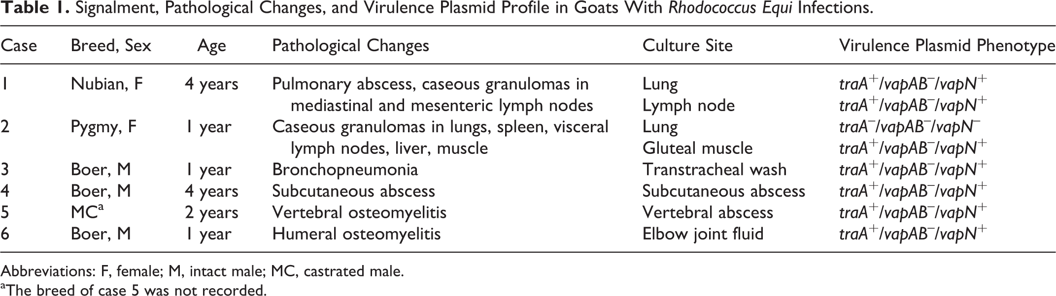
Abbreviations: F, female; M, intact male; MC, castrated male.
aThe breed of case 5 was not recorded.
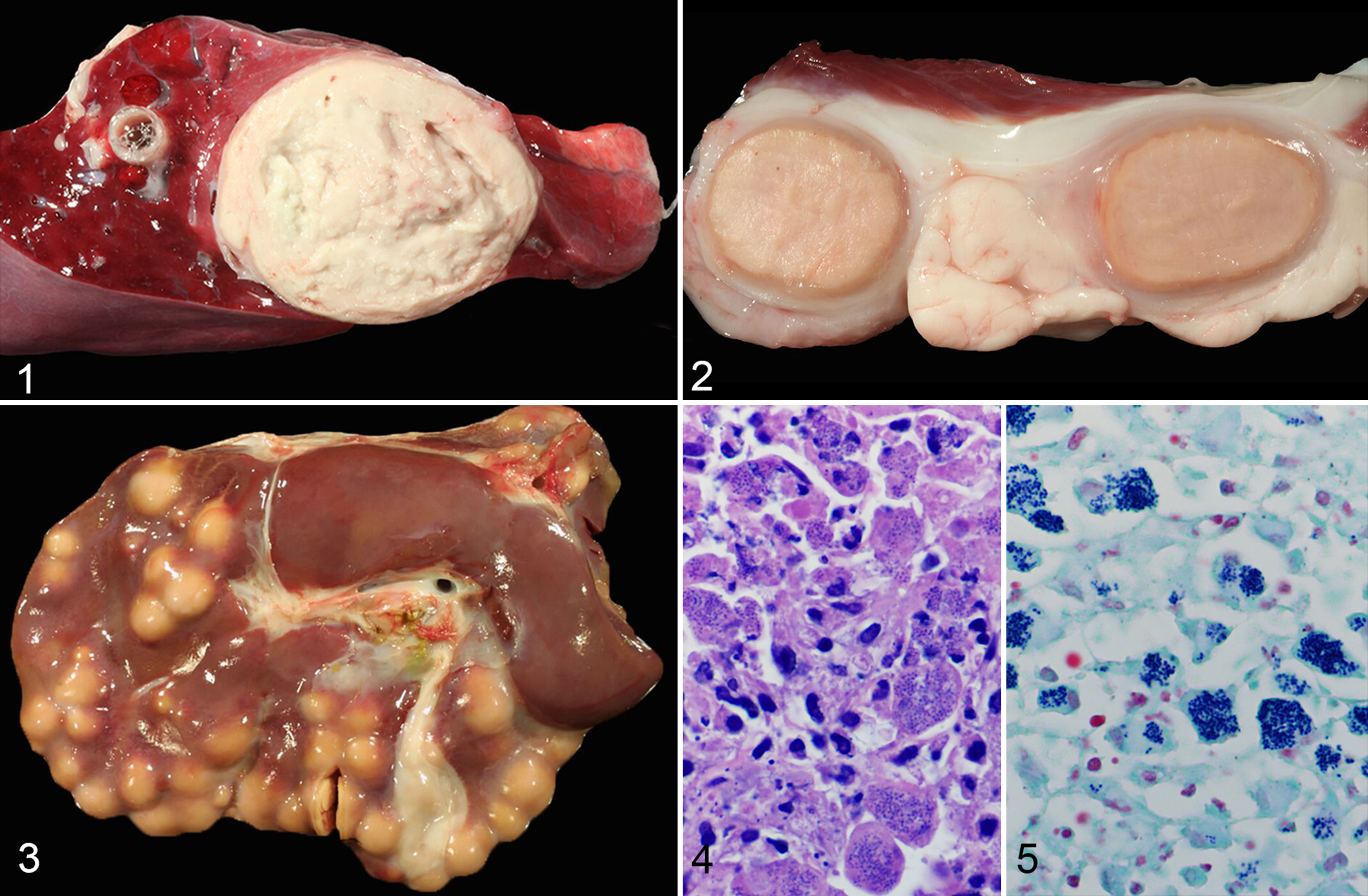
Two of the goats were euthanized and submitted for complete necropsies (cases 1 and 2), and 1 was necropsied by the referring veterinarian with select organs submitted for histopathologic evaluation (case 5). Sections of major organs were fixed in 10% neutral buffered formalin and processed routinely for histopathologic examination by hematoxylin and eosin and Hucker-Twort Gram’s stain. Gross lesions attributable to R. equi infection were variable. Case 1 had a pulmonary abscess (Fig. 1) and caseous granulomas composed of concentric layers of tan, friable material within a mediastinal lymph node and numerous abdominal lymph nodes (Fig. 2). The abdominal lymph nodes were firmly adhered to one another and adjacent abdominal viscera, resulting in the mass effect detected on ultrasound. Gross lesions in case 2 included caseous granulomas within the left gluteal muscles, lungs, spleen, and liver (Fig. 3) and mediastinal and mesenteric lymph nodes as well as ulcerations of the jejunal mucosa. In case 5, a well-demarcated abscess affecting the fifth lumbar vertebral body compressed the spinal cord. Histologically, the lesions in all cases were composed predominantly of numerous foamy macrophages and degenerate neutrophils admixed with necrotic cellular debris and surrounded by dense fibrous connective tissue. In cases 1 and 2, myriad intracytoplasmic, gram-positive coccobacilli were observed within macrophages (Figs. 4 and 5). No bacteria were seen histologically in case 5, and histology was not available for cases 3, 4, and 6.
Aerobic culture (see supplemental material) of the lung and mesenteric lymph node of case 1 and the lung and gluteal muscle of case 2 yielded 4+ and 3+ growth of R. equi, respectively. Unspecified numbers of R. equi were cultured from the tracheal wash fluid of case 3, an aspirate of the subcutaneous nodule in case 4, the vertebral abscess in case 5, and the elbow joint fluid in case 6. Isolates from all cases were evaluated via polymerase chain reaction (PCR) for the R. equi virulence plasmid genes, as previously described: traA, vapA, vapB, 9 and vapN. 3 Isolates from all of the goats were positive for vapN and negative for vapA and vapB by PCR (traA+/vapAB–/vapN+), while the lung isolate from case 2 had the avirulent plasmid phenotype (traA–/vapAB–/vapN–).
Seven R. equi isolates (88%) were vapN positive. Two isolates of R. equi from 1 goat (case 2) exhibited different virulence profiles: one was vapN-positive and the other was considered avirulent as it carried no virulence plasmid genes and was traA-negative. To the authors’ knowledge, this is the first report of pVapN detection in goats. Virulence plasmids are essential for the pathogenicity of R. equi. In horses, a single gene deletion in vapA results in an inability to proliferate in macrophages and survive in vivo in mice. 8 Virulence plasmids also appear to play a key role in host tropism, with a high prevalence of vapA in equine isolates and of vapB in porcine isolates. 10 Until the characterization of the virulence plasmid, pVapN, it was thought that bovine isolates did not carry a virulence plasmid as all isolates were negative for vapA and vapB. 10 However, bovine isolates have since been found to carry the traA gene, and in a recent study, 95% of bovine isolates were positive for pVapN. 16 The VapN plasmid is a linear plasmid that, as with pVapA, is essential for intracellular proliferation of R. equi in macrophages. Plasmid-cured bovine isolates lose the ability to replicate in mouse and human macrophages in vitro and are cleared from the lungs of mice at a much higher rate than the parent strain. 16
R. equi isolates from human cases have been shown to carry pVapA, pVapB, or no virulence plasmids, suggesting that infections in humans are opportunistic and non–host adapted. 12 This observation has also led to some speculation that infection with R. equi might be zoonotic, with infection potentially resulting from exposure to farming environments or contaminated meat. 16,17 This hypothesis is further supported by the higher proportion of traA + A – B + “pig-type” and traA + AB – “bovine-type” isolates in human infection in comparison to traA+A+B– “equine type” and the frequency—as high as 26%—with which R. equi can be recovered from lymph nodes of apparently healthy swine at slaughter. 17 Interestingly, the presence of pVapN has never been documented in human-derived isolates of R. equi, and it is possible that some previously documented traA + AB – “bovine-type” isolates were pVapN-positive. Human cases of R. equi infection predominantly occur in HIV/AIDS patients and in those undergoing immunosuppressive therapy. 12 Dogs appear to follow a similar pattern, with one recent article describing 5 cases of R. equi infection in dogs, 4 of which showed evidence of immunosuppression in the form of severe endocrinopathy, autoimmune disease, or treatment with immunosuppressive medications. 3
The rarity of reported cases of R. equi infection in goats and other domestic species apart from horses indicates that most animals are relatively resistant to infection and that predisposing factors such as stress, illness, or immunosuppression may be required for infection to occur. 12 Proposed underlying factors include young age, coccidiosis, and pregnancy. 5,15 In the goats of the present report, conditions that could potentially alter immune status included hemonchosis (cases 1, 2, 6) and severe copper deficiency (case 5). Previous investigation into virulence plasmids carried by R. equi isolates from goats has generally demonstrated a lack of pVapA and pVapB, although pVapA has been identified in a single goat isolate. 15 In the present report, most of the R. equi isolates were positive for pVapN, with 1 strain classified as avirulent. The lack of pVapN carriage in the lung isolate of case 2 may represent spontaneous loss of the plasmid or mixed genotype R. equi infection, as has been reported in a foal. 2 This suggests that infection in goats can be associated with virulent and avirulent strains, and the diversity of virulence phenotypes present in R. equi strains from goats supports their role as an opportunistic pathogen in this species.
Pneumonia is the most common reason for presentation in infected foals, with some exhibiting dissemination to the colon, mesenteric lymph nodes, and other sites. 13 In cattle, R. equi is associated with granulomas in respiratory lymph nodes, 16 while most infections in goats are disseminated with frequent involvement of the liver, 4,5,7,9,11,14,15 often with concurrent pulmonary abscessation and/or lymphadenitis. 5 The extensive involvement of the liver in case 2 suggests spread via the portal circulation from the gastrointestinal tract with hematogenous dissemination to other organs. However, the involvement of the lungs in case 1 with no lesions in the liver or gastrointestinal tract suggests a respiratory route of infection with subsequent hematogenous dissemination. Case 4 likely represents wound infection, a less common but frequently reported route of entry of R. equi in multiple domestic species. 12 The routes of entry for cases 4, 5, and 6 are unclear as full necropsies were not performed. In cases 1 and 2, Corynebacterium pseudotuberculosis was originally suspected as the cause of the disseminated caseous granulomas, highlighting the importance of culture in distinguishing this more frequent infection from disseminated R. equi infection. This distinction can have great significance for a herd as caseous lymphadenitis, caused by C. pseudotuberculosis, is highly contagious and can result in severe economic losses resulting from decreased production efficiency and carcass condemnation. 6
All of the goats were infected by an R. equi isolate that carried the VapN-associated plasmid, while an additional isolate from 1 goat was classified as avirulent. This is the first report of pVapN in R. equi isolates from goats, and it indicates that infection in goats is not always associated with an avirulent phenotype, as previously thought. The rarity of reported cases of R. equi infection in goats suggests that R. equi is an opportunistic pathogen with possible predisposing factors for infection. The importance of the VapN plasmid in human infections has yet to be investigated.
Footnotes
Acknowledgements
The authors thank the technical staff of the Texas A&M Veterinary Medical Teaching Hospital and the Texas Veterinary Medical Diagnostic Laboratory Clinical Microbiology Laboratories for their efforts to culture and preserve the bacterial isolates used in this study. The authors also thank Emily Stranahan for her assistance in assembling the figure.
Declaration of Conflicting Interests
The authors declared no potential conflicts of interest with respect to the research, authorship, and/or publication of this article.
Funding
The authors received no financial support for the research, authorship, and/or publication of this article.
Supplementary material for this article is available online.
References
Supplementary Material
Please find the following supplemental material available below.
For Open Access articles published under a Creative Commons License, all supplemental material carries the same license as the article it is associated with.
For non-Open Access articles published, all supplemental material carries a non-exclusive license, and permission requests for re-use of supplemental material or any part of supplemental material shall be sent directly to the copyright owner as specified in the copyright notice associated with the article.