
Abstract
A 6-year-old Nubian goat with a history of progressive weight loss and cough was presented for necropsy. The goat tested negative for antibodies to caseous lymphadenitis and caprine arthritis and encephalitis by hemagglutination inhibition assay and enzyme-linked immunosorbent assay, respectively. Postmortem examination revealed marked enlargement and, with histopathology, a fibrinopurulent necrotizing lymphadenitis of a tracheobronchial lymph node, with an appearance similar to that reported in cases of caseous lymphadenitis. An organism characterized by molecular methods as Actinomyces hyovaginalis was isolated together with Staphylococcus spp. and Streptococcus spp. from the lesion. No Corynebacterium pseudotuberculosis was recovered. To the authors' knowledge, this is the first isolation of A. hyovaginalis from a goat. Although the exact contribution of A. hyovaginalis to the lesion remains to be established, this case demonstrates that A. hyovaginalis should be considered in cases of caseous lymphadenitis–type lesions, especially when C. pseudotuberculosis has been excluded.
In small ruminants, enlargement of the internal and superficial lymph nodes has been commonly reported with Corynebacterium pseudotuberculosis infection, which causes the disease caseous lymphadenitis (CLA). Although CLA is not a federally reportable disease, owners often choose to cull affected animals because the disease causes severe and progressive ill thrift and is highly contagious (Canadian Department of Justice, 1990: Reportable Diseases Regulations (SOR/91-2). In: Health of Animals Act. Available at: http://laws.justice.gc.ca/en/showtdm/cr/SOR-91-2//?showtoc=&instrumentnumber=SOR-91-2. Accessed October 3, 2008). Because of the economic impact of this disease, accurate diagnosis is important. In the present report, a case of CLA-like disease in a goat from which Actinomyces hyovaginalis was isolated is described. This organism is uncommonly found in species other than pigs, and, until now, it has not been identified in goats.
A 6-year-old female Nubian, nonpregnant goat was presented to Tufts Ambulatory Service (Woodstock, CT) for evaluation. The goat was one of a herd of 11 pet Nubian goats; none of the other goats had evidence of disease. The animal had lost weight over a 3-month period and was severely emaciated despite having a good appetite and adequate nutrition. During this period, the animal had a history of cough. A blood sample was also submitted for competitive enzyme-linked immunosorbent assay antibody detection for caprine arthritis encephalitis (CAE) and a serum hemagglutinin inhibition assay for caseous lymphadenitis. Both tests were negative.
The animal's condition deteriorated and it died. The goat was necropsied at the Connecticut Veterinary Medical Diagnostic Laboratory (Storrs, CT) 2 months after initial examination. On gross examination, the animal was thin. One of the tracheobronchial lymph nodes was significantly enlarged (17 cm X 13 cm X 15 cm) and was compressing the right cranial lung lobe. Its wall varied in thickness from 1–3 cm. On cut section, there was a large amount of flocculent yellow material, green-brown fluid, and soft tan material (fibrin) within the lymph node (Fig. 1). The outer wall of the node adhered to the pericardial sac and trachea, and the heart was displaced caudally and to the left.
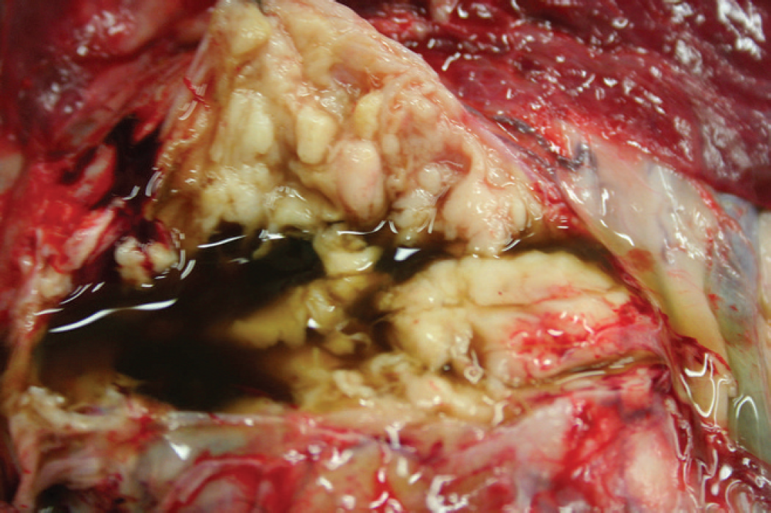
Enlarged tracheobronchial lymph node with tan to yellow caseous material, green-brown fluid, and fibrin stands. Fibrin was more copious in other areas.
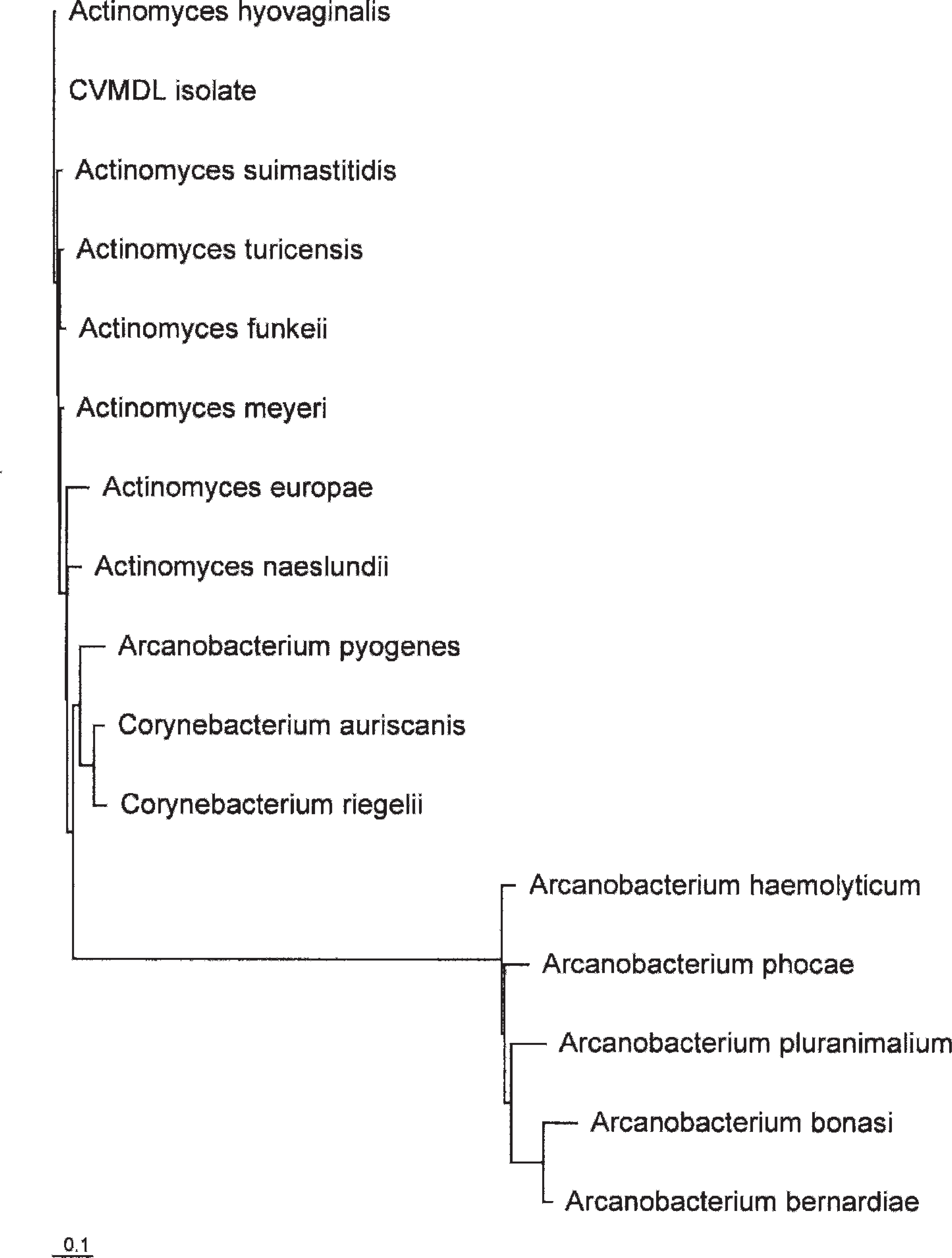
Neighbor-joining phylogenetic analysis of 16S ribosomal DNA from Actinomyces, Corynebacterium, and Arcanobacterium bacteria and from the Connecticut Veterinary Medical Diagnostic Laboratory (CVMDL) isolate biochemically identified as a Corynebacterium-like species. Multiple-sequence alignments were done with ClustalW 8 (BioEdit version 7.0.0 e ).
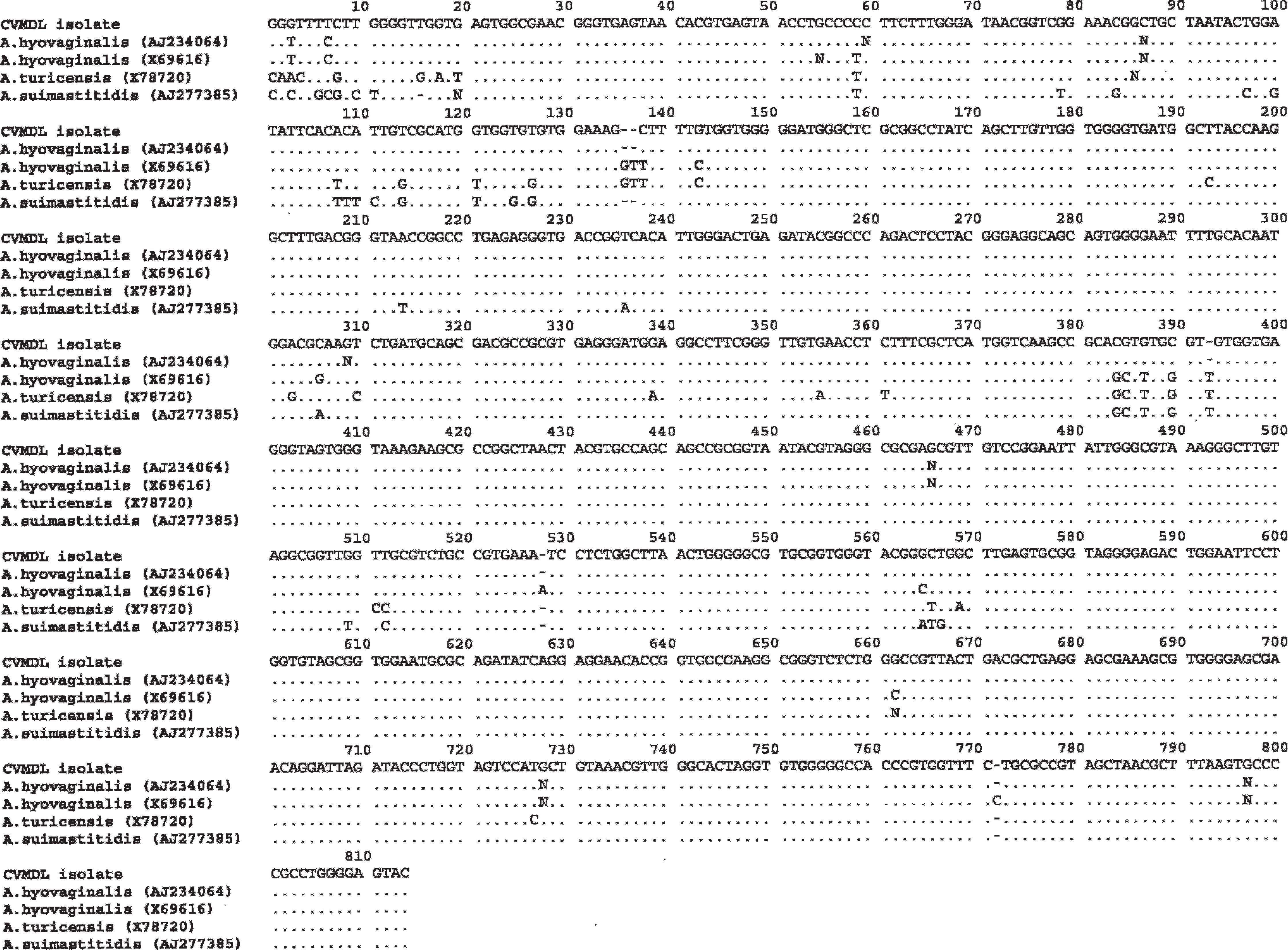
Multiple-sequence alignment comparison of 810 nucleotides of the 16S ribosomal DNA of the Actinomyces hyovaginalis–like organism Connecticut Veterinary Medical Diagnostic Laboratory (CVMDL) isolate with A. hyovaginalis (AJ234064), Actinomyces turicencis (X78720), and Actinomyces suimastitidis (AJ277385). The GenBank accession numbers are indicated in parentheses. Sequence alignment was performed with ClustalW 6,8 (BioEdit version 7.0.9.0 e ).
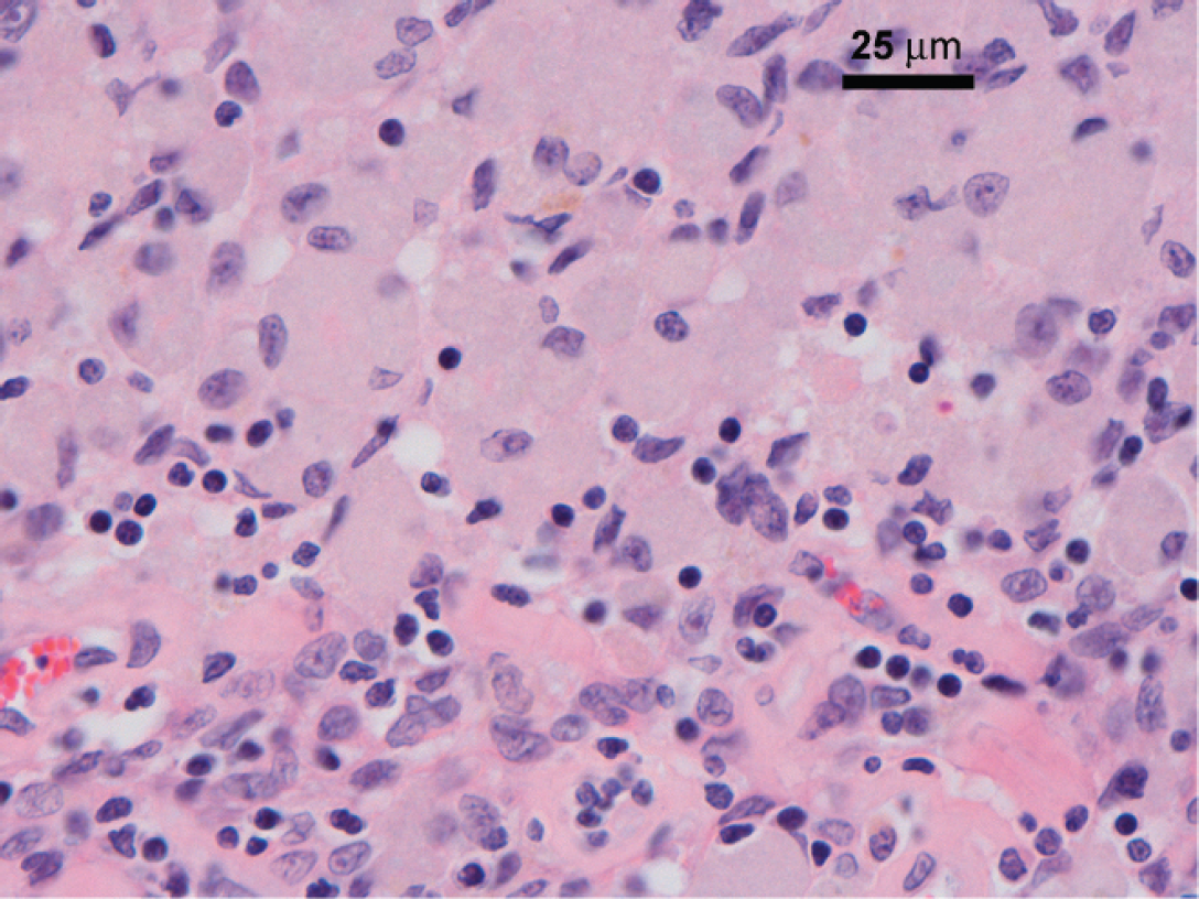
Tracheobronchial lymph node, Nubian goat. There is effacement of normal nodal structure by sheets of macrophages. Hematoxylin and eosin. Bar = 25 μm.
Based on gross lesions, CLA was suspected. The internal surface of the lymph node was sliced with a sterile blade and swabbed with a sterile cotton-tipped applicator stick, which was streaked onto 5% blood agar, incubated at 37°C under microaerophilic conditions, and examined after 24 and 48 hr. Identification of the organisms recovered was based on cell and colonial morphology, growth characteristics, and biochemical tests. Culture yielded a mixed culture of Streptococcus sp., Staphylococcus sp., and small colonies with morphology similar to C. pseudotuberculosis (i.e., creamy white, dry, waxy colonies). However, the colonies were dissimilar to C. pseudotuberculosis in that they were small pinpoint colonies, as opposed to the medium-sized colonies of C. pseudotuberculosis. 10,14 Biochemical testing revealed that this particular organism was catalase negative, which is inconsistent with C. pseudotuberculosis. A Biolog microbial identification system a assay yielded inconclusive results.
Colonies of this organism were resuspended in bacterial RLT (guanidine isothiocyanate) b lysis buffer containing 1 % vol/vol of 2-mercaptoethanol, c and DNA was extracted with a commercial kit b according to the manufacturer's instructions. Eubacterial 16S ribosomal RNA (rRNA) gene was polymerase chain reaction (PCR) amplified using the universal primer set fD1: 5′-AGAGTTTGATCCTGGCTCAG-3′/rD1: 5′-AAGGAGGTGATCCAGCC-3′. 17 Amplified DNA was resolved in a 1% wt/vol agarose gel, and a band of the expected size of 1,500 bp was purified and sequenced with primers fD1 and rD1. Sequence data were assembled with Sequencher d and compared with those in the GenBank nucleotide collection database c using BLAST (http://www.ncbi.nlm.nih.gov/blast/Blast.cgi). The sequence revealed 99% identity to A. hyovaginalis. Phyloge-netic analysis of Connecticut Veterinary Medical Diagnostic Laboratory (CVMDL) isolate with 16S ribosomal DNA of Actinomyces, Corynebacterium, and Arcanobacterium species (Fig. 2) further confirmed the observed similarity with A. hyovaginalis. Multiple sequence alignments (Fig. 3) comparing the CVMDL isolate with closest Actinomyces organisms revealed 99% identity with A. hyovaginalis (AJ234064), 97% identity with A. hyovaginalis (X69616), and 95% identity with A. turicensis (X78720) and A. suimastitidis (AJ277385).
Multiple tissues, including the affected tracheobronchial lymph node and adjacent lung, were fixed in 10% neutral buffered formalin. The affected tissues were processed routinely for histologic examination and stained with hematoxylin and eosin, Brown and Brenn's modified Gram stain, Fites stain, Masson's trichrome stain, phosphotungstic acid hematoxylin stain, and Ziehl–Neelsen acid-fast stain.
Histologically, the tracheobronchial lymph node had a thick fibrous capsule, with thick fibrous connective tissue septae extending from the capsule. The normal nodal structure was effaced by sheets of macrophages, lymphocytes, and collagen (Fig. 4). Fibrinous deposits were sometimes present adjacent to the necrotic area. No bacteria were detected in the examined sections with any of the stains, although only the outer wall of the lesion was examined histologically. These histologic lesions were consistent with those described for CLA. 7 Other histologic findings included pulmonary interstitial fibrosis and periportal and subcapsular fibrosis in the liver.
In the distal trachea and main stem bronchi, there was loss of cilia and a moderate lymphocytic plasmacytic infiltrate subepithelially and in periglandular tissue. There was mild hemorrhage in the lamina propria and the submucosa was very edematous with a few scattered but moderately dense infiltrates of neutrophils. There were no inflammatory infiltrates in the gastrointestinal tract indicative of Johne's disease or other causes of chronic weight loss. Corynebacterium pseudotuberculosis is a commonly reported cause of lymphadenitis in small ruminants. 5 Corynebacterium pseudotuberculosis is a Gram-positive bacterium that causes chronic, purulent infections in a number of other species. Other causes of lymph node enlargement in small ruminants include other bacterial infections, CAE, and lymphosarcoma. In goats, the disease melioidosis or pseudoglanders, caused by Burkholderia pseudomallei, may have a similar presentation 16 or, as described in the present report, A. hyovaginalis may be involved in producing similar lesions.
Actinomyces hyovaginalis is an actinomycete bacterium. Actinomycetes are Gram-positive, polymorphic, non–acid-fast, slow-growing bacteria that cause pyogranulomatous lesions. Actinomyces hyovaginalis was first described as a novel organism in 1993 4 and has been isolated mainly from pigs, in particular from purulent lung lesions, vaginal discharge, and nasal conchae. 2,9,13,15 It is an important cause of disseminated necrotic lung lesions in slaughtered pigs and is believed to be acquired via inhalation. 1 In swine, A. hyovaginalis causes well-circumscribed lesions with a core of necrotic debris and bacteria, surrounded by an inner zone of neutrophils and a broad outer zone of macrophages surrounded by fibroplasia. 1,11 In infections with A. hyovaginalis in pigs, as well as other actinomycosis, clubs may form around the bacterial clumps. 12 No clubs were seen in the present case. The source of A. hyovaginalis infection in swine is unknown, although Actinomyces species relevant to human and bovine disease are commensals of the oral cavity of healthy carriers. Actinomyces hyovaginalis has previously been isolated in mixed culture from a chest wall abscess in a sheep, 3 but to the authors' knowledge, this infection has not been reported in goats. The current case suggests that A. hyovaginalis should be considered in cases of caseous lymphadenitis–type lesions, especially when C. pseudotuberculosis has been excluded. In the present case, A. hyovaginalis was found in mixed culture with Streptococcus sp. and Staphylococcus spp. The pathogenesis of the lesion may have involved interactions between the various bacterial species recovered.