
Abstract
Many previously unrecognized fungi are emerging as potential pathogens. One such group is dematiaceous fungi of the Chaetomiaceae family (phylum Ascomycota, class Sordariomycetes). These fungi are rare causes of opportunistic, neurotropic phaeohyphomycosis in humans but are not known to cause similar infections in animals. The aims of this study were to investigate equine hyphal mycotic encephalitis, characterize key histopathologic features, and classify causative organisms with molecular diagnostic techniques. Seven cases were evaluated by histopathology. Panfungal PCR targeting the ribosomal RNA large subunit coding region and the noncoding internal transcribed spacer-2 region was performed on DNA extracted from formalin-fixed, paraffin-embedded sections of affected brain, and the resulting sequences were queried against published fungal genomes. Affected animals ranged from 8 to 22 years of age and presented with neurologic signs. Macroscopic lesions within affected brains included multifocal hemorrhage, focal swelling of the thalamus with red and yellow discoloration, and focal cerebral malacia. Major histologic findings included multifocal discrete foci of necrosis, neutrophilic to granulomatous inflammation, vasculitis, and intralesional fungal hyphae variably affecting the cerebrum, thalamus, and brainstem. DNA sequences in 4 cases showed > 98% homology with species within the Chaetomiaceae family, including Acrophialophora fusispora, Acrophialophora levis, and Chaetomium strumarium. Histomorphologically, Chaetomiaceae fungi were 7 to 10 μm wide, septate, parallel walled, and nonpigmented, with dichotomous branching in affected horses. This case series is the first report of equine mycotic encephalitis caused by members of the Chaetomiaceae family, previously reported as rare emerging pathogens in humans.
Fungi are a rare cause of encephalitis in horses. Reported equine mycotic encephalitis cases include 4 reports of aspergillosis, 17,18,24 1 of which had a concurrent Mucor sp. infection, 38 and 1 report of cryptococcosis. 39 Across species, Cryptococcus spp. and Aspergillus spp. are the most common causes of nervous system mycosis. 16,19,34 Other less common causes include opportunistic pathogens such as Fusarium spp., Mucor spp., Rhizopus spp., and pigmented fungi causing phaeohyphomycoses. 34 Infrequently, horses fed moldy corn containing the mycotoxin fumonisin, produced by Fusarium moniliforme, as well as other Fusarium spp., can develop leucoencephalomalacia. 23 Fungal organisms, however, are not observed in affected cases, as these lesions are produced by microcirculatory damage from the mycotoxin, fumonisin B1. 23
Phaeohyphomycosis is an infection caused by dematiaceous (pigmented) fungi, which in culture produce pigmented molds and in tissue produce variably pigmented fungal elements. 8 These fungi are a heterogeneous group composed of more than 100 species and 60 different genera, 30 and although they are generally considered to be opportunistic pathogens, increasing evidence suggests that some may be true pathogens. 30,34,35 Melanin and melanin-like products confer the pigmentation of dematiaceous fungi, and melanin protects the fungal organisms against environmental pressures and host immune response and contributes to the pathogenicity of these fungi. 6,10,11,30,35 Phaeohyphomycosis has been observed in humans and across veterinary species and most commonly affects immunocompromised individuals. 7,28,29,30,35 In humans, the most commonly reported neurotropic pigmented fungi are Cladophialophora bantiana, Ramichloridium mackenziei, and Ochroconis gallopavum, 30,32 but other species are emerging as rare opportunistic pathogens. 30 –32 Similarly, phaeohyphomycosis in animals is most commonly reported to be caused by Cladophialophora bantiana, Ochroconis spp., and Alternaria alternata, and infections involving the central nervous system exhibit similarities to human cerebral phaeohyphomycosis. 35
The Chaetomiaceae family is one such group of opportunistic fungi. These dematiaceous, saprophytic, and coprophytic fungi are commonly found in plant debris and play a significant role in plant degradation. 16 Whereas these fungi most commonly cause cutaneous infections and onychomycosis, 15,36 Chaetomium strumarium, Chaetomium perlucidum, Chaetomium atrobrunneum, Acrophialophora levis, and Acrophialophora fusispora are reported to cause cerebral abscesses in immunocompromised humans. 1 –5,14,16,22 These cerebral infections are markedly neurotropic and have a high mortality rate. These fungi have yet to be recognized as neurotropic pathogens in veterinary species.
Although mycotic infections are a rare cause of encephalitis in horses, the advent of new molecular techniques is resulting in the identification of many previously unrecognized pathogens. The objective of this study was to histologically characterize cases of equine mycotic encephalitis caused by hyphal fungi and to classify the causative organisms using DNA sequencing and phylogenetic analysis.
Materials and Methods
Case Selection and Histopathology
Four equine cases of encephalitis caused by fungal hyphae were identified at Texas A&M Veterinary Medical Diagnostic Laboratory in 2014 and 2015. To find additional cases, databases were searched at Texas A&M University and Oklahoma State University. Histologic sections were examined. Central nervous system lesions were characterized on hematoxylin and eosin (HE) stain, Grocott’s methenamine silver stain with light green counterstain, and Fontana-Masson stain.
Molecular Analysis
Samples were analyzed by polymerase chain reaction (PCR) and sequence analysis of the internal transcribed spacer (ITS) region and ribosomal RNA large subunit (LSU) coding region. The methods used for the ITS PCR have been previously published. 25 Similar methods were also followed for the LSU PCR. Briefly, 50-μm-thick sections were obtained from paraffin blocks. Total DNA was extracted using the Mo Bio BiOstic Formalin-Fixed Paraffin-Embedded (FFPE) DNA Isolation Kit (Qiagen), and fungal DNA was amplified by PCR targeting the ITS region using ITS3 (5’-GCATCGATGAAGAACGCAGC-3’) and ITS4 (5’-TCCTCCGCTTATTGATATGC-3’) primers. 25,41 PCR targeting the LSU (28 S) was performed using LR0 R (5’-ACCCGCTGAACTTAAGC-3’) and LR5F (5’-TCCTGAGGAAACTTCG-3’) primers. 33 All PCR products were separated on a 2% agarose gel and bands were excised and purified using the EZNA kit (OMEGA). 25 DNA sequences were queried against the GenBank database (NCBI) using the basic local alignment search tool (BLAST). Sequences were assessed quantitatively with a quality score given in Sequencher® and qualitatively by inspection of the electropherogram. Sequences with a quality score of less than 70% and those with a majority of overlapping base peaks were classified as poor quality and remained phylogenetically unclassified. 25 Fungal classification was determined for a match greater than 98% identity. For those sequences matching more than 1 organism, the organism with the highest score was chosen. If the sequences had equal identity, both were reported.
For LSU neighbor-joining analyses, a reference database was constructed using LSU sequences for members of the Chaetomiaceae family, order Sordariales (Chaetomium globosum JX280688, Chaetomium subfimeti FJ666354, Chaetomium fimeti FJ666358, A. levis KM995849, A. fusispora KM995871, Acrophialophora nainiana KM995865, Chaetomium malaysiense AF286409, Chaetomium seminudum AF286410, Thielavia subthermophila JX280760). 33 Closely related genera from the order Sordariales (Madurella pseudomycetomatis JX280752, Madurella fahalii JX280751, Madurella mycetomatis JX280743, Neurospora terricola FR774282) were also included in the database, as well as an outgroup from the Sphaeriales order (Sordaria fimicola AY681160). The database was then combined with the LSU sequences for case Nos. 1, 2, and 3. The unformatted FASTA sequences were aligned using MUSCLE. 9 Neighbor-joining analysis using the Tamura-Nei model 37 in Molecular Evolutionary Genetics Analysis version 7.0 20 was performed on the sequence alignment with a bootstrapping of 1000 replications.
Results
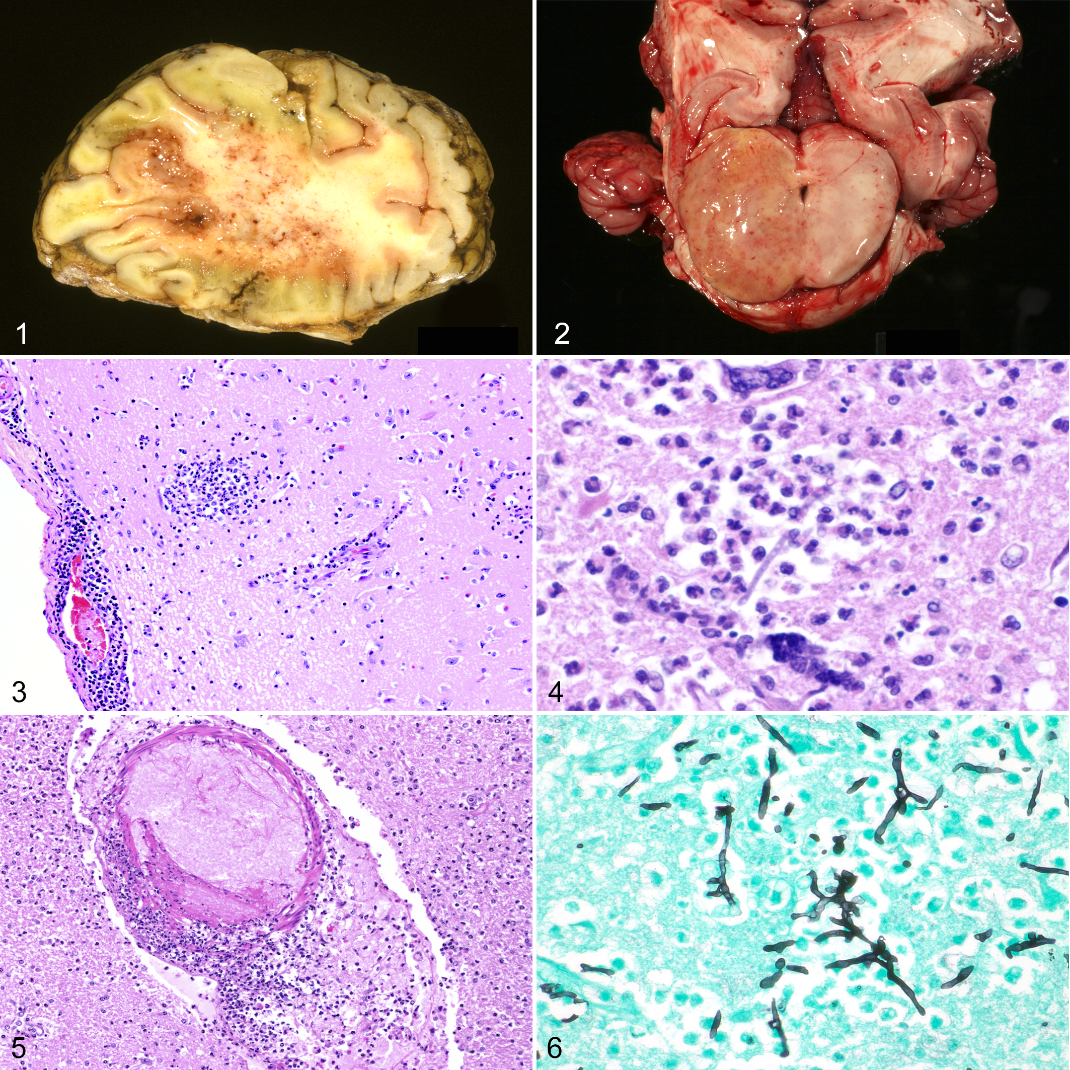
Seven cases were identified at the 3 institutions, including 6 cases with a complete necropsy and 1 case with only the brain submitted for diagnostic testing. A summary of the clinical histories, macroscopic lesions, and additional testing is provided in Table 1. Six of the 7 cases were from Texas, and 1 was from Oklahoma. Six cases were adults between 8 and 22 years of age, and 1 was a foal. The neurologic signs were variable and typically progressive. In the 6 cases in which necropsies were performed, 3 had lesions isolated to the brain and 3 had systemic infections. In case No. 7, Aspergillus flavus was cultured from the lung; in case No. 4, an unidentified saprophyte colony was cultured from the brain; and in case No. 1, branching hyphae were observed on cytology of the lung. Two of the cases had known underlying immunosuppression or concurrent infections. Case No. 1 aborted 1 week prior to the onset of clinical signs for unknown reasons, and Streptococcus zooepidemicus, Enterobacter sp., and Klebsiella pneumoniae were cultured from the lungs. Case No. 5 was a 3-week-old foal with a history of maladjustment syndrome, surgical correction of a patent urachus, and bronchopneumonia. The route of infection could not be definitively determined in these cases.
Clinical and Laboratory Findings in 7 Horses With Mycotic Encephalitis.
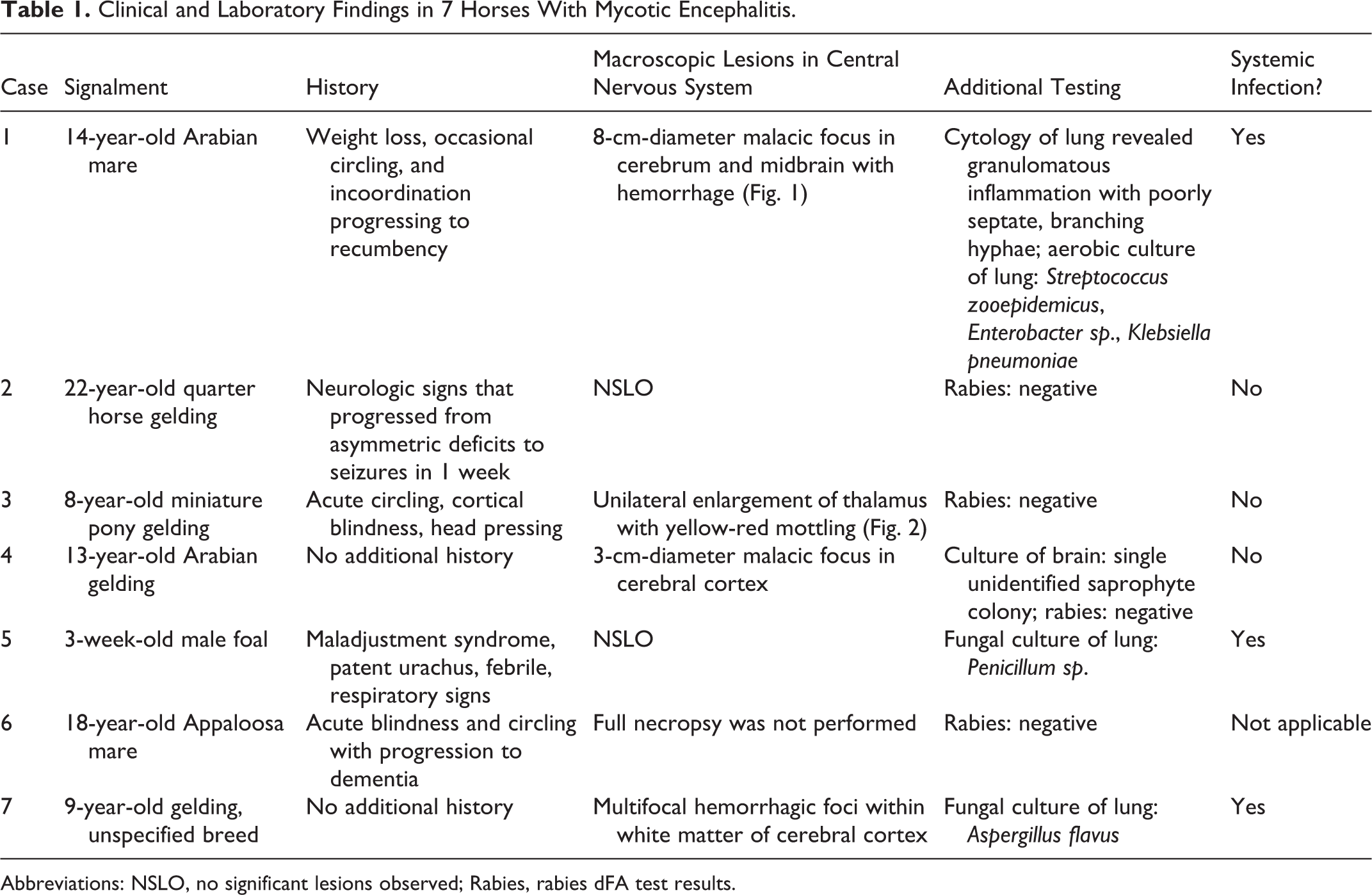
Abbreviations: NSLO, no significant lesions observed; Rabies, rabies dFA test results.
Central Nervous System Histopathology
Histologically, the inflammatory lesion was similar in all cases, characterized by varying degrees of pallor and rarefaction (edema and necrosis) (7/7) with multifocal accumulations of neutrophils, macrophages, multinucleated giant cells, and fewer lymphocytes (7/7) within both the gray and white matter, forming suppurative to pyogranulomatous foci within the parenchyma (Figs. 3 and 4). Inflammatory infiltrates expanded Virchow-Robbins’ space and extended into the parenchyma (7/7). Vasculitis was often prominent with vascular walls obscured by neutrophils, macrophages, necrotic cellular debris, serum protein, and fibrin (7/7) (Fig. 5). Neuronal necrosis was frequent in affected gray matter (5/7; case Nos. 1–4 and 6). In case No. 7, scattered dilated myelin sheaths were within affected white matter. Multifocal hemorrhage ranged from mild (case Nos. 4 and 5) to moderate (case No. 2) to severe (case Nos. 1, 3, 6, and 7) within the areas of inflammation. Marked gliosis with increased numbers of astrocytes and microglia was within the affected parenchyma (7/7). In case Nos. 1, 2, and 3, similar inflammatory infiltrates extended into the meninges. Affected areas of the brain included the cerebrum (6/7; case Nos. 1–3 and 5–7), thalamus (2/7; case Nos. 3 and 4), hippocampus (1/7; case No. 6), and pons (1/7; case No. 5). Inflammatory cells extended into the lateral ventricle in 1 case (case No. 6). Fungal hyphae were often observed in HE-stained sections with variable ease in 6 of 7 cases (case Nos. 1–3 and 5–7) and were visible in all cases with Grocott’s methenamine silver stain within the inflammatory infiltrates, often penetrating vessel walls and infiltrating the adjacent parenchyma (Fig. 6). The hyphae in all cases were septate and nonpigmented and displayed dichotomous branching, although branching was rare in case No. 7. Hyphae were 7 to 10 μm wide in 5 of 7 cases (case Nos. 1–4 and 6), whereas they were 10 to 14 μm wide in case Nos. 5 and 7. Hyphae were parallel walled in all cases except case No. 5. None of the fungal hyphae stained with Fontana-Masson. The number of fungal hyphae was low in 3 of 7 cases (case Nos. 4–6), moderate in 2 of 7 cases (case Nos. 2 and 7), and high in 2 of 7 cases (case Nos. 1 and 3).
ITS-2 and LSU PCR and Sequencing
Of the 7 cases, all were ITS-2 PCR positive, and none were negative. The ITS PCR positive cases yielded a single clear band in 5 of 7 cases, and 2 of 7 cases had multiple bands. The average length of PCR product was around 350 base pairs. Good quality DNA sequences were obtained from 4 of the 7 samples with single strong bands, and none having multiple bands. Poor overall quality DNA sequences were not queried against the NCBI database and were excluded from further evaluation (case Nos. 5–7). Good overall quality DNA sequences from case Nos. 1–4 matched the following published fungal genomes in the NCBI database with the corresponding homology: A. fusispora (99% homology, case No. 1), A. levis and Paecilomyces sp. (100% homology, case No. 2), Chaetomium sp. (99% homology, case No. 3), and C. strumarium (100% homology, case No. 4) (Table 2). Of the 7 cases, 4 were LSU PCR positive, and 3 were negative, and all positive cases yielded a single clear band. The average length of PCR products was 900 base pairs. Good quality DNA sequences were obtained from 3 of the 4 positive cases. Poor overall quality DNA sequences were not queried against the NCBI database and were excluded from further evaluation (case No. 7). Good overall quality DNA sequences matched the following published fungal genomes in the NCBI database with the corresponding homology and GenBank accession number: A. fusispora (100% homology, KM995873.1, case No. 1), A. levis (100% homology, KM995862.1, case No. 2), Chaetomium sp. (99% homology, KP336837.1, JX280739.1, JX280710.1, JX280700.1, KP970647.1, DQ368628.1, JX280736.1, case No. 3) (Table 2).
Histopathologic Findings and Results of Fungal DNA Sequencing in 7 Horses With Mycotic Encephalitis.a
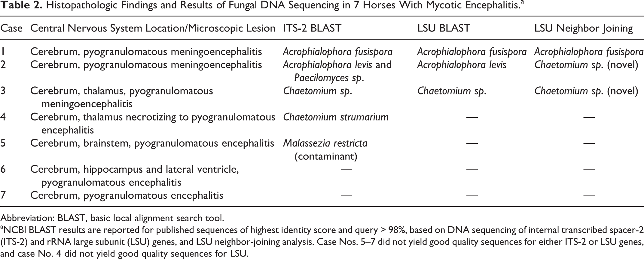
Abbreviation: BLAST, basic local alignment search tool.
aNCBI BLAST results are reported for published sequences of highest identity score and query > 98%, based on DNA sequencing of internal transcribed spacer-2 (ITS-2) and rRNA large subunit (LSU) genes, and LSU neighbor-joining analysis. Case Nos. 5–7 did not yield good quality sequences for either ITS-2 or LSU genes, and case No. 4 did not yield good quality sequences for LSU.
LSU Neighbor-Joining Analysis
LSU sequences from case Nos. 1, 2, and 3 were phylogenetically placed within the Chaetomiaceae family using neighbor-joining analysis (Fig. 7). Case No. 1 clustered with A. fusispora and A. nainiana (reclassified as A. fusispora2), which corroborated the ITS-2 and LSU BLAST results for this case. Case Nos. 2 and 3 appeared to represent a potentially novel species of Chaetomium as they clustered separately from all other Chaetomium and Acrophialophora species with a bootstrap support value of 100. The ITS-2 BLAST, LSU BLAST, and LSU neighbor-joining results were consistent for case No. 3; however, the ITS-2 and LSU BLAST results for case No. 2 identified the causative agent as the closely related A. levis (Table 2).

Discussion
This study reviewed and histologically characterized 7 cases of encephalitis in horses caused by hyphal fungi and successfully classified the fungi in 4 of 7 cases. Similar to previously reported cases in horses and reports in other species, lesions ranged from well-demarcated foci of suppurative inflammation to more disseminated pyogranulomatous encephalitis. 17,18,24,38 As is expected in fungal infections, vasculitis was a prominent feature. Determining the full distribution of the lesions was not possible, as the study was limited to archival cases; however, the most common central nervous system location was the cerebrum.
Molecular techniques improve diagnostic accuracy when fungal culture is not available. We were able to successfully classify the intralesional fungi in 4 of the 7 cases using panfungal PCR targeting the ITS and LSU regions on FFPE tissues. The ITS-2 region is often useful due to its hypervariability among fungi and has been shown to distinguish between some hyphal fungi such as Aspergillus sp. 21,25,27 Of the 4 successfully classified cases, the fungi in 2 were classified to the species level as A. fusispora and C. strumarium, and the fungi in the third case were classified to the genus level as Chaetomium sp. The fourth case had equal homology to A. levis and Paecilomyces sp., reflecting the limitations of the ITS-2 region at differentiating these 2 fungi. The LSU PCR primers amplified longer sequences and were able to classify the fungi in 3 of the cases with similar results, and in the case with equal identity for A. levis and Paecilomyces sp., the LSU PCR primers more specifically classified the fungi to the species level as A. levis. Finally, the interpretation of the phylogenetic tree reflects a similar phylogenetic relationship of these genera within the Chaetomiaceae family as previously reported. 33 It is interesting that the LSU neighbor-joining results classified 2 of the cases as a potentially novel Chaetomium species. One of these cases (case No. 2) was classified as A. levis with the LSU sequence queried against published genomes in the NCBI database. This may reflect an area of potential research to determine the overall genetic relationships between the genera within the Chaetomiaceae family. Over the past several years, many fungi within this family have already been reclassified based on such genetic analysis. 12,13,33,40,42
Although encephalitis caused by fungi within the Chaetomiaceae family has not been previously reported in horses, these fungi are well recognized as opportunistic neurotropic pathogens in immunocompromised people. 1 –5,14,22 Affected human patients are most frequently reported to be adults, as were the 4 affected horses, and only 1 person survived infection. 2 In human cases, the lesions were limited to the central nervous system in 66% of cases, 1,3,4,22 as were 3 of the 4 equine cases infected with Chaetomium spp. and Acrophialophora spp. All of the infected humans had underlying immunosuppression, indicating that the Chaetomiaceae fungi are opportunistic pathogens. Only 1 equine case had clear evidence of immunosuppression.
The lesions and hyphal morphology of Chaetomiaceae fungi in the equine cases described here are similar to those reported in human cases. In humans, the reported macroscopic lesions ranged from discrete abscesses to widespread encephalitis with necrosis and edema. 2,3,5,16 One case had a grossly visible brown lesion in the pons. 1 The histologic description of hyphal morphology in reported human cases was brief and nonspecific, indicating only that hyphae were septate, had acute angle branching, and had occasional bulbous swellings. 1,2,4,5 A single case had lightly pigmented hyphae, 3 but pigmentation was not mentioned in the other cases. 1,2,4,5,16 A few cases reported Fontana-Masson as a potentially useful diagnostic aid, as it lightly stained some of the hyphae. 1,5 Unlike these human cases, the cases in this series did not stain positively with Fontana-Masson. Although the presence of pigmentation of fungal elements on histopathology is confirmatory for phaeohyphomycosis, poor pigmentation of dematiaceous fungi is well documented. 8 For these poorly pigmented fungal elements, stains such as Fontana-Masson can be used to accentuate the presence of melanin or melanin-like substance. 1,5,8,30,35 Minimal or a lack of apparent pigmentation, therefore, should not preclude a diagnosis of phaeohyphomycosis if supported by other diagnostics such as culture or molecular diagnostics.
An important consideration with performing PCR on archived paraffin-embedded tissues is the potential for contamination that may confound efforts to classify causative organisms. Fungal contamination of paraffin blocks has been reported, 26 and such contamination could impede the accuracy of molecular diagnostics. The likelihood of this in the current study is considered minimal, as previous reports describe visible mold on the blocks, 26 which was not observed in any of the collected samples. Furthermore, these 4 cases came from 2 different institutions, stored in different rooms, and 2 had PCR curls collected within a few months of accessioning the cases. Finally, each of the positive results was a unique sequence, further minimizing the potential of contamination.
In conclusion, this case series of mycotic encephalitis in horses introduces A. fusispora, A. levis, and C. strumarium as novel, neurotropic equine pathogens. Characteristic histopathologic findings include a pyogranulomatous meningoencephalitis with vasculitis and necrosis. An important diagnostic feature of these cases is the unexpected lack of fungal pigmentation on histologic evaluation despite being caused by dematiaceous fungi.
Footnotes
Acknowledgements
We would like to thank Dr Melanie Breshears for assistance in acquiring cases, and Drs Joanne Mansell, Ralph Storts, Timothy Snider, and Jay Hoffman for making initial diagnoses in these cases.
Declaration of Conflicting Interests
The author(s) declared no potential conflicts of interest with respect to the research, authorship, and/or publication of this article.
Funding
The author(s) received no financial support for the research, authorship, and/or publication of this article.