
Abstract
Articular osteochondrosis (OC) often develops in typical locations within joints, and the characterization of OC distribution in the pig tarsus is incomplete. Prevalence of OC is high in domestic pigs but is presumed to be low in wild boars. Postmortem and computed tomography (CT) examinations of the talus and distal tibia from 40 domestic pigs and 39 wild boars were evaluated for the locations and frequencies of OC, synovial fossae, and other articular indentations, and frequency distribution maps were made. All domestic pigs but only 5 wild boars (13%) had OC on the talus. In domestic pigs, OC consistently affected the axial aspect of the medial trochlea tali in 11 (28%) joints and the distomedial talus in 26 (65%) joints. In wild boars, all OC lesions consistently affected the distomedial talus. On the articular surface of the distal tibia, all domestic pigs and 34 wild boars (87%) had synovial fossae and 7 domestic pigs (18%) had superficial cartilage fibrillation opposite an OC lesion (kissing lesion). Other articular indentations occurred in the intertrochlear groove of the talus in all domestic pigs and 13 wild boars (33%) and were less common on the trochlea tali. The prevalence of tarsal OC in wild boars is low. In domestic pigs and wild boars, OC is typically localized to the distomedial talus and in domestic pigs also to the medial trochlea tali. Further investigations into the reasons for the low OC prevalence in wild boars may help in developing strategies to reduce OC incidence in domestic pigs.
Keywords
Articular osteochondrosis (OC) is one of the most common joint lesions 24,25 in the domestic pig (Sus scrofa domestica) and is an important cause of economic loss in the pig industry. 34 It is defined as a focal ischemic necrosis of the epiphyseal growth cartilage, initiated by necrosis of cartilage canal blood vessels (OC latens) that causes a focal delay in endochondral ossification in growing pigs, with a subsequent focal cartilage retention into the subchondral bone (OC manifesta). 18,51 The most serious manifestation of OC is osteochondrosis dissecans (OCD), in which clefts and defects develop in the articular cartilage overlying the soft necrotic growth cartilage, with subsequent synovitis and sometimes loosening of an osteochondral fragment. 51
Inherited traits are important predisposing factors for the development of OC in domestic pigs. 2,31,34,36,51 In the few studies that have been done, the wild boar (Sus scrofa ferus), the closest ancestor to the domestic pig, is suggested to have no or only a very low frequency of articular OC. 11,30 The etiological factors that contribute to such a potentially large difference in OC prevalence between members of the same species are poorly understood. Further comparative studies of OC in wild boars and domestic pigs may fill this knowledge gap and hence contribute to the development of strategies to lower the prevalence of OC in domestic pigs.
The talus is reported to have one of the highest frequencies of OC in domestic pigs. 12,22,39,47 There are currently no published studies that show and compare both the location and frequency of OC lesions on the talus in both domestic pigs and wild boars. Intrinsic joint factors may affect joint dynamics and subsequently also the frequency distribution of OC. Such factors include the location of synovial fossae (naturally occurring cartilage-free depressions found in the joint surface of some synovial joints) 6,42 and the presence of other articular indentations on the joint surfaces. Thus, the locations of these features may be important.
Computed tomography (CT) has been described as a promising, noninvasive method for evaluating OC in pigs. 2,37 However, a limitation of CT is that synovial fossae and other articular indentations in the joint surface may have a similar appearance to OC. These structures must therefore be differentiated from OC through macroscopic and, if inconclusive, microscopic evaluation.
The aims of this study were to characterize the frequency, location, and morphology of OC lesions, synovial fossae, and other articular indentations on the articular surfaces of the talus and the distal tibia, in both wild boars and domestic fattening pigs. Our hypothesis was that domestic pigs and wild boars would show different patterns of the lesions and features evaluated on the articular surfaces and that wild boars would have a lower prevalence of OC.
Materials and Methods
The study used right hind legs from 40 cross-bred (Hampshire boars, Yorkshire × Landrace sows) domestic fattening pigs and 40 wild boars. The material from the domestic pigs originated from an earlier study approved by the Gothenburg Ethical Committee (Dnr. 56/12). 13 In brief, 100 organic piglets at 12 weeks of age were moved to a fattening farm where they were raised in free-range housing consisting of 2 identical 90-m2 indoor pens (feeding and resting areas), two 26-m2 outdoor concrete paddocks, and an approximately 2500-m2 pasture. The pigs were kept in accordance with the European Union regulations on organic farming and fed following the Swedish University of Agricultural Sciences standards for pigs. 8,14 At a live weight of 95–110 kg and 6 months (±2 weeks) of age, the pigs were slaughtered at a public slaughterhouse. The legs were frozen at –20°C, and 40 right hind legs were selected randomly for the study.
The hind legs of the wild boars were from wild animals shot in Sweden during autumn and early spring over a 3-year period (2012–2015). The hunters used fur color, body size, and postmortem dental age assessment 44 to estimate the age of each animal. Since OC develops in growing animals and wild boars grow more slowly and reach puberty later than domestic pigs, 38 wild boars of both sexes estimated to be between 6 and 18 months of age were included in the study. The right leg of each wild boar was disarticulated at the hip joint and cut transversally through the diaphysis of the metatarsal bones, which resulted in intact knee and hock joints. The legs were transported and frozen at –20°C. Prior to the CT imaging and pathologic examinations, the legs were thawed overnight at room temperature.
CT Acquisition of Talus and Distal Tibia
The legs were imaged with a 64-slice CT scanner (Definition AS, Siemens Medical Systems, Erlangen, Germany). The joints were positioned with the hock joint in extension and the plantar surface facing the table top. A helical protocol was used to acquire images from the distal metaphysis of the femur to the proximal metatarsal region, with the following acquisition parameters: tube voltage, 120 kVp; tube current, 100 mA; exposure time, 1 second; focal spot, 0.7; pitch factor (spiral), 0.8; and matrix, 512 × 512 pixels. Sagittal plane reconstructions were made of the hock joint regions from the raw CT data using the Definition AS workstation and with the following image parameters: slice thickness, 0.6 mm; slice increment, 0.2 mm; sharp convolution kernel, B70f; and reconstruction diameter, 79–88 mm.
Pathologic Examination
The hock joints were opened, and the joint surfaces of the distal tibia and the proximal and distal joint surfaces of the talus, as well as 3- to 4-mm thick sagittal bone slabs, were evaluated for OC and abnormal depressions in the joint surfaces. Photographs of the examined joint surfaces and tissue slabs were taken with a digital camera (Canon Rebel XTi model DS126151, DC 8.1 V). The details of the pathologic examination and the criteria of the OC scoring system followed a published method. 13
Synovial fossae were identified on the distal joint surface of the tibia according to their macroscopic appearance as cartilage-free depressions in the articular cartilage covered by varying amounts of synovial tissue. 6,42 All other abnormal depressions noted in the articular cartilage of the talus, or the distal articular surface of the tibia, that did not fulfill the criteria for OC or synovial fossae were classified macroscopically as other articular indentations. Other indentations present in the intertrochlear groove of the talus were further subcategorized as intertrochlear indentations. When definitive classification was not possible from macroscopic evaluation, histologic examination was performed.
After macroscopic examination, the bone slabs were fixed in 10% buffered formalin. Specimens that required histologic examination were decalcified in 15.5% formic acid, trimmed, embedded in paraffin, and cut into 4-μm sections. These were stained with hematoxylin and eosin and toluidine blue, and examined with light microscopy.
Evaluation of Tarsal Joint CT Images
The sagittal plane reconstructions of the hock joint region were used for image analysis of the distal tibia and the talus. CT images were viewed using a bone window (window level, 300 Hounsfield units; window width, 1400 Hounsfield units), and further processing of the images was done using Digital Imaging and Communications in Medicine image processing software (OsiriX version 5.8.5. 64-bit; Pixmeo, Geneva, Switzerland). The distal tibial physeal growth plate in wild boars starts to close at approximately 18 months of age. 7 If this growth plate was closed, the age of the individual was regarded as being older than 18 months, and the leg of the individual was excluded from the study.
The preparation of the CT images was performed in 4 separate steps: (1) separately marking OC lesions, other articular indentations, intertrochlear indentations, and synovial fossae in the sagittal plane CT images with point regions of interest (ROIs); (2) manual segmentation of the talus and distal tibia; (3) display of changes marked in the sagittal plane images on 3-dimensional volume rendering (3DVR) CT images; and (4) manual orientation of these images to standard views. Details of these 4 steps are in Supplement 1, and Figs. 1–6 illustrate how pathologic examination and CT image reconstructions were applied to identify and mark an OCD of the medial trochlea of the talus of a domestic pig.
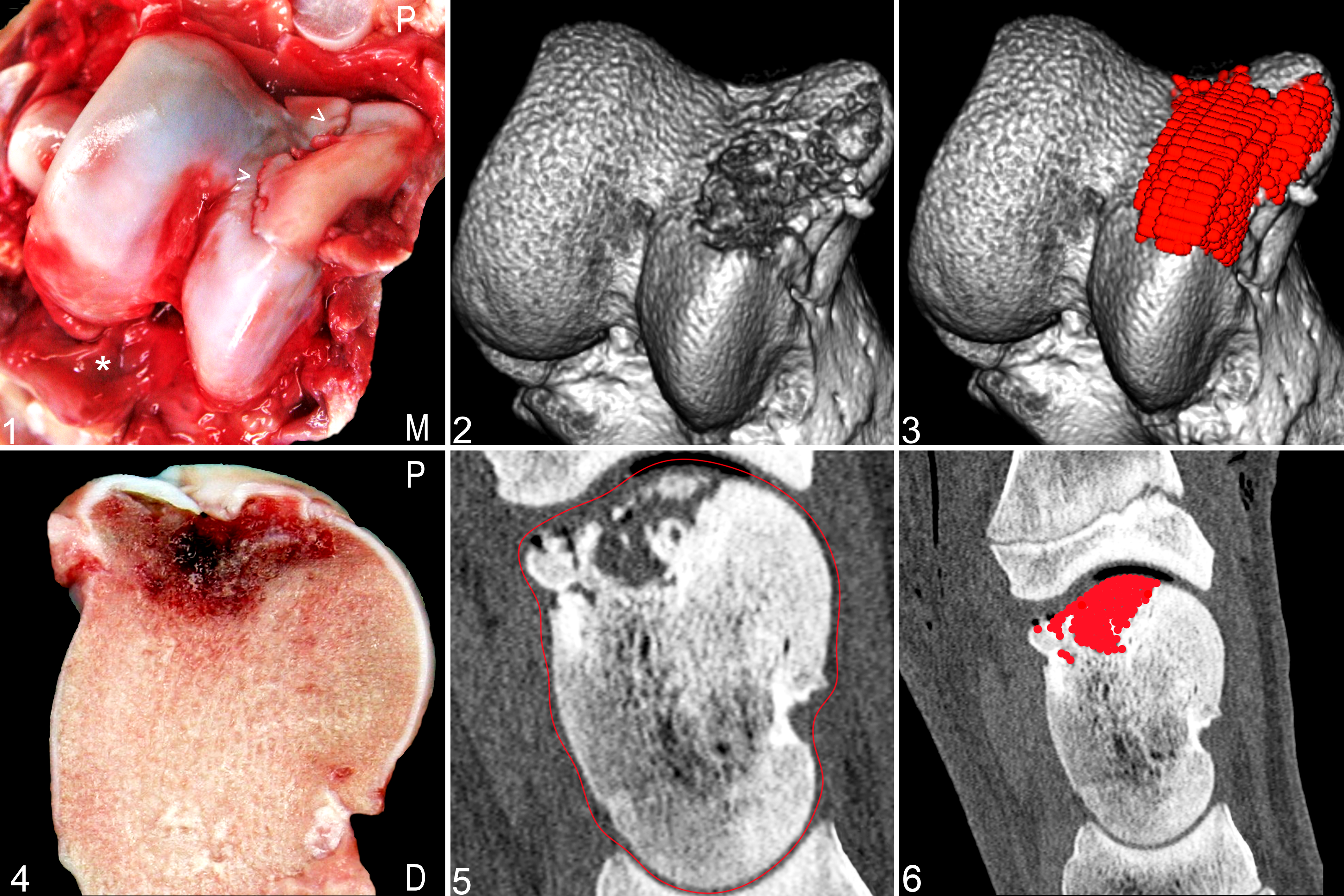
Osteochondrosis dissecans (OCD), right talus, domestic pig.
Frequency Distribution Maps
Image analysis was used to create a mean shape from the 3DVR images, so that all point ROIs for each type of change could be mapped onto the standard views of one 3DVR image for the domestic pigs and several views of one 3DVR image for the wild boars. Using image analysis software (Matlab, MathWorks, Natick, MA), a reference 3DVR image shape was compared with all other 3DVR images from the same view, and an affine alignment was used to generate a mean talus or distal tibia shape. The reference shape for the domestic pig group was the same as the reference images used to guide the 3DVR image preparation. The reference shape for the wild boar group was selected from the joints that did not have any lesions or detectable synovial fossae and had a typical shape without obvious anatomical variation. Groups of images marked with ROIs were then co-registered onto the mean joint shape, 45 and the resulting co-registered images were fused to form a frequency distribution map of the ROIs. Frequency distribution maps showed frequency distribution of OC lesions, synovial fossae (distal tibia only), and other articular indentations in the joint surface. A Gaussian filter was applied to the summed values of the ROIs in the fused image. Summed ROI values were displayed in a red, green, blue look-up table, in a frequency distribution map that was superimposed on the mean talus or distal tibia 3DVR image. Each map was presented with a color scale bar specific for that map. The highest value of the scale bar was represented as dark red, and the areas in the map with this color were the region(s) where the highest numbers of ROIs were located. The lowest color value possible in the maps was 1, which was represented by dark blue areas. Areas where no ROIs were located were transparent. Values in between dark red and dark blue received, in descending order, light red, orange, yellow, green, and light blue color shades.
Statistical Analysis
Numerical frequencies were calculated in Microsoft Excel. Statistical analysis of the frequency differences between the domestic pigs and the wild boars were calculated in Minitab version 17.1.0 using a log-likelihood contingency chi-square to test the interaction (statistical relationship) between sex and group. When there was no interaction between group and sex, the difference in results between the younger (6–9 months) and the older (10–18 months) wild boars, and between the wild boars and the domestic pigs, were tested with Barnard’s unconditional P value for comparing 2 independent proportions, using SMP version 2.1. 4,5
The response (measured) variables used were frequency on the whole talus of 1, OC lesions; 2, other articular indentations; and 3, intertrochlear indentations. The same model was also applied to the response variables of the distal articular surface of the tibia, which were frequency of synovial fossae and frequency of other articular indentations. Significance was defined in all analyses as P ≤ 0.05.
Results
One leg from a wild boar had a closed growth plate of the distal tibia and was excluded from the study. Thus, 19 hind legs from wild boars aged approximately 6–9 months (none of the deciduous teeth had been replaced) and 20 legs from wild boars aged approximately 10–18 months were included in the study. Twenty of the legs from wild boars were from gilts and 19 from boars. Twenty-six of the legs from the domestic pigs were from gilts and 14 from castrated boars. Sex did not have a significant effect on the results, and there was no significant interaction between sex and groups (domestic pigs versus wild boars) for any of the examined response variables.
The frequencies of OC, other indentations, and synovial fossae on the talus and the distal tibia of the domestic pigs and the wild boars are shown in Fig. 7. No OC lesions were detected on the distal articular surface of the tibia. All evaluated features were visible in CT images, whereas, depending on the feature type, macroscopic evaluation recognized between 57% and 100% of the features (Supplement 2, Table 1).
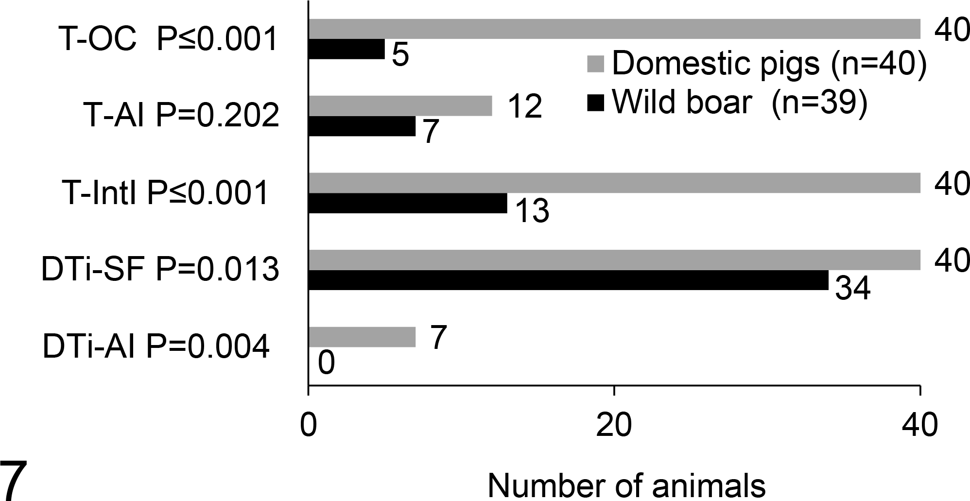
Frequency of osteochondrosis (OC), other articular indentations (AI), intertrochlear indentations (IntI), and synovial fossae (SF) on the right talus (T) and the distal tibia (DTi) in the domestic pigs and the wild boars. Significance defined as P ≤ 0.05.
In 9 domestic pigs and 16 wild boars, macroscopic and CT examination of the talus and/or distal tibia was followed up by histology for final classification of the findings.
There were no significant differences between the younger and the older age groups of the wild boars for any of the response variables. Therefore, only the data for the domestic pigs compared with the wild boars, but not within the 2 age categories of the wild boars, are presented here.
Osteochondrosis
Osteochondrosis was more frequent (Fig. 7) and occurred over a larger proportion of the joint surface in the domestic pigs (Figs. 8–10) compared with the wild boars (Figs. 11–13). The difference in OC frequency distribution between the pig groups was most evident on the trochlea and the distolateral aspect of the talus, where the domestic pigs commonly had OC lesions but wild boars did not. Details of the frequency of OC on the lateral and medial aspects of the trochlea and on the distal talus in the domestic pigs are in Supplement 3 (Fig. S1). The distomedial aspect of the distal talus was the only location where OC lesions were detected in the wild boars (Figs. 11–12 and 14–16).
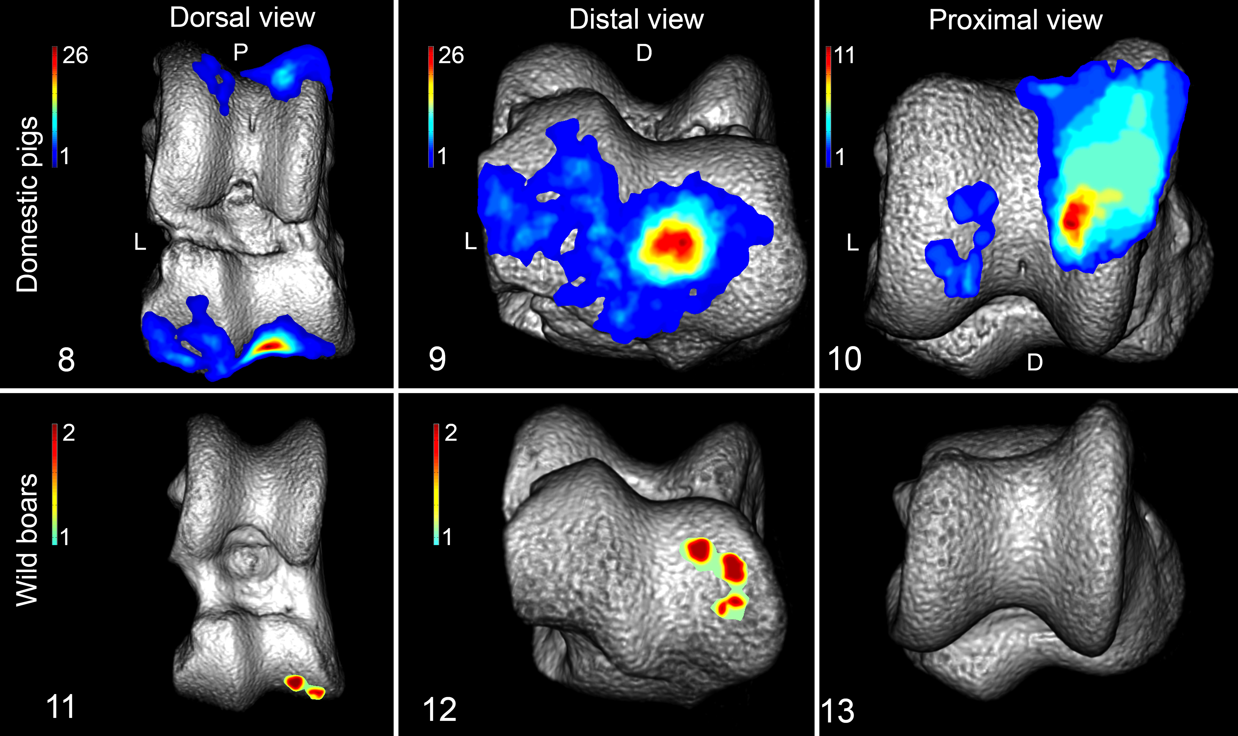
Frequency distribution maps of osteochondrosis (OC) on the right talus. OC locations are shown as colored regions, and the color bars (upper left corner of the figures) show the minimum–maximum number of OC lesions according to location. D, dorsal aspect; L, lateral aspect; P, proximal aspect.
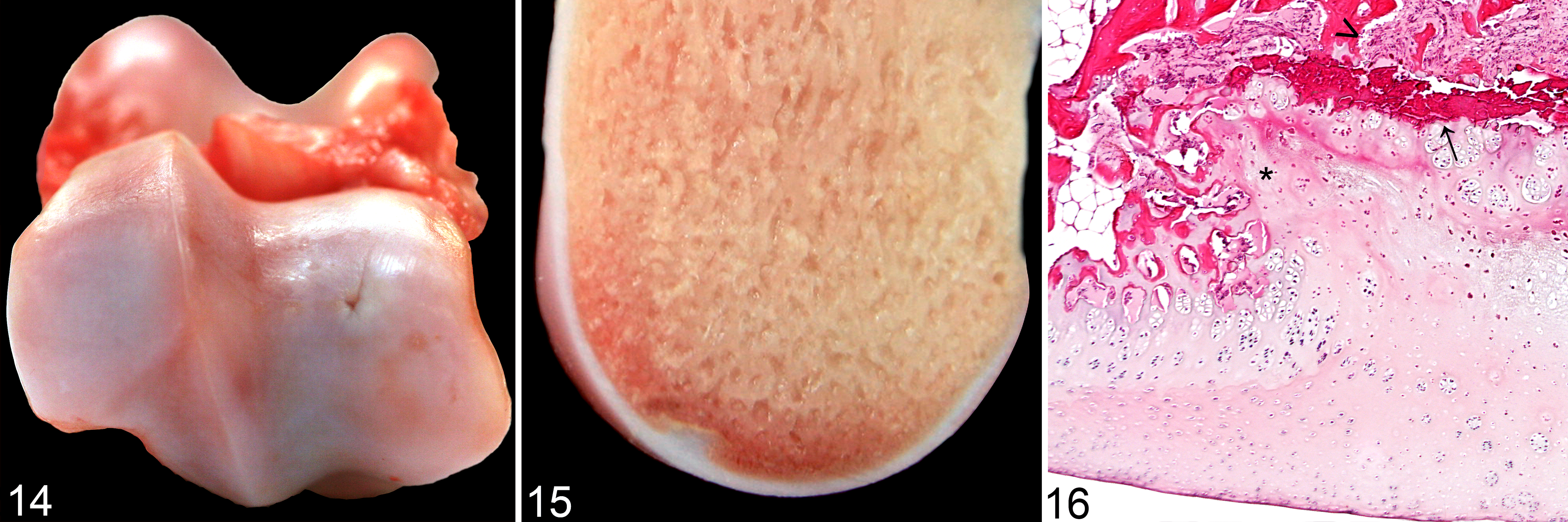
Osteochondrosis (OC), right talus, wild boar.
The OC lesions of the domestic pigs had a maximum surface diameter of 16 mm, and the largest lesions were on the medial trochlea. The OC lesions in wild boars had a maximum surface diameter of 4 mm. Six of the 40 OC lesions on the medial trochlea in the domestic pigs were OCD, and OCD was not seen in the wild boars. Small OC lesions were more obvious on CT than on macroscopic examination of slabs. In some cases, very small OC lesions were not detected at the initial pathologic examination, but these lesions were identified in CT images and in subsequent macroscopic and histologic examinations of stored bone slabs.
Other Articular Indentations on Talus
Prevalence of other articular indentations on the talus was low, and there was no significant difference between the prevalence in domestic pigs and wild boars (Fig. 7). The locations of other articular indentations were similar in the domestic pigs (Figs. 17–19) and the wild boars (Figs. 20–22).
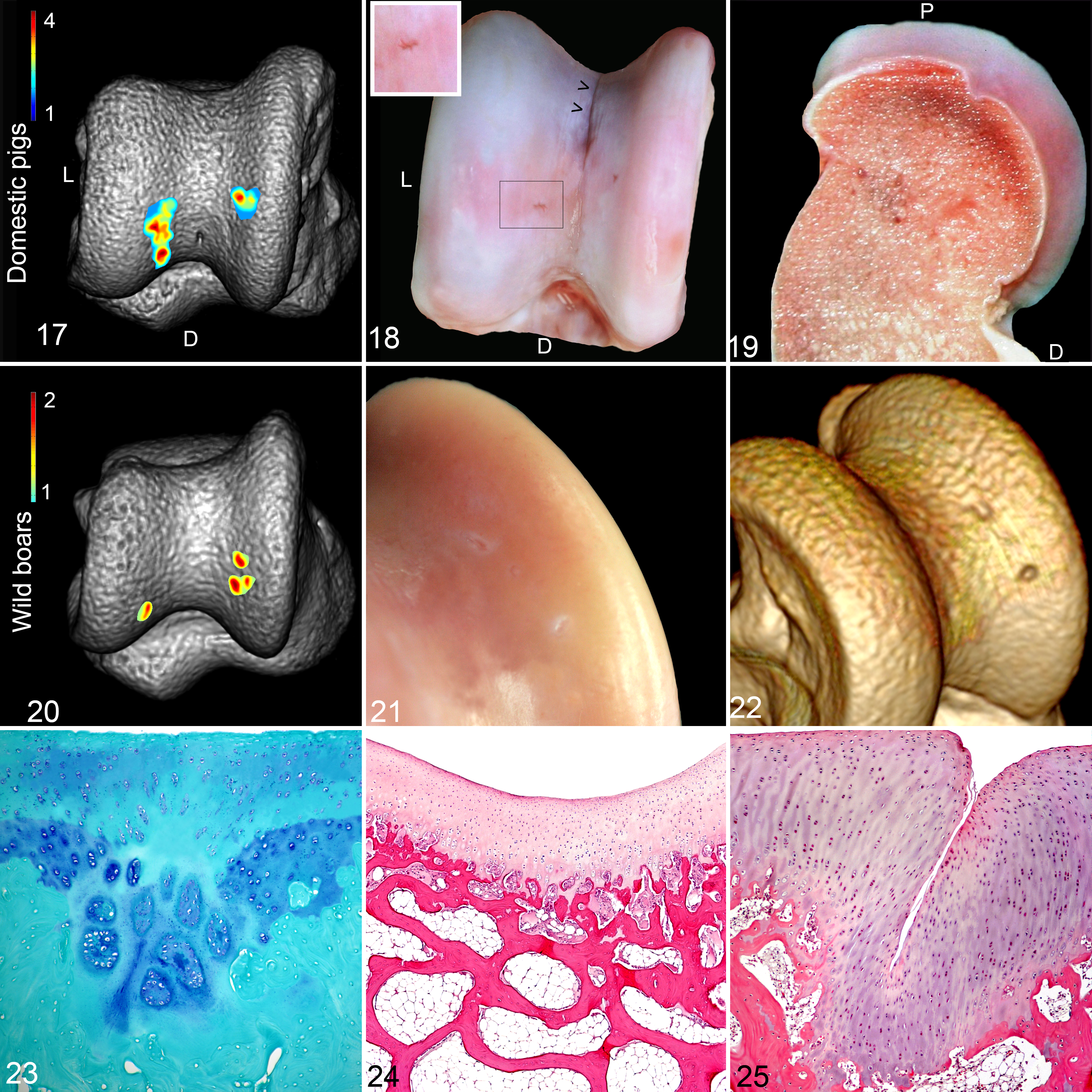
Other articular indentations, right talus.
The macroscopic appearance of the other articular indentations varied (Figs. 18 and 21). The indentations had a maximum articular surface diameter of 3 mm and were most easily detected in the CT images (Fig. 22). Histologically, 3 types of other articular indentations were identified: (1) focal areas of cartilage necrosis with clusters of chondrocytes. In some cases, such clusters were present in mineralized cartilage and subchondral bone, causing disruption of the osteochondral junction (Fig. 23). (2) Minor indentations in the joint surface with normal histological appearance of the articular cartilage and the subchondral bone (Figs. 19 and 24), (3) Folds or invaginations of thickened articular cartilage (Fig. 25).
Intertrochlear Indentations
Other articular indentations located in the intertrochlear groove region of the talus were called intertrochlear indentations. Intertrochlear indentations occurred in all domestic pigs, whereas a significantly lower prevalence was recorded in the wild boars (Fig. 7). The intertrochlear indentations occurred over a larger proportion of the joint surface in the domestic pigs (Figs. 26–28) compared with the wild boars (Figs. 29–31).
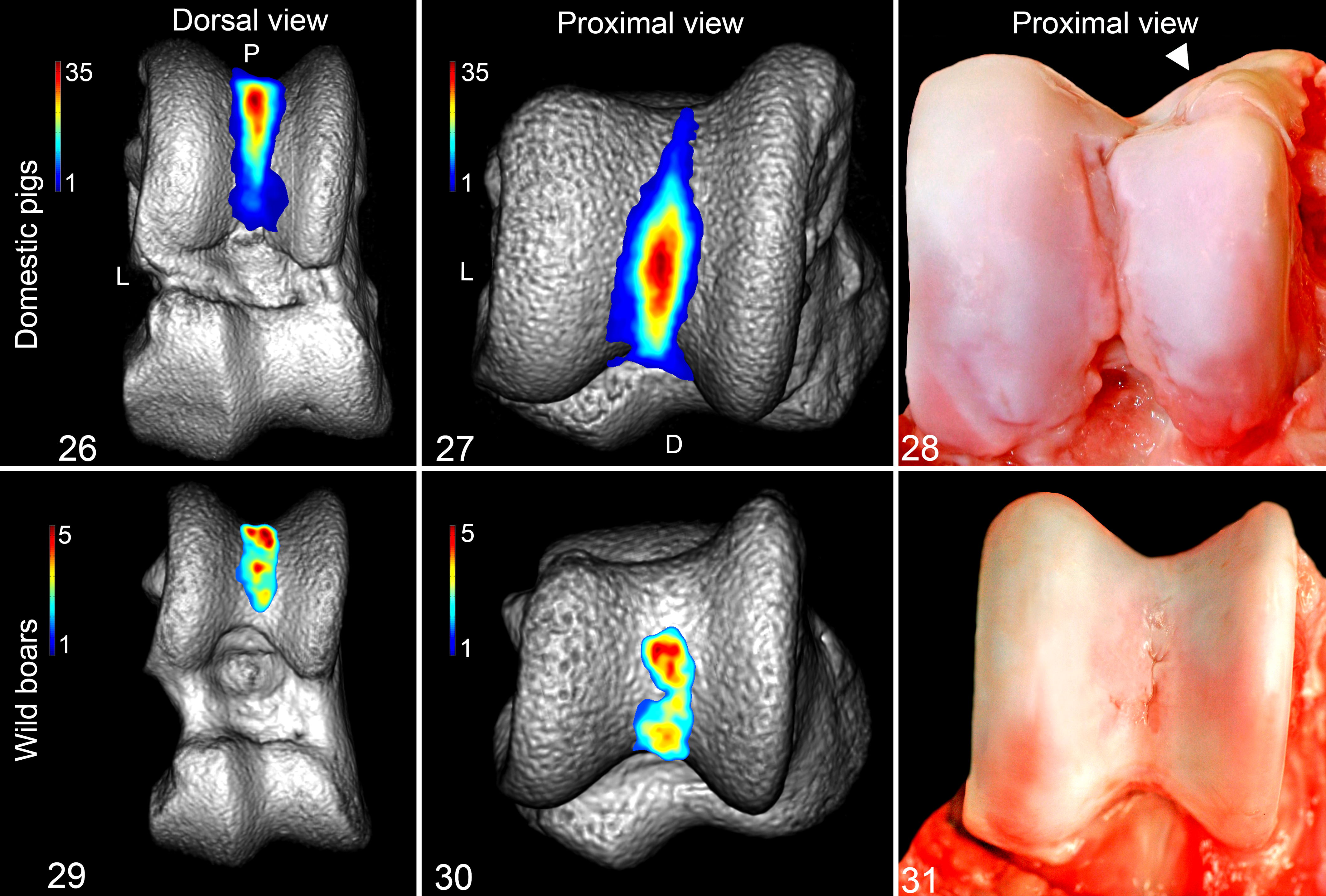
Intertrochlear indentations, right talus.
The size and shape of the intertrochlear indentations in both the domestic pigs and the wild boars varied considerably. Some indentations were linear (Fig. 18) while others had a multifocal distribution (Fig. 31) with indentations joining to form 1 or more larger indentations in the intertrochlear groove. The linear indentations were histologically characterized as invaginations of a normal articular cartilage. Many of the intertrochlear indentations of the domestic pigs were large (long, deep, and wide), particularly in pigs that had OCD on the medial trochlea (Figs. 1 and 28). Some of the indentations were partially covered by a synovial membrane that extended from the synovial membrane present distal to the trochlea of the talus. Other intertrochlear indentations had focal areas of necrotic, often thickened, mature hyaline cartilage not covered by synovial tissue. In these indentations, a variable amount of fibrovascular connective tissue (Fig. 32) or fibrocartilage (Fig. 33) could be found in the osteochondral junction and subchondral bone.
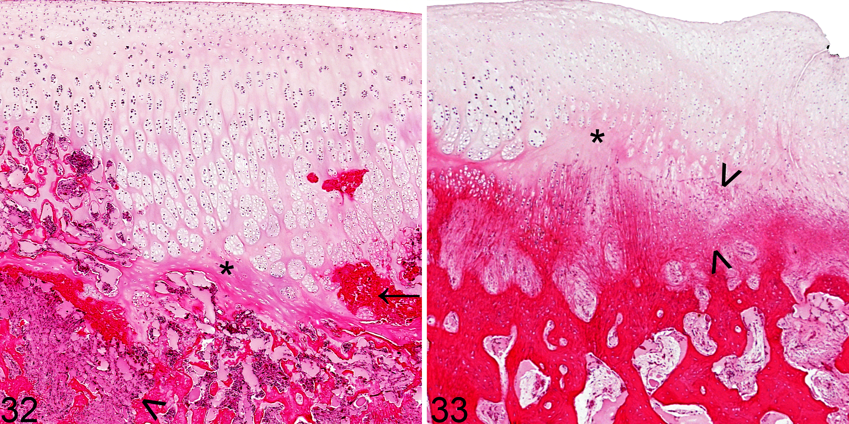
Intertrochlear indentations, right talus. Hematoxylin and eosin.
Other Articular Indentations of the Distal Tibia
Other articular indentations on the distal tibial joint surfaces had low prevalence in domestic pigs and were never detected in wild boars (Fig. 7). Based on the macroscopic appearance and the consistent location on the craniomedial aspect of the distal tibial joint surface (Suppl. 4, Fig. S2) opposite OCD of the medial trochlea, all these articular indentations were considered to be kissing lesions. The kissing lesions were characterized by fibrillation of the articular cartilage, sometimes with erosions and ulcerations that partly disclosed the underlying subchondral bone.
Synovial Fossae of the Distal Tibia
Synovial fossae on the joint surfaces of the distal tibia were present in all domestic pigs and covered a larger area compared with the wild boars (Suppl. 5, Figs. S3, S4). The prevalence of these synovial fossae was significantly higher in the domestic pigs (Fig. 7).
Discussion
This study demonstrates that OC was more prevalent, and was found in more locations, in the domestic pigs compared with the wild boars. The intertrochlear indentations of the talus and the synovial fossae on the distal tibia occurred more frequently, and were larger, in the domestic pigs compared with the wild boars, but the presence of other articular indentations on the medial and lateral trochlear ridges of the talus did not differ.
The frequency distribution maps are unique in how they portray the location and frequency of OC, other articular indentations, and synovial fossae in the hock joints of these 2 porcine subspecies. The map information can be used to identify specific high-frequency locations within joint regions, which can assist in focusing future investigations into the causes of these joint features and lesions.
Osteochondrosis
Wild boars may have metaphyseal OC 15,30 but no cases of articular OC in wild boars have been verified. 11,15,30 Another study reported that 100% of the domestic pigs and 93% of wild boars have OC on the distal tibial surface; however, no distinct criteria for OC are described, and the lesions appear to be compatible with synovial fossae of the distal tibia. 43 The current study is, as far as we are aware, the first to document articular OC in wild boars and the first to use CT to compare the frequency distribution of OC between wild boars and domestic pigs.
Previous studies report that domestic pigs have a high prevalence of OC on the medial trochlea of the talus 10,13,27,37,47 and on the distomedial aspect of the talus, 18,39 and this was confirmed in the current study. Apart from one study, which involved some of the pigs from the current study, 13 we found no recent reports describing the distal talus OC. In the current study, the prevalence for OC on the distal talus of the domestic pigs (98%) was much higher than the prevalence on the trochlea (45%). Thus, examining the distal aspect of the talus is very important when performing OC screening and research in pigs.
Many studies have found that OC has a predilection for medial joint locations, 17,22,47 and this was clearly illustrated in the maps of both the domestic pigs and the wild boars in the current study. Although OC occurred most commonly on the medial trochlea region in the domestic pigs, it was the axial aspect of the medial trochlea (red area of Fig. 10) that was consistently most often affected. A similar pattern was noted on the distal aspect of the talus in domestic pigs, namely, that OC lesions occurred most commonly on the axial region of the distal joint surface. However, the few OC lesions in the wild boars were not found in an axial location but were rather located in a mid to abaxial location on the distomedial aspect of the talus. These findings strongly suggest that most OC lesions initiate in these focal areas, and this gives support to proposals that there are local features that make some areas more vulnerable to OC than others. 51 Several studies speculate that a locally high load of biomechanical forces, 13,33,51,52 alone or in association with an “unfortunate” joint conformation, 13,19,51 may cause microtrauma to the cartilage canals of the articular epiphyseal cartilage and that this may promote development of OC in specific regions of joints. Differences in anatomic and biomechanical features between the domestic pigs and the wild boars may be one reason for the difference in distribution and prevalence of OC between these 2 porcine groups.
The wild boars differ in many respects from the domestic pigs, and one of these is size. At 6 months of age, a domestic boar weighs approximately 110–120 kg, whereas a wild boar weighs approximately 45 kg at 6 months and 65 kg at 12 months of age. 50 In dogs, OC is reported only in large breeds, 20,41 and in horses, OC is very seldom reported in (small) pony breeds. 21 It is suggested that in ponies, this may be due to a thinner cartilage and fewer vessels in articular epiphyseal cartilage complex, making them less susceptible to OC. 21 These studies 20,21,41 may indicate that the size of an animal matters, and the difference in size may hence have contributed to a lower OC frequency in wild boars compared with the domestic pigs.
A genetic association between muscle mass and the prevalence of OC has also been shown in domestic pigs. 28 A larger body mass combined with a relatively higher muscle mass may result in a heavier load on the joint surfaces of domestic pigs than in the smaller wild boars. This may have a role in explaining the higher OC frequency of the domestic pigs. Future comparative studies of the anatomical conformation and load distribution within the joints of the domestic pigs and the wild boars could further clarify these issues.
The etiology of OC is complex and regarded as multifactorial, 36,51 and so factors other than biomechanics and conformation are likely to have contributed to the development of OC in the 2 porcine groups. The reported effects of growth rate on the development of OC are not consistent, but some studies suggest that rapid daily gain (during specified periods of growth) and genetic factors 2,26,31,34,41 are associated with OC in pigs. Wild boars and domestic pigs live in quite different environments, and this is another factor that may affect the development of OC. Wild boars must roam for their food, are hunted, and have predators, 23 so healthy joints and good locomotion are vital for their survival. Domestication and confinement have strongly reduced the role of the locomotor system in the survival of domestic pigs. Since the main breeding focus in domestic pigs for many decades has been enhancing growth rate and meat percentage (dressing percentage), 1,29 selection for these production traits may also have resulted in an unintentional selection for undesirable traits, 1 such as selection of pigs with a genetic predisposition for OC. 28 Thus, the domestic pigs may have acquired anatomic and genetic traits that increase the risk for OC development, while the wild boars’ low frequency of OC may be attributed to an evolutionary pressure favoring healthy joints that are well adapted to physical activity.
Breeding programs aimed at improving traits for leg and joint soundness in domestic pigs commenced in the 1970s and 1980s, 16,31 and breeding companies now strive to reduce OC in domestic pigs. 1 Further investigations into the reasons for low OC prevalence in wild boars may help develop strategies to reduce OC prevalence in domestic pigs. To promote a more robust locomotor system in domestic pigs, wild boars could be considered for inclusion in alternative (research) breeding programs. The mode of OC inheritance is, however, still being explored, and a positive outcome for such studies is not necessarily given. Two studies reported that the offspring of sows crossed with wild boars had no 41 or much lower 3 prevalence of OC, and 2 other studies reported that offspring between wild boars and domestic pigs (F1 and/or F2 generation) did not have a significantly lower prevalence of OC compared with purebred pigs. 46,48
Other Articular Indentations on Talus
The indentations on the trochlear ridges of tali that had focal areas of necrotic cartilage and chondrocyte clusters disrupting the osteochondral junction (as in Fig. 23) resembled OC. However, as they occurred in mature articular cartilage, caused visible indentations on the surface (not normally associated with OC latens or minor OC manifest lesions), and were not associated with vascular necrosis or inflammatory reactions, these indentations were not considered to be OC. Furthermore, the wild boars had a low prevalence of OC elsewhere on the talus, and these other indentations were equally present in a limited area of the axial aspects of the trochlea in both wild boars and domestic pigs, supporting that these indentations are not OC lesions.
An alternative possibility is that all of the other articular indentations represent remnants of, or healed, OC lesions. It sometimes takes months for OC lesions to develop, and during this development period, some OC lesions heal. 37 Healing of an area of necrotic cartilage caused by OC may, for example, result in the collapse of overlying cartilage, leaving only granulation tissue and mildly disorganized trabecular bone structure rather than a typical OC lesion. To reveal the true nature of these lesions, longitudinal studies describing the development of the cartilage canals and their lesions from birth to cessation of growth are required.
Intertrochlear Indentations
Even though the indentations in the intertrochlear groove of the talus were seen in all domestic pigs, we found only rare literature reports that describe these indentations or their etiology in pigs. Intertrochlear indentations including areas of cartilage necrosis, fibrovascular connective tissue, or fibrocartilage may indicate active pathologic processes and are histologically difficult to differentiate from OC. This observation is supported by a study on fatteners that interpreted intertrochlear “depressions” as OC. 27 However, histologic changes consistent with the intertrochlear indentations in the current study have also been described as normal developmental stages of synovial fossae in the intertrochlear groove of the talus in calves. 49 Although the latter authors report that OC lesions did rarely occur in the developing synovial fossae of a few calves, they describe the transition from normal articular cartilage to synovial fossae in the calves.
The fact that the intertrochlear indentations occurred in all domestic pigs supports that many of these indentations represent different developmental phases of normal synovial fossae. However, it raises the question as to why they were detected in only 33% of the wild boars. One explanation could be that synovial fossae are not present at birth but develop with age. 6,9,42,49 Wild boars mature at an older age than do the domestic pigs, 41 and hence, they may develop the synovial fossae later in life. However, all of the wild boars in this study were older than any of the domestic pigs, and there was no difference in the results between the younger and the older wild boars. Therefore, we consider it unlikely that differences in age are responsible for the difference in prevalence.
The suggestion that the intertrochlear indentations may be normal synovial fossae is not supported by the results in a thesis that states that the proximal taluses of both young and adult pigs completely lack synovial fossae. 42 However, another report referred to a linear groove, described as “an invagination of articular cartilage extending into subchondral bone” between the medial and the lateral condyle of the distal humerus, as being normal. 9 The description of this linear groove is similar to the linear intertrochlear indentations seen in the talus of some of the domestic pigs (Fig. 18) in the current study. In contrast, the humeral intercondylar groove has been reported as a pathologic change associated with “arthrosis.” 42 The disagreement between the studies indicates that more research on talus intertrochlear indentations in pigs is needed, to establish whether or not they should be regarded as normal age-related structures or are OC lesions or remnants.
Synovial Fossae
The synovial fossae present on the distal tibia are recognized as normal anatomical structures. 42 Studies on domestic pigs have indicated that the size and development of synovial fossae are not only age dependent 42 but also can be associated with pathologic joint changes. 10 This study reports that the size of synovial fossae is larger in joints with synovitis and osteoarthritis (OA). Osteochondrosis dissecans will cause a secondary inflammation in the joint, resulting in OA. 40 Hence, the larger synovial fossae present in the domestic pigs compared with the wild boars may be related to OA caused by OC/OCD. Another study reported that lambs isolated in small pens developed larger synovial fossae on the metacarpus than did those who were kept in larger pens. 32 This was interpreted as less-active lambs needing larger synovial fossae to ensure sufficient nourishment of the joint cartilage. 32 These arguments may also apply to our results, suggesting that less exercise and a higher prevalence of OCD in the domestic pigs may be reasons why the domestic pigs had more numerous and larger synovial fossae than the wild boars.
CT Imaging as a Tool for Diagnosing Joint Disease
All cases of articular cartilage indentations detected in this study involved the mineralized tissues of the ossification front and hence could be seen on CT images. The CT examinations resulted in detailed 3D images of the mineralized tissues of the joints, and evaluation of these images revealed more joint surface lesions and features than did the macroscopic evaluation of opened joints and slab sections. This observation supports recent studies that concluded that CT has an important role in the evaluation of OC in pigs. 37 Recognition of small indentations is important for the correct description and understanding of joint-related diseases. The methodology applied and described in this study may also be useful for other studies that seek to evaluate lesion locations within joints.
Conclusions
Our hypothesis that the frequency distribution of OC, other articular indentations, and synovial fossae in the hock joint would differ between the domestic pigs and the wild boars was largely confirmed. The domestic pigs displayed more numerous OC lesions in more locations of the talus than did the wild boars, while the highest frequency location of OC on the talus of both groups was the distomedial aspect. In both the domestic pigs and the wild boars, there were focal areas in which the OC frequency was much higher than elsewhere, strongly suggesting that these areas may be sites where OC lesions initiate. The etiology of the other articular indentations on the trochlea, and in the intertrochlear groove of the talus, remains unclear. The intertrochlear indentations on the talus, and the synovial fossae on the joint surface of the distal tibial joint surface, were more frequent and covered larger areas in the domestic pigs than in the wild boars, which may be related to differences in age and prevalence of joint disease. The use of CT imaging as a diagnostic tool enhanced the evaluation of osteochondral lesions and indentations within the hock joints, and we recommend that future investigations into porcine joint health should include CT evaluation. More research on OC in crossed offspring between wild boars and domestic pigs may help elucidate further which role genetic and anatomical factors play in OC development and so contribute to improving joint health in domestic pigs.
Footnotes
Supplemental material for this article is available on the Veterinary Pathology website at ![]() .
35
.
35
Acknowledgements
We are sincerely grateful to Ola Schulzberg and Kaj and Yvonne Rehnholm for assistance in collecting the legs from the wild boars, Agneta Boström and Christina Nilsson at the Swedish University of Agricultural Sciences (SLU) for laboratory assistance, Lars Hammarsten at the National Veterinary Institute (SVA) for his assistance with disarticulation and sawing of joints, and Christina Larsson at SLU for performing the CT scans. The authors are also grateful to David Morrison at Uppsala University for guidance and comments on the statistics and to Björnar Ytrehus at the Norwegian Institute for Nature Research for comments on the manuscript.
Declaration of Conflicting Interests
The author(s) declared no potential conflicts of interest with respect to the research, authorship, and/or publication of this article.
Funding
The author(s) disclosed receipt of the following financial support for the research, authorship, and/or publication of this article: This investigation was supported by grants from The Swedish Research Council FORMAS (Dnr. 221-2013-317). Additional support was provided by grants from Hjärrefonden and Gerhard Forssells stipendiestiftelse.
References
Supplementary Material
Please find the following supplemental material available below.
For Open Access articles published under a Creative Commons License, all supplemental material carries the same license as the article it is associated with.
For non-Open Access articles published, all supplemental material carries a non-exclusive license, and permission requests for re-use of supplemental material or any part of supplemental material shall be sent directly to the copyright owner as specified in the copyright notice associated with the article.