
Abstract
Haemophilus parasuis is a recognized pathogen in domestic pigs; the pathogen has been also isolated from healthy wild boar (Sus scrofa). In the current report, a case of fatal H. parasuis infection in a wild boar piglet from central Spain is described. The affected animal presented severe pneumonic lesions, inflammation in tarsal joints with presence of fibrinous deposits, and epidural hemorrhage in the atlanto-occipital joint. Pure growth of H. parasuis was obtained from lungs and tarsal joints. The current case illustrates the susceptibility of wild boar to this agent. The gross pathology results were similar to that described in domestic pigs, but there were no fibrinous deposits on serosal surfaces.
Haemophilus parasuis is the causative agent of Glässer’s disease, an inflammatory condition featured by serofibrinous to fibrinopurulent exudates at single or multiple serosal surfaces (porcine polyserositis and arthritis). 1 Polyarthritis is usually more severe in the atlanto-occipital and large limb joints. 19 Haemophilus parasuis can also be involved in swine pneumonia, acting as a primary pathogen, secondary invader, or predisposing agent.9,12,21
Haemophilus parasuis is considered an early colonizer of the respiratory tract of domestic pigs, being one of the most significant disease-causing bacteria that affect swine. 14 The pathogenic potential and the development of the disease vary according to the different strains and serovars, 2 individual host resistance, 4 age and colostral protection, 5 and herd health status and origin. 13
The pathological features of H. parasuis infection have been widely described in domestic pigs, 16 but there is little knowledge about the infection in wild boar (Sus scrofa). Antibodies to H. parasuis have been detected in wild boar in Slovenia, 22 but similar serosurveys carried out in southern Spain were negative. 23 The microorganism has been detected by molecular methods in Germany 17 and isolated from hunted wild boar in Spain. 15 The pathogenic potential of H. parasuis isolated from wild boar has been demonstrated by experimental infection in domestic pigs. 2 However, it appears that no disease has been reported to be caused by this agent in wild boar, so the impact of the agent on these animals is unknown. In the current report, a case of fatal H. parasuis infection in a wild boar piglet from central Spain is described.
The affected animal came from a game estate located in the “Montes de Toledo” area in the province of Toledo (central Spain), a region with a continental thermo-Mediterranean climate (hot and dry summers and mild and moderately wet winters). The estate is completely fenced, enclosing approximately 1,000 hectares. The dominant vegetation consists of holly oaks (Quercus ilex) accompanied by a large amount of shrub, mainly heather (Erica sp.) and rock rose (Cistus sp.) in sunny areas, and arbutus (or, strawberry tree, Arbutus unedo) in shady areas. Approximately 20% of the property is occupied by crops of oat (Avena sativa) and vetch (Vicia sp.), providing food for the estimated 300 wild boar that live in the estate. The wild boar share the habitat with a small population of Western roe deer (Capreolus capreolus), with no other artiodactyls being present on the estate.
The subject of the current study was a young male wild boar approximately 5 months old, caught by the manager of the farm during a routine patrol. The animal was walking with difficulty behind his mother, showing a noticeable respiratory distress and weakness. In other observation areas, more animals were observed with similar symptoms, but it was impossible to capture these individuals due to the difficult terrain. After capture, the animal died naturally, and the body was chilled. Necropsy was carried out less than 24 hr after capture, to allow a complete pathological and microbiological study.
The animal showed poor body condition and generalized lymphadenopathy. The conjunctiva and other mucosal surfaces were pale, and a great amount of mucus was seen around the snout. There was an evident asymmetry in the hind paws, with unilateral increase of the size of the tarsal joint (Fig. 1). The bursa presented an evident inflammation, together with fibrin and wall thickening. The lungs showed pneumonic foci (Fig. 2), well delimited and hyperemic, and the liver and spleen appeared enlarged and congestive. Finally, epidural hemorrhage was observed in the atlanto-occipital joint (Fig. 3).
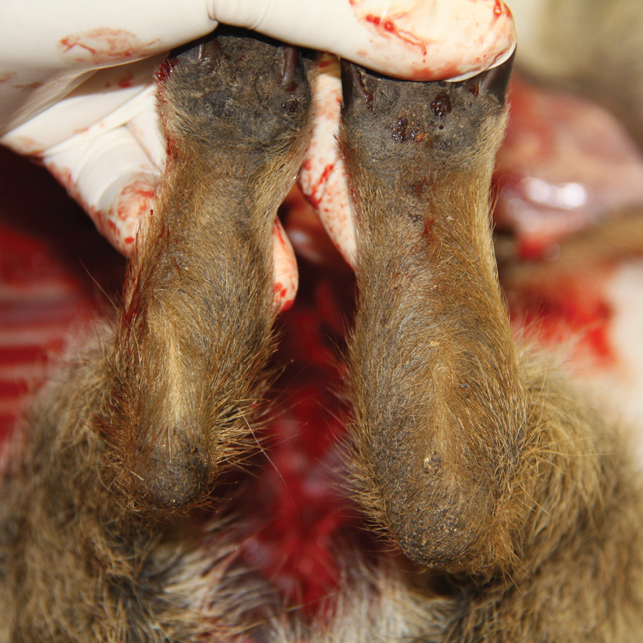
Wild boar (Sus scrofa); hind paws. Note the asymmetry and unilateral increase of the thickness of the joint.
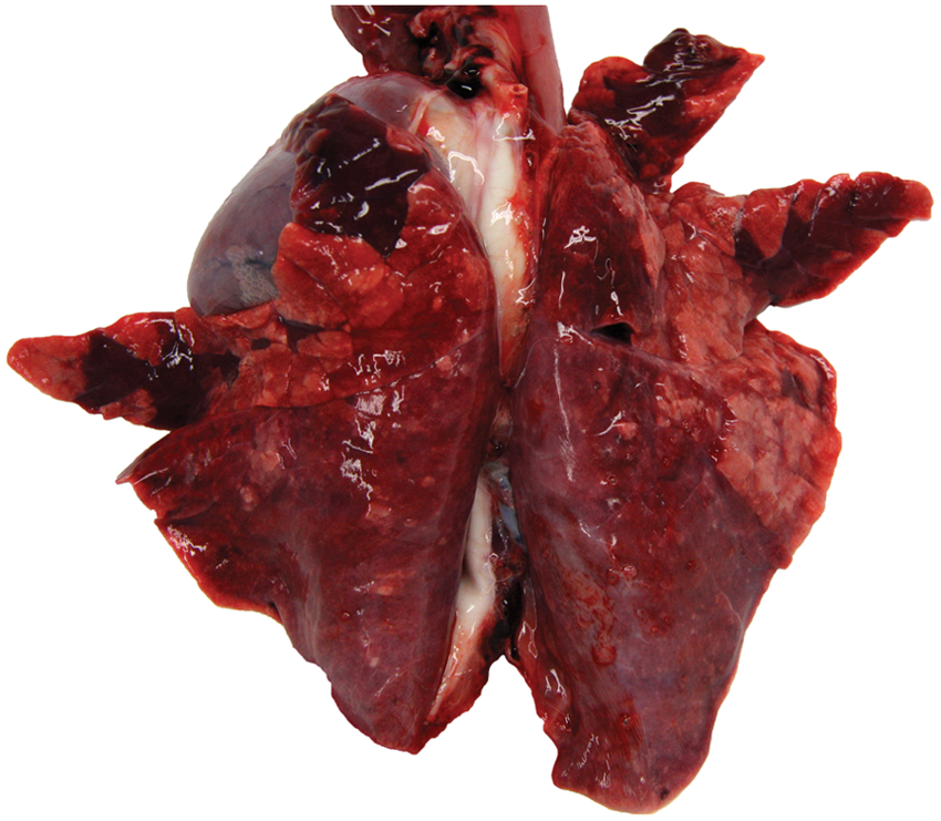
Wild boar (Sus scrofa); lungs. Note the well-delimited and hyperemic pneumonic foci distributed throughout the parenchyma.
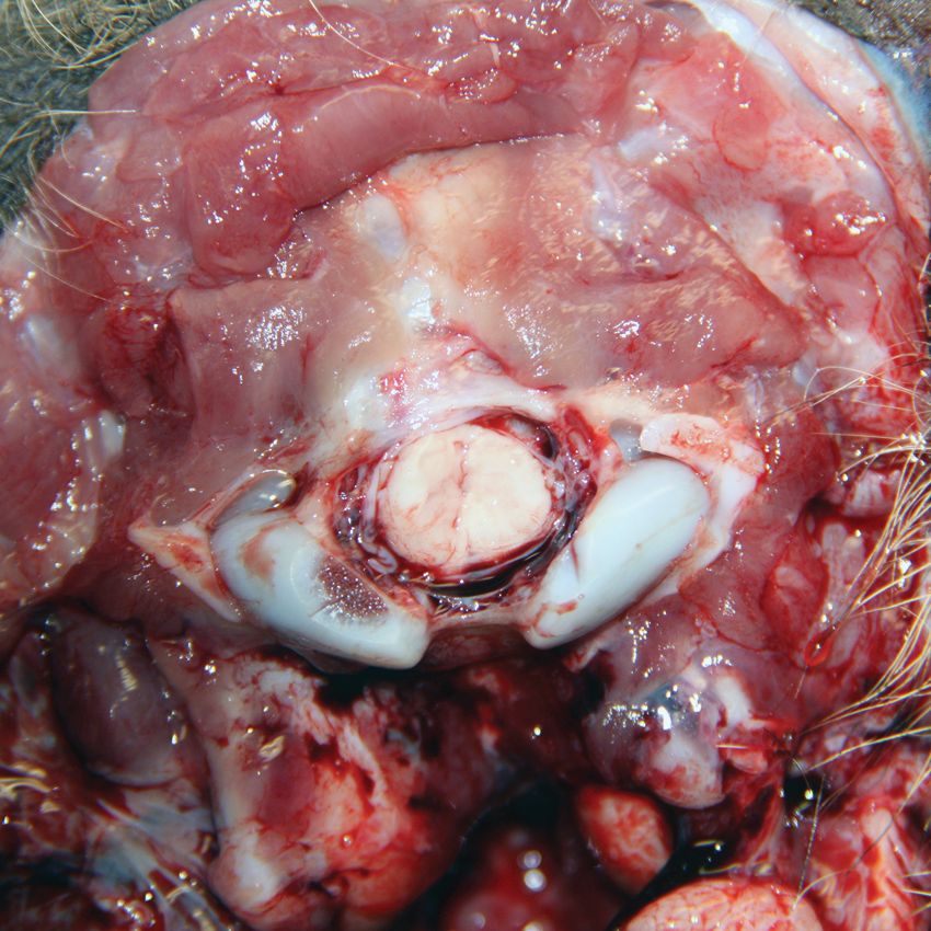
Wild boar (Sus scrofa). The atlanto-occipital joint showing epidural hemorrhage.
Samples from tarsal joint, lungs, liver, spleen, kidneys, and nervous system were fixed in 3.5% buffered formalin, processed, and stained with hematoxylin and eosin. a The tarsal joint presented a slight inflammatory infiltration, associated to fibrin. The lung samples showed an interstitial pneumonia progressing to exudative bronchopneumonia (Fig. 4). The kidneys showed interstitial nephritis and some hemorrhages. Tubulonephrosis was also present. The nervous system showed mild inflammation and slight meningitis.
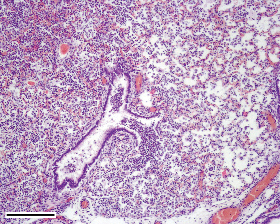
Wild boar (Sus scrofa); lungs. The lungs showing bronchopneumonia, and purulent exudates composed by neutrophils, pyocites, and cellular debris. The exudate extends from terminal bronchioles to alveoli. Hematoxylin and eosin stain. Bar = 200 µm.
Tissue samples from liver, spleen, kidney, lung, tarsal joint, and heart were cultured on blood agar, b chocolate agar, b and MacConkey agar plates. c The plates were incubated for at least 24 hr at 37°C under aerobic conditions. A pure growth of grayish colonies approximately 1–1.5 mm in diameter was obtained from the lungs and the joints on chocolate agar plates after 24 hr incubation, while all other plates and all other tissues failed to yield any growth at all after 72 hr incubation. Microscopically, the organisms appeared as small Gram-negative rods that formed occasional chains of coccobacilli. By standard phenotypic tests, 3 the isolate was identified as H. parasuis. To confirm the identification, a species-specific polymerase chain reaction (PCR) for H. parasuis was carried out essentially as previously described 14 except that a different Taq enzyme d was used. The isolates gave positive results. A field strain of H. parasuis obtained from a pig affected by Glässer’s disease was used as the positive control and a reference strain of Pasteurella multocida as the negative control in the PCR, with controls giving the expected positive or negative result, respectively. Furthermore, to assess the presence of other respiratory pathogens, specific PCR assays for Mycoplasma hyopneumaniae 6 and Porcine circovirus-2 7 were carried out on DNA extracted from lungs, yielding negative results.
It is known that domestic pig, European wild boar, and their relatives are susceptible to a similar range of pathogens, 11 such as Suid herpesvirus 1 (Aujeszky disease virus), 10 Erysipelothrix rhusiopathiae, 18 and Mycobacterium spp. 8 The current case indicates that H. parasuis can be added to the list of shared pathogens.
The animal examined in the current study showed lesions such as unilateral thickening of the joint, inflammation of the bursa, with a small amount of fibrin and hemorrhage in the atlanto-occipital joint, all of which are typical lesions seen in Glässer’s disease. 19 However, inflammation in other serous membranes and large accumulations of fibrin, common findings in Glässer’s disease,19,20 were not observed.
Although there has been controversy about the pathogenicity of H. parasuis in wild boar, 17 the finding of a fatal pneumonic process associated with isolation (in pure culture) and identification of the pathogen casts doubt on the susceptibility of wild boar to H. parasuis infection. It is possible that the lack of reports on pathogenic cases may be due to differences in the pathogenicity of H. parasuis strains. 2
Only 1 case of H. parasuis infection was confirmed in the estate, suggesting a low mortality rate with this pathogen. However, other animals were seen with similar signs, thus the mortality rate could have been higher. Unfortunately, these other possible cases could not be confirmed as the finding of dead animals in this kind of estate is very difficult due to the terrain.
The affected animal came from a semifree population living in a fenced estate but emulating natural conditions with enough surface and resources to avoid the need for supplementary feeding. This suggests that H. parasuis can be a cause of mortality in natural wild boar populations. However, as diseases caused by H. parasuis in the domestic pig have been commonly related with factors such as stress, 14 it is possible that H. parasuis is more important in intensive wild boar farming than in true wild populations.
The current study demonstrates the susceptibility of wild boar to H. parasuis, as has been previously reported with other common pathogens of the pig.8,10,18 However, the pathological features present in the animal examined in the current study were slightly different to that observed in domestic pigs affected by Glässer’s disease, as no large deposits of fibrin on the serous membranes were found. The difficulties of studying this kind of wild animal can result in an underestimation of pathogenic potential of H. parasuis as a cause of disease or death within similar enclosed environments.
Footnotes
a.
Isokit, Bio Optica Milano S.p.A., Milan, Italy.
b.
Oxoid Ltd., Cambridge, UK.
c.
Scharlau SL, Barcelona, Spain.
d.
Kapa Taq, Kapa Biosystems Inc., Woburn, MA.
Declaration of conflict interests
The author(s) declared no potential conflicts of interest with respect to the research, authorship, and/or publication of this article.
Funding
The author(s) disclosed receipt of the following financial support for the research, authorship, and/or publication of this article: This work was supported by the Ministerio de Ciencia e Innovación (Gobierno de España) PS0900513 and Gobierno de Extremadura GRU10142. J. Cuesta Gerveno has a grant from Fundación Valhondo Calaff. D. Risco has a FPU grant from the Ministerio de Ciencia e Innovación (AP2009-0704).