
Abstract
An unknown-aged adult female wild boar (Sus scrofa) was brought to Kyungpook National University for postmortem examination. Gross examination revealed gallbladder agenesis. Histologically, the liver was cirrhotic and had intrahepatic cholelithiasis, the choleliths were yellow, brown, gray, and black, and had coffin-lid and pyramidal appearances. Fourier-transform infrared spectroscopy analysis revealed that the components were 80% struvite and 20% calcium oxalate monohydrate. Chronic inflammatory cell infiltration was observed, with hyperplastic hepatocellular nodules characterized by large nuclei, prominent nucleoli, and scant cytoplasm with frequent binucleation, surrounded by thick fibrous septa. The epithelium of intrahepatic bile ducts that contained choleliths had undergone gallbladder-like metaplasia, which might have been induced by chronic irritation from the stones or by the accompanying chronic bacterial infection that was observed in Gram stains.
Choleliths, or gallstones, are solid masses of cholesterol crystals, mucin, calcium bilirubinate, proteins, bile pigments, or bile acids.2,6,9,14,16,21 Cholelithiasis is one of the most prevalent diseases associated with the digestive tract in humans and is a serious condition given its economic burden on individuals and society. 15 Moreover, cholelithiasis can also be a critical threat to individual health because it is a risk factor for pancreatitis and for the development of gallbladder, pancreatic, and colorectal cancer.15,20 The most common cause of cholelithiasis is the supersaturation of cholesterol in bile, decreased gallbladder motility, decreased bilirubin glucuronide conjugation leading to biliary infection, and increased biliary bilirubin load. 13
Occasional cases of cholelithiasis have been reported in veterinary medicine.7,17,22,23 Cholelithiasis is important in veterinary medicine because it can cause life-threatening extrahepatic biliary tract obstruction. 10 In pigs, choleliths composed of cholesterol, bile pigments, calcium, and bile acids have been reported.14,21
In intrahepatic cholelithiasis, or hepatolithiasis, choleliths accumulate in the intrahepatic bile ducts. 11 In veterinary medicine, hepatolithiasis has been associated with cholecystolithiasis and extrahepatic bile duct obstruction caused by choledocholithiasis, many of these cases have been reported in domestic animals, particularly in dogs and cats. 7 We retrieved no cases of intrahepatic cholelithiasis in wild boars in a search of Google, PubMed, Web of Science, and Scopus, suggesting that this condition has not been reported in wild boars. Intrahepatic cholelithiasis has not been well described in veterinary medicine. We report here a case of intrahepatic cholelithiasis with gallbladder-like metaplasia of the hepatic bile ducts in a wild boar.
A 70-kg, unknown-aged, free-living, adult female wild boar that had been shot was brought to the Department of Pathology, Kyungpook National University (Daegu, Republic of Korea) for autopsy. Hemothorax was noted, plus epistaxis and froth in the trachea. The liver was enlarged, extremely firm, and had many nodules on its surface, suggestive of cirrhosis. Agenesis of the gallbladder was also noted (Fig. 1A). Hepatic parenchyma was displaced by intrahepatic choleliths that filled the biliary tree (Fig. 1B, 1C). The intrahepatic stones varied in size, individual stone component was up to 3 mm in diameter, the stones were yellow, black, brown, and gray (Fig. 1D). Representative samples of the liver were fixed in 10% neutral-buffered formalin and processed routinely, 4-µm sections were stained with H&E and Masson trichrome stains.
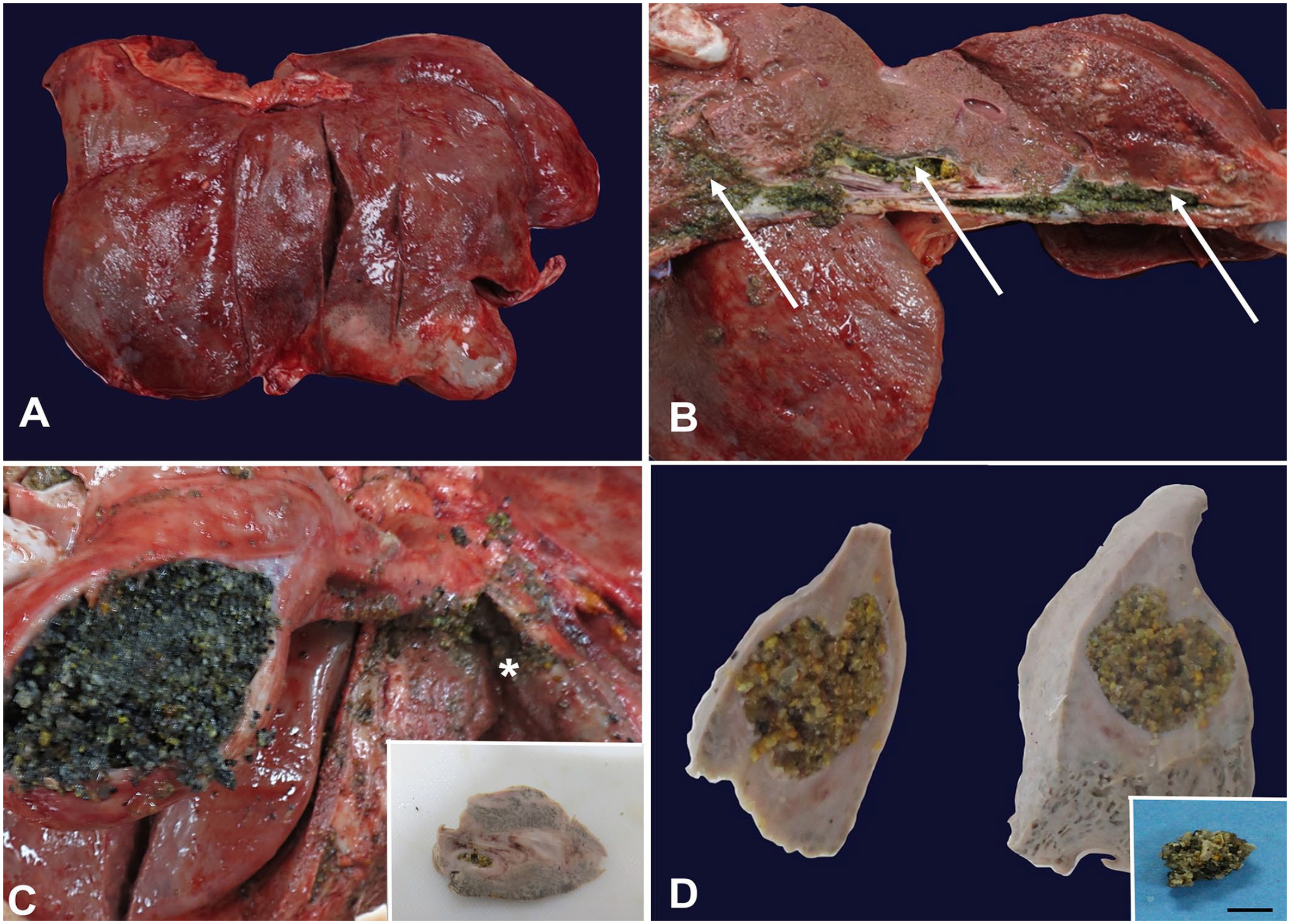
Gallbladder agenesis and intrahepatic cholelithiasis in a wild boar.
Histologically, the liver had severe cirrhotic changes characterized by thick dense collagen septa that formed pseudolobules and replaced the normal hepatic structure (Fig. 2A, 2B). A few regenerative nodules of hepatocytes were characterized by hepatocytes with large nuclei, prominent nucleoli, and scant cytoplasm with frequent binucleation, surrounded by thick fibrotic septa (Fig. 2A, 2B). Moreover, gram-positive bacilli and gram-negative cocci were observed in the sites of intrahepatic cholelithiasis by Gram staining (Fig. 2B). A striking pathologic finding was metaplasia of the intrahepatic bile ducts into gallbladder-like structures (Fig. 2C, 2D), which were composed of a mucosal layer including a simple columnar epithelium with striated border, lamina propria, and crypts. These structures were surrounded by thick fibrous tissue (Fig. 2C, 2D).
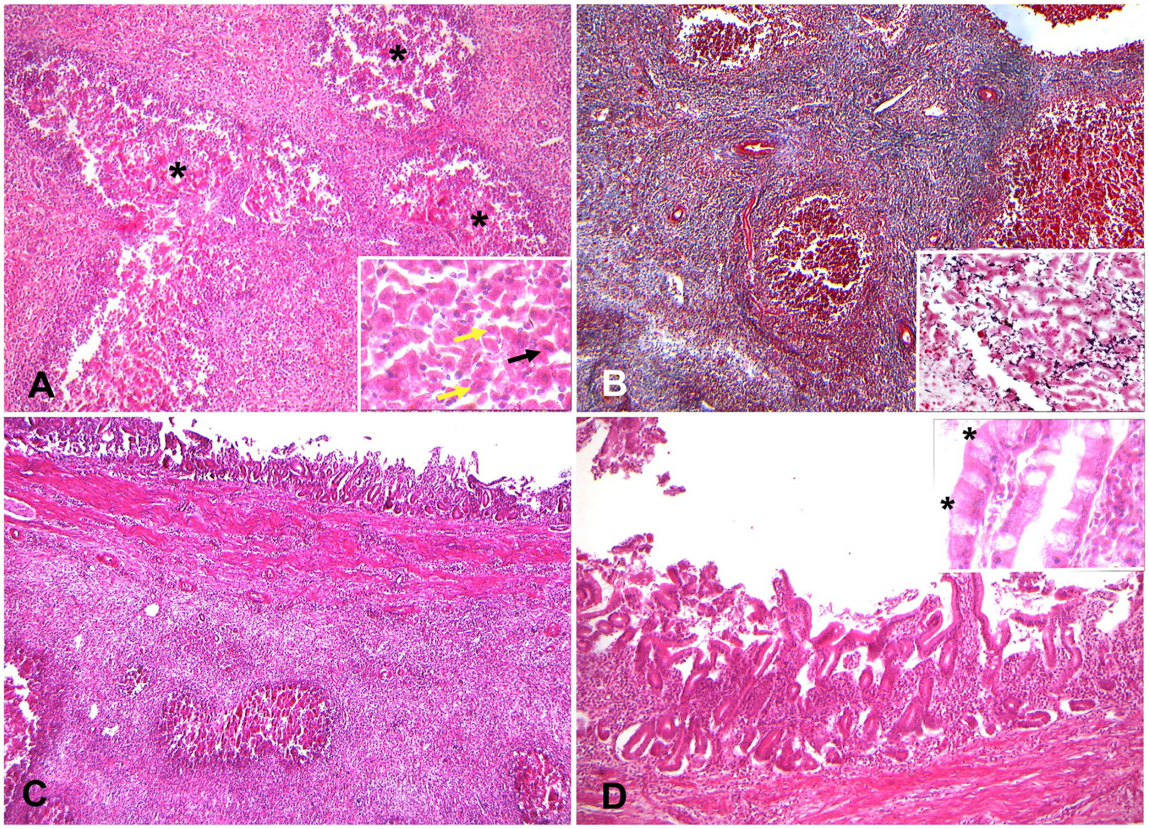
Representative microscopic features of the hepatic lesions in the wild boar, moderate autolysis.
Choleliths were submitted for Fourier-transform infrared spectroscopy, the stones were composed of 80% struvite and 20% calcium oxalate monohydrate (Fig. 3). Struvite (magnesium ammonium phosphate) and calcium oxalate monohydrate stones are commonly observed in the kidney.3,18 The main cause of struvite formation in the kidney is infection by bacteria, such as Proteus, Staphylococcus, Pseudomonas, Providencia, and Klebsiella. 1 However, struvite cholelithiasis is extremely rare, even in humans. 19

Fourier-transform infrared spectrum of a wild boar cholelith compared to a struvite reference spectrum.
Bile is secreted by hepatocytes, passes through the intrahepatic bile ducts, and is concentrated and stored in the gallbladder. 12 Agenesis of the gallbladder could result in the continuous release of unconcentrated bile into the small intestines, 12 which may have occurred in our case. If the amount of bile produced exceeded the capacity of the intrahepatic bile ducts, obstruction and cholestasis may have occurred. Retention of bile in the bile ducts could have initiated cholelithiasis, which may have caused secondary irritation, ascending infection of the biliary tree, and inflammation that resulted in the transformation of the intrahepatic bile duct epithelial cells into gallbladder-like epithelial cells.
Metaplasia is the reversible transformation of an adult cell type to another to adapt to abnormal stimuli. 5 Pseudopyloric gland metaplasia of the intrahepatic bile ducts as a result of intrahepatic cholelithiasis has been reported in humans. 8 Intestinal metaplasia of bile ducts has also been reported after cholecystectomy in humans. 4 The common cause of metaplasia was inflammation. 4 Metaplasia of the bile ducts might have occurred as a result of irritation, inflammation, and infection in our case of intrahepatic cholelithiasis.