
Abstract
The aim of this study was to elucidate the cause of a neurological syndrome characterized by stridor in adult goats with clinical signs of copper deficiency. The main clinical signs consisted of apathy, emaciation, pale mucous membranes, mucous nasal discharge, dyspnea, severe achromotrichia, diffuse alopecia, torpor, ataxia, and stridor. When the goats were forced to move, the stridor increased. In a herd of 194 Toggenburg goats, 10 adult goats with clinical signs of copper deficiency were removed from the herd and divided into 2 groups: group 1, which consisted of 4 nannies and 1 buck with stridor, and group 2, which consisted of 4 nannies and 1 buck without stridor. Group 3, used as a control, consisted of 5 adult goats from another flock without any clinical signs of disease. The mean serum copper concentrations were 1.3 ± 0.3 μmol/L in group 1, 8.1 ± 1.1 μmol/L in group 2, and 11.3 ± 2.2 μmol/L in group 3. The mean serum iron concentrations were 42.3 ± 14.2 μmol/L in group 1, 39.1 ± 8.2 μmol/L in group 2, and 20.6 ± 6.1 μmol/L in group 3. The main histological lesions in goats from group 1 were axonal degeneration of the recurrent laryngeal nerves and atrophy of the muscles of vocal folds and of the dorsal cricoarytenoid and right and left cricothyroid muscles. Goats with ataxia had neuronal degeneration and necrosis of cerebellar Purkinje cells and of the cranial cervical ganglion. We concluded that the stridor was caused by axonal degeneration of the recurrent laryngeal nerves due to the severe copper deficiency.
Copper is an essential trace mineral in the functioning of the nervous system and plays a critical role in a variety of metabolic functions essential for the activity of numerous enzymes, cofactors, and reactive proteins. 28 It functions in oxidation and phosphorylation reactions as part of the cytochrome oxidase system and is a cofactor for activity of enzymes, including copper-superoxide dismutase, dopamine-β-monooxygenase, ceruloplasmin, cytochrome C oxidase, monoamine oxidase, lysyl oxidase, and tyrosinase. 3,5,9,28
Copper deficiency in animals is classified as primary or secondary. 32,33 Primary copper deficiency occurs mainly in monogastric animals and is due to inadequate dietary copper intake. In ruminants, secondary copper deficiency commonly occurs due to the reduced copper absorption, mainly caused by the excess intake of Mo, S, Ca (such as calcium carbonate), Zn, Fe, Mn, Co, Pb, and Cd. 2,3,5,10,33 Selenium may also react with dietary copper, reducing its absorption from the gastrointestinal tract. 23,28
Neurologic diseases in offspring due to copper deficiency in their dams have been found in newborns of sheep, goat, cattle, red deer, guinea pig, pigs, and rats. 8,18,19,30 General clinical signs are tremors, incoordination, paralysis, and death. 33 Recently, copper deficiency was associated with peripheral neuropathy, leading to acute blindness, paresthesia, urinary incontinence, and loss of nociception in the extremities. 4,14,15,25 In Brazil, it is thought that copper deficiency influences the development of an uncharacterized disease referred to as “snoring” or “roaring” disease in adult cattle, whose main characteristic is stridor when the animals are forced to move. 27 Similarly, peripheral neuropathy due to molybdenosis has been described in copper-deficient moose (Alces alces) and yaks (Bos grunniens). 7,31 Laryngeal paralysis, most commonly attributed to the syndrome of recurrent laryngeal neuropathy (RLN), is long recognized as the most important equine upper airway disease. 6 Nevertheless, copper deficiency is not associated with RLN. 6,32
The neurologic consequences of copper deficiency in sheep and goats are mainly characterized by the development of enzootic ataxia, which occurs in neonates up to 180 days of age. 18,19,22,30 In adult animals, neurodegenerative diseases associated with copper deficiency are poorly understood, and in goats with severely low copper status, peripheral neuropathies have not been reported. The aim of this study was to investigate the cause of a neurological condition characterized mainly by stridor in adult goats in a semiarid region of northeastern Brazil and to describe the epidemiological, clinical, and pathological aspects of this disease.
Materials and Methods
Animal experiments were approved by the ethics committee of the Federal University of Pernambuco (Process Number 23082.019750/2014-71).
This study was performed on a herd of 194 Toggenburg goats showing signs of severe copper deficiency in the semiarid region of Pernambuco, northeastern Brazil (07° 52′ 29′′ S 35° 27′ 01′′ W). Ten adult goats were removed from the herd and distributed among 2 groups: group 1, formed by 4 nannies and 1 ram exhibiting stridor and clinical signs of copper deficiency, and group 2, formed by 4 nannies and 1 ram unaffected by stridor but exhibiting clinical signs of copper deficiency. A third group (group 3), used as control, comprised 5 adult goats from another flock without any clinical signs of disease. The average age of goats in the 3 groups was 4.6 years.
The goats of the 3 groups were examined in detail as to their general condition, behavior, attitude, mental status, coordination, head posture, movement, appetite, mucous membrane color, rectal temperature, heart and respiratory rates, abdominal morphology, reticulum-rumen motility, and the physical appearance of their feces, urine, and skin. 20 Neurological exams were performed according to a previous description. 16
A complete blood count was performed to determine the packed cell volume using the micro-hematocrit technique, hemoglobin level was assessed by the cyanomethemoglobin method, and red blood cell (RBC) count was assessed using a Neubauer chamber. To determine the plasma protein level, blood samples were collected with EDTA at 10%, heated at 57°C for 3 minutes, centrifuged, and analyzed by refraction. 11
Mineral Analysis
Twenty samples of Buffel grass (Cenchrus ciliaris) were collected and processed according to the methodology proposed by the Association of Official Analytical Chemists (AOAC) 1 for determining the levels of copper, iron, molybdenum, and zinc.
In addition, serum samples were analyzed by mass-coupled atomic absorption spectrometry to characterize their mineral content. Samples were diluted from 6.0× to 20.0× in ultrapure water (Milli-Q, Merck Millipore, California, USA). 24 The samples were weighed on an analytical balance, placed in borosilicate tubes containing nitric perchloric acid (4:1 v/v), and maintained at rest for 12 hours. The tubes were then placed in the digestion system (microwave X-Plus Mars, CEM Microwave Technology Ltd, Buckingham, UK) at 150°C. At the end of the digestion, 10 ml hydrochloric acid 0.1 N was added, and the samples were sent for analytical procedures. 26 The elements copper, molybdenum, iron, and zinc were assessed by mass-coupled atomic absorption spectrometry (ICP-OES) using a SpectrAA L 200 device (Varian, Ringoes, NJ, USA). 13
Pathology
Three goats from each group (2 females and 1 male) were euthanized by xylazine (2%) and sodium pentobarbital (5%) overdose. The entire central nervous system (CNS) and samples of the principal abdominal and thoracic organs, the vocal folds, and the laryngeal muscles (ie, cricothyroid, cricoarytenoid, hyoepiglottic and thyroarytenoid) were fixed in 10% buffered formalin. After fixation, samples of the CNS were obtained from the cerebrum, brainstem, cerebellum, diencephalon, and spinal cord. Samples of the peripheral nervous system (PNS), including the glossopharyngeal, vagus, accessory, hypoglossal, cranial laryngeal, caudal laryngeal, and recurrent laryngeal nerves, and the cranial cervical ganglia were fixed in 20% buffered formalin. All samples were processed routinely and were stained with hematoxylin and eosin (HE). In addition, the CNS and PNS samples were stained using a luxol fast blue stain.
Therapeutics
After the initial visit, goats in the herd were subcutaneously injected with a commercially available copper solution (5.5 mg copper lactobionate + 3.1 mg copper gluconate + 980 mg copper octadecanoate) at a dose of 0.1 ml/kg of body weight. This treatment was repeated after 30, 60, 90, and 120 days.
Statistical Analysis
Data were tested for normal distribution and analyzed by Tukey test using the software ASSISTAT version 7.7 beta, UFCG/Paraíba/Brazil.
Results
Epidemiology and Clinical Signs
The disease occurred on a small farm of approximately 10 hectares in the semiarid region of Pernambuco, Brazil. This region is part of the semiarid region of the country and experiences rainfall not exceeding 295 mm during the rainy season and 25 mm during the dry season. The goats were raised in a semi-intensive system. During the morning, the goats were allowed to graze in a paddock with Buffel grass (C. ciliaris). Late in the morning, they were transferred to a corral, where they were fed commercial food, mineral salt for sheep (350 mg/kg of copper), and chopped grass. Water obtained from a well was provided ad libitum. During inspection of the pasture, it was noted that a neighboring property was used for the production of ceramic bricks and did not control its waste release.
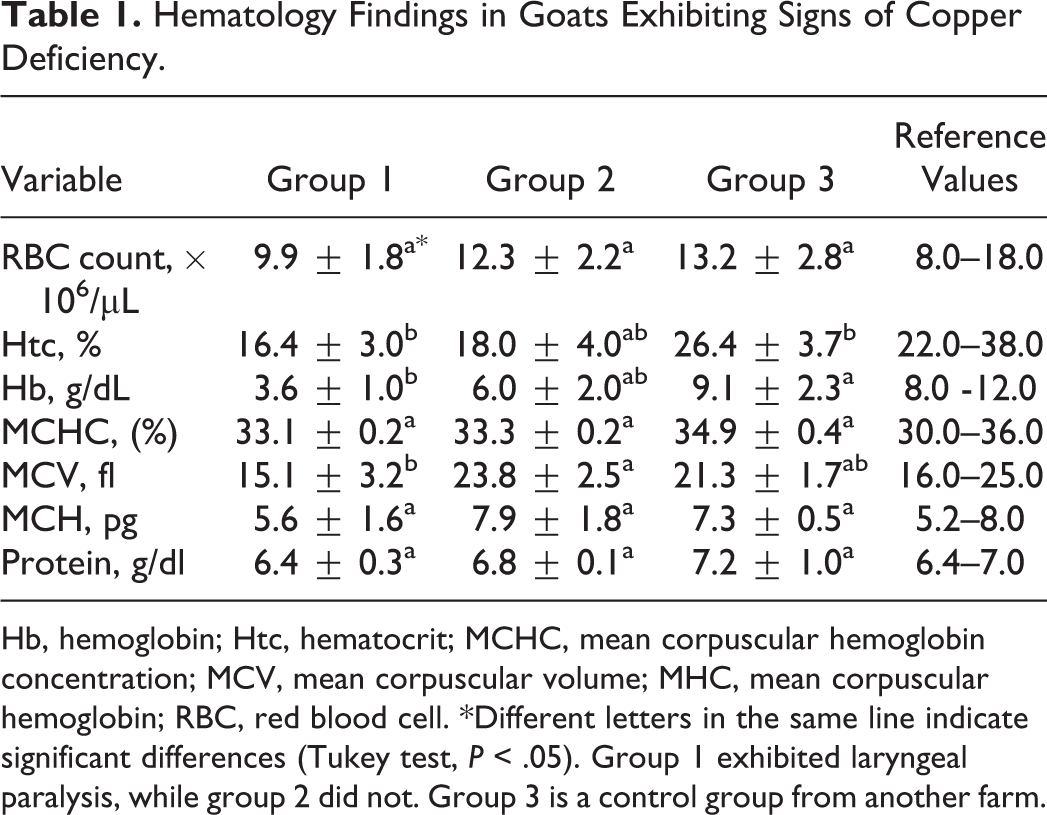
The farmer reported goats with difficulty breathing, respiratory noises, and high neonatal mortality throughout the last year, regardless of the season. The herd consisted of 194 Toggenburg goats, including 8 bucks, 150 nannies, and 36 young goats of different ages. Twelve (7.6%) adult goats exhibited apathy, anorexia, emaciation, pale mucous membranes, mucus nasal discharge, dyspnea, severe achromotrichia, diffuse alopecia, ataxia, torpor (ie, a slow and unsteady gait), reluctance to move, prolonged recumbency, and stridor (Figs. 1, 2; a video is available as Supplemental Material). The stridor was heard at rest but worsened when the animals were forced to walk or run. Less severe signs of copper deficiency without stridor or ataxia were observed in 111 goats (69.6%). Blood-tinged diarrhea was noted in both groups but only at the beginning the disease.
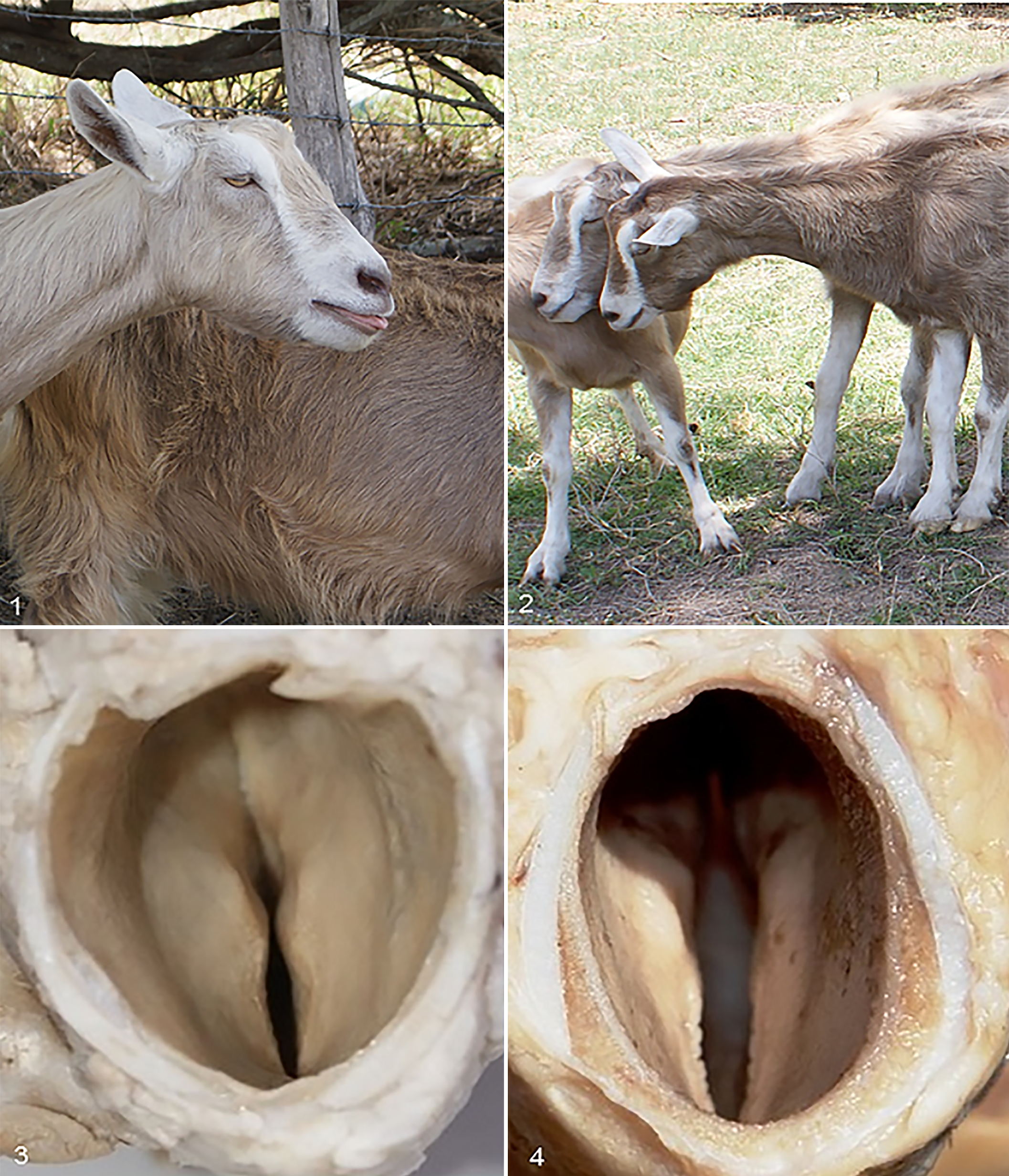
Laryngeal paralysis associated with copper deficiency, adult goats, group 1.
Blood Analysis and Determination of Mineral Content of Serum and Forage
The average hematocrit and hemoglobin levels were markedly reduced in goats from group 1 and were near the lower limits in goats from group 2. Both groups had microcytic normochromic anemia. There were no changes in the average total plasma protein. The results for the goats in group 3 were within normal limits (Table 1). 11
Hematology Findings in Goats Exhibiting Signs of Copper Deficiency.
Hb, hemoglobin; Htc, hematocrit; MCHC, mean corpuscular hemoglobin concentration; MCV, mean corpuscular volume; MHC, mean corpuscular hemoglobin; RBC, red blood cell. *Different letters in the same line indicate significant differences (Tukey test, P < .05). Group 1 exhibited laryngeal paralysis, while group 2 did not. Group 3 is a control group from another farm.
The average copper concentration in serum of goats from group 1 was markedly lower than for group 2 or 3, whereas the iron serum content was significantly increased. In group 2, the average copper concentration was near the lower limit, while the average iron content was increased. Serum concentrations of molybdenum and zinc in groups 1 and 2 were within the reference values. The average serum concentrations of copper, iron, molybdenum, and zinc in group 3 goats were within normal limits (Table 2). 12
Serum Concentrations μmol/L of Copper, Iron, Molybdenum, and Zinc in Goats Exhibiting Signs of Copper Deficiency.
Different letters in the same line indicate significant differences (Tukey test, P < .05). Group 1 exhibited laryngeal paralysis, whereas group 2 did not. Group 3 is a control group from another farm.
In forage samples, the average copper content was within the reference range, whereas the average iron values were high. The average values of zinc and molybdenum were within the reference parameters (Table 3). 28
Copper, Zinc, Molybdenum, and Iron Concentrations (mg/kg) in the Forage of Goats Exhibiting Signs of Copper Deficiency.
Therapeutics
The goats in group 1 did not recover from the disease after treatment with subcutaneously injected copper solution and died between 6 and 8 months after the onset of clinical signs. The goats in group 2 and the remaining goats of the herd showed an improvement in clinical status after 60 days of treatment and had completely recovered by 90 and 105 days after the onset of treatment.
Pathology
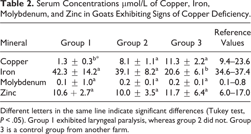
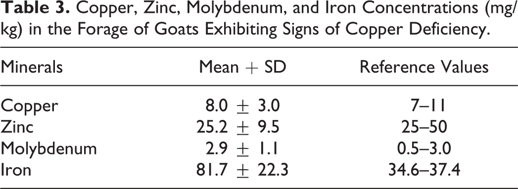
In goats in group 1, the main gross lesions consisted of atrophy of the vocal folds (Figs. 3, 4) and pallor of the dorsal cricoarytenoid and cricothyroid muscles. In 2 goats with less severe muscle atrophy, the lesion was more pronounced on the left side. Three goats exhibited focal dark red areas in the accessory and cranial lobes of the lung and in the ventral portion of the caudal lobe. The mediastinal and mesenteric lymph nodes were swollen and enlarged. There were no significant changes in the other organs examined. In group 2, no lesions were observed in the CNS, PNS, or larynx. The lungs and lymph nodes showed similar lesions to those described for group 1. Gross lesions were not observed in any of the goats in group 3.
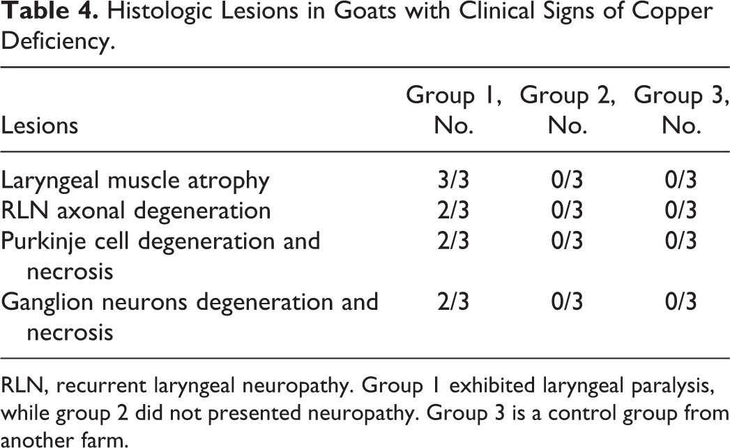
Histologically, in adult goats with stridor, the muscles of the vocal folds, the dorsal cricoarytenoid, and the right and left cricothyroid muscles were atrophic (Figs. 5, 6). The atrophy was characterized by reduction in myofiber diameter with an angular contour of shrunken myofibers. Atrophic fibers were interspersed with nonatrophic fibers, without evidence of clustering, resulting in marked variation in fiber size. In these muscles, we observed a minimal increase of satellite cells, infiltration of lymphocytes and macrophages, and replacement of the fibers by connective tissue (Figs. 7, 8). In the recurrent laryngeal nerve, there was axonal degeneration with myelin sheath expansion and the presence of vacuoles, usually in chains and containing axonal debris or macrophages (Figs. 9, 10). On the transverse section of these nerves, the fragmentation and loss of myelin and axons were more evident (Figs. 11, 12). In the nervous systems of goats with ataxia (group 1), there was Purkinje cell degeneration with swollen perikarya, axons, and dendrites. Purkinje cells with shrunken perikarya and pyknotic nucleus were also observed with less intensity. Neuronal necrosis was also observed in the cranial cervical ganglia, where some neurons exhibited irregular Nissl staining, whereas others had red perikarya and marginal and pyknotic nuclei (Table 4). No lesions were observed in other CNS or PNS samples. In the lungs, the lesions consisted of hyperemia and interstitial edema with infiltration of mononuclear polymorphonuclear leukocytes. Goats without stridor (groups 2 and 3) did not exhibit any laryngeal or neurologic lesions. The lung lesions in these goats consisted mainly of bronchopneumonia similar to that for group 1.
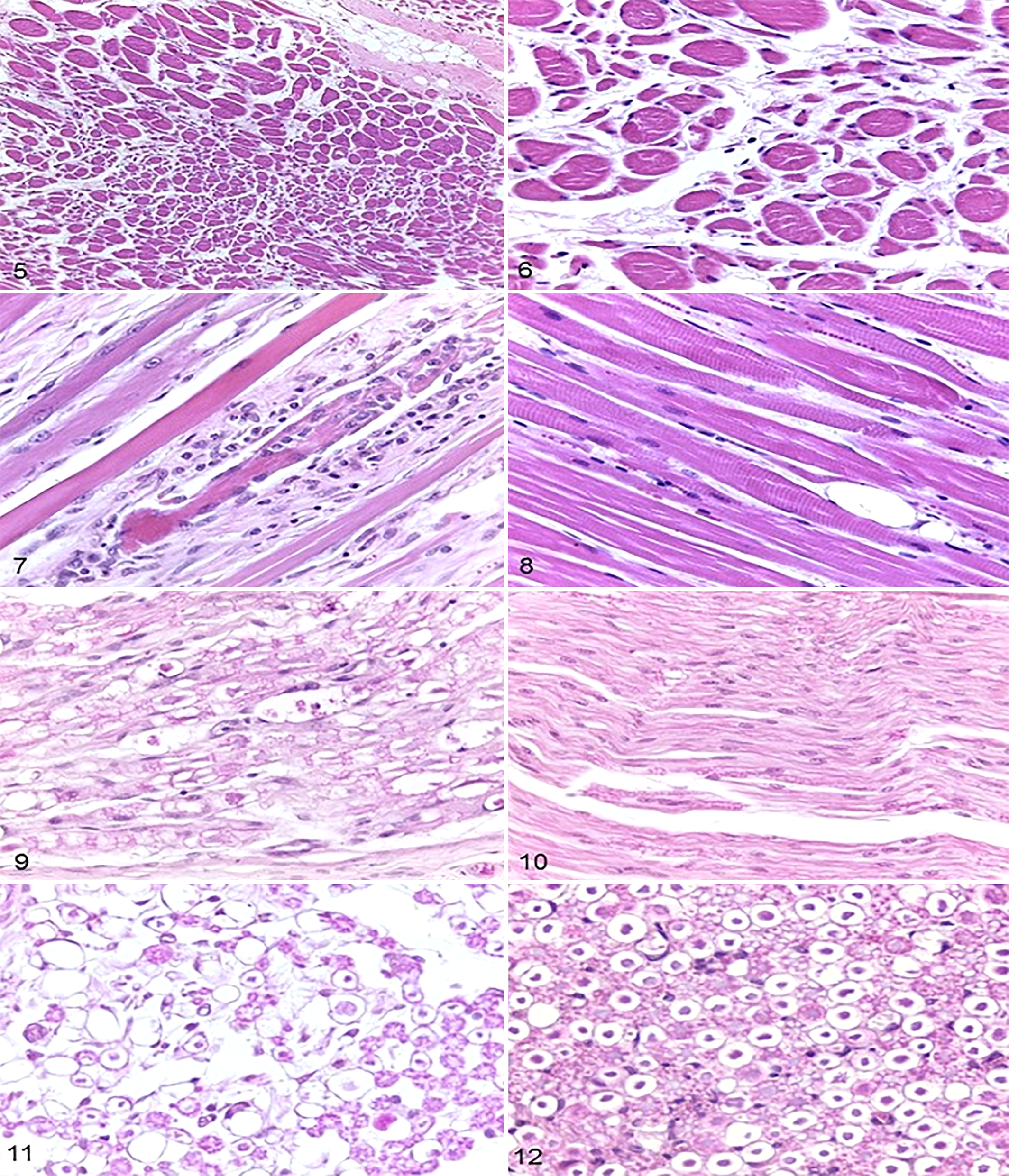
Denervation atrophy, dorsal cricoarytenoid muscle, adult goats.
Histologic Lesions in Goats with Clinical Signs of Copper Deficiency.
RLN, recurrent laryngeal neuropathy. Group 1 exhibited laryngeal paralysis, while group 2 did not presented neuropathy. Group 3 is a control group from another farm.
Discussion
This study describes a novel condition in a group of adult goats characterized mainly by apathy, achromotrichia, alopecia, ataxia, anemia, stridor, laryngeal neuropathy with muscle atrophy, and neuronal necrosis of cerebellar Purkinje cells and the cranial cervical ganglion. Serum copper concentrations in affected goats were 1.36 ± 0.39 μmol/L, lower than those reported previously for goats with copper deficiency, which ranged from 3 to 9 μmol/L. 12,21,23,24,28 Normal values range from 9.4 to 23.6 μmol/L. 9,21,23,24,28 These results indicate that stridor in this group of goats is associated with severe copper deficiency.
Serum iron concentrations were higher than the reference limits for the species (34.6–37.45 μmol/L). 24 Values higher than 39 μmol/L were considered excessive. The plasma zinc and molybdenum levels fell within the reference values for goats, which ranged from of 6.0 to 17.0 μmol/L for zinc and 0.1 to 0.8 μmol/L for molybdenum. 9,12 These data and the high iron concentrations of the forage suggest that iron may be responsible for the severe copper deficiency observed in this herd. 10 Environmental contamination by the neighboring ceramic industry could be responsible for the high concentration of iron in the forage; the principal refuse compounds of this industry are iron oxides and hydroxides. 17
Notwithstanding the trace minerals investigated in this research, it is necessary to remember that others are associated with reduction of copper absorption in the ruminant gastrointestinal tract (eg, calcium carbonate, S, Mn, Co, Pb, and Cd). 33 In relation to copper deficiency secondary to iron overload, it is well established that higher iron intake may antagonize copper absorption through the production of iron sulfide (FeS) in the intestine, which binds with copper to form insoluble iron compounds, and through the utilization of nonspecific transporters of multiple metals by soluble iron. 8,10,21,23
In this outbreak, wide variation in the clinical signs of adult animals was observed, including anemia, weight loss, achromotrichia, alopecia, and diarrhea at the onset of clinical signs. Ataxia, torpor, aspiration pneumonia, and stridor were observed in the more severely affected animals. This variation in signs should be taken into account when diagnosing severe copper deficiency. The cause of stridor in the goats of group 1 was paralysis of one or both arytenoid cartilages due to axonal degeneration in the recurrent laryngeal nerve and neurogenic atrophy of the fibers of the cricoarytenoid and cricothyroid muscles. In a previous study, a secondary experimental copper deficiency due to molybdenosis was investigated in goats, and no peripheral nervous lesions were observed. 2
Copper is required for the catalytic activity of enzymes that play an essential role in neurobiology and disease, including cytochrome C oxidase for electron transport in the mitochondrial respiratory chain, Cu-Zn superoxide dismutase for antioxidant defense, dopamine β-hydroxylase for catecholamine biosynthesis, and ceruloplasmin for brain iron homeostasis. 29 It is now generally accepted that a low copper content in the nervous system leads to a deficiency of COX and a decreased synthesis of phospholipids. 33 These are proposed as the main mechanisms of nervous system dysfunction reported in this study.
In this study, adult goats with severe copper deficiency associated with high iron intake developed severe clinical signs, including anemia, weight loss, achromotrichia, alopecia, and diarrhea at the onset of clinical signs; ataxia and stridor are also observed in more severely affected animals. The stridor was presumed to result from laryngeal paralysis, caused by degeneration of the recurrent laryngeal nerves, leading to neurogenic atrophy of the dorsal cricoarytenoid and right and left cricothyroid muscles.
Footnotes
Declaration of Conflicting Interests
The author(s) declared no potential conflicts of interest with respect to the research, authorship, and/or publication of this article.
Funding
The author(s) disclosed receipt of the following financial support for the research, authorship, and/or publication of this article: the National Council for Scientific and Technological Development (Process 309725/2015-1) provided financial support.
References
Supplementary Material
Please find the following supplemental material available below.
For Open Access articles published under a Creative Commons License, all supplemental material carries the same license as the article it is associated with.
For non-Open Access articles published, all supplemental material carries a non-exclusive license, and permission requests for re-use of supplemental material or any part of supplemental material shall be sent directly to the copyright owner as specified in the copyright notice associated with the article.