
Abstract
Wooden breast (WB) myopathy of broiler chickens is a myodegenerative disease of an unknown etiology and is macroscopically characterized by a hardened consistency of the pectoralis major muscle. Our aim was to describe the development and morphology of WB over the growth period in broilers. Additionally, the effect of restricted dietary selenium on the occurrence of WB was examined by allocating the birds in 2 dietary groups: restricted and conventional level of selenium. The experiment included 240 male broilers that were euthanized at ages of 10, 18, 24, 35, 38, or 42 days and evaluated for WB based on abnormal hardness of the pectoralis major muscle. The severity and the distribution of the lesion and presence of white striping were recorded. The first WB cases were seen at 18 days; 13/47 birds (28%) were affected and the majority exhibited a mild focal lesion. In subsequent age groups the WB prevalence varied between 48% and 73% and the lesion was usually diffuse and markedly firm. White striping often coexisted with WB. Histological evaluation performed on 111 cases revealed a significant association of myodegeneration and lymphocytic vasculitis with WB. Vasculitis and perivascular cell infiltration were restricted to the veins. Restricted dietary selenium did not affect the occurrence of WB (P = .44). Our results indicate that WB starts focally and spreads to form a diffuse and more severe lesion.
Keywords
Wooden breast (WB) myodegeneration is an emerging disease in broiler chickens, macroscopically recognized as hardness of the pectoralis major muscle, often accompanied with pale color and white striping. 27 In slaughter-age broilers, that is, approximately 35 days and older, the main histological lesions of the breast muscle consist of chronic myodegeneration with regeneration and interstitial edema, accumulation of loose connective tissue, or fibrosis. 27 Replacement of the severely degenerated muscle fibers with connective tissue may partly account for the muscle hardness, but the hardened consistency seems to also occur in cases without excessive fibrosis. 27 Clinical signs have not been reported with WB and the lesion is usually detected postmortem. Although affected birds can exhibit diffuse and severe chronic myodegeneration of the pectoralis major muscle, it remains controversial whether the condition is painful. 27
The cause and pathomechanism of WB are currently unknown. There are several causes of myodegeneration in animals, such as deficiency of selenium or vitamin E, an excess of their antagonists, ionophore toxicity, exertional myopathy, hypoxia, and muscular dystrophies. 14,27,29 However, those are not associated with the substantial hardness of the muscle tissue, which is the key feature and the current basis for the diagnosis of WB. In addition, many of the previously described causes also affect other organs, whereas WB seems to be restricted to the skeletal muscle. 14,29
Selenium deficiency as a cause of oxidative damage leading to degenerative myopathy has been described in many animal species including broiler chickens. 7,12,14 Grain is the usual content in the diet of broilers; the variation in the natural selenium content of the soil and the use of fertilizers devoid of supplemented selenium are potential causes for insufficient selenium content of broiler feed. Recently, organic selenium has been shown to have a better bioavailability and antioxidative capacity than the inorganic selenium that traditionally has been used as a feed additive. 5,9,23
In addition to WB, there are other myodegenerative conditions of unknown etiology, with a recent emergence in the broilers. White striping is a condition affecting the breast and thigh muscles of broilers and it occurs alone or concurrently with WB. 17,27 It is macroscopically characterized by pale striations up to a few millimeters wide parallel to the muscle fibers. Histologically it shares many features with WB, such as degeneration, regeneration, interstitial inflammatory infiltrates, and fibrosis. 17,25,27 A dorsal myodegeneration, very similar to WB both macroscopically and histologically, has been reported in the anterior latissimus dorsi (ALD) muscles of broilers in Brazil. 31 Both the ALD myopathy and white striping have been associated with the heaviest birds or fillets of the flock or increased growth rate. 3,15,16,31
The objective of this study was to characterize the time of onset of WB, its progression over the growth period, and the associated morphological features. Additionally, we tested the effect of reduced selenium content of the feed on the occurrence of WB.
Materials and Methods
Two hundred and forty 1-day-old male Ross 508 broiler chicks were divided into 24 pens, 10 birds per each, in 1 room of an experimental facility. They received no medical treatments or vaccinations; their feed, however, included a narasin-based coccidiostat at the level of 0.07% (Monteban, Elanco, IN). The maintenance and clinical observation were performed on a daily basis. Due to heating lamps, the lighting was continuous for the first 7 days, which followed a daily 6-hour darkness period. The bedding consisted of wood chip litter on rubber mats on a concrete floor. Other husbandry conditions were adjusted according to the recommendations for conventional breeding. 1 Experimental procedures were not performed during life, and the birds were euthanized at the ages of 10, 18, 24, 35, 38, or 42 days by mechanical cervical dislocation, followed by phlebotomy of the jugular veins. The selection of birds for euthanasia was as follows: 2 birds per pen were randomly selected for the first and second age-group (10 and 18 days), 1 random bird per pen for the third age-group (24 days), and then all remaining birds from 10 pens at 35 days, 10 pens at 38 days, and the last 4 pens at 42 days (Table 1). This arrangement aimed at mimicking the density of birds per square meter near to that used in the conventional breeding. However, birds that died between the set age points were excluded from the study, which affects the number of birds per age-group. For each age-group, a balanced number of birds were chosen from both the dietary treatments, which are described in detail below. Before euthanasia, each bird was weighed individually. Ethical permission for the experiment was issued by the Research Animal Resources Committee of the University of Helsinki (Decision number KEK12-045).
Prevalence, Severity, and Location of the Macroscopic Wooden Breast Lesion in Broiler Chickens With Wooden Breast (WB) Myopathy.
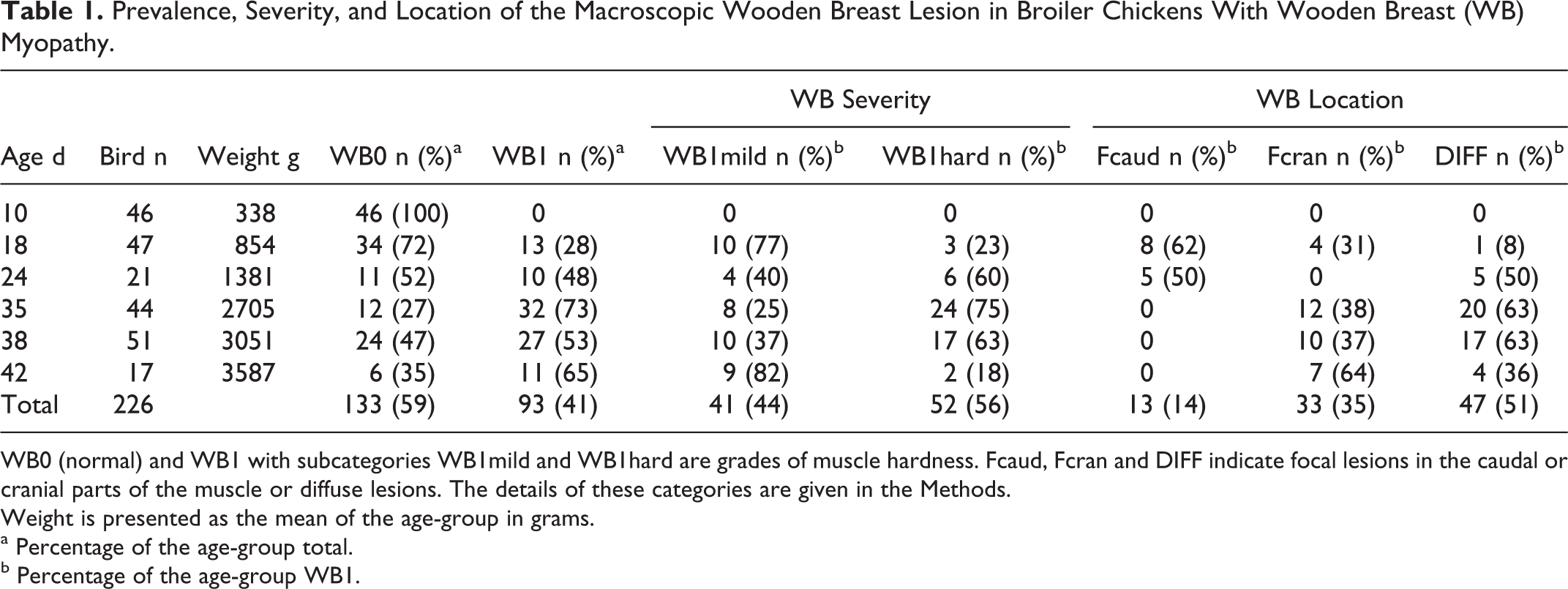
WB0 (normal) and WB1 with subcategories WB1mild and WB1hard are grades of muscle hardness. Fcaud, Fcran and DIFF indicate focal lesions in the caudal or cranial parts of the muscle or diffuse lesions. The details of these categories are given in the Methods.
Weight is presented as the mean of the age-group in grams.
a Percentage of the age-group total.
b Percentage of the age-group WB1.
Dietary Treatment
The birds were assigned for 2 ad libitum dietary treatments, SeLow and SeNorm, 12 pens each by random allocation. SeLow feed contained inorganic selenium (sodium selenite, Na2SeO3; Anima Ltd., Krakow, Poland) 0.11 mg/kg for the first 7 days and then 0.13 mg/kg until the end of the experiment. SeNorm feed contained organic selenium yeast (Selsaf; Lesaffre Feed Additives, Marcq-en-Baroeul, France) 0.32 mg/kg for the first 7 days and then 0.30 mg/kg until the end of the experiment. 2 The selenium content of the feed was analyzed by the inductively coupled plasma–mass spectrometry (Nordic Committee on Food Analysis Method 161:1998, International Organization for Standardization 17294–2). The feed was based on wheat and soybean. With the exception of selenium, the levels of other nutrients were adjusted according to recommendations and were similar for both groups. 1
Necropsy and WB Diagnosis
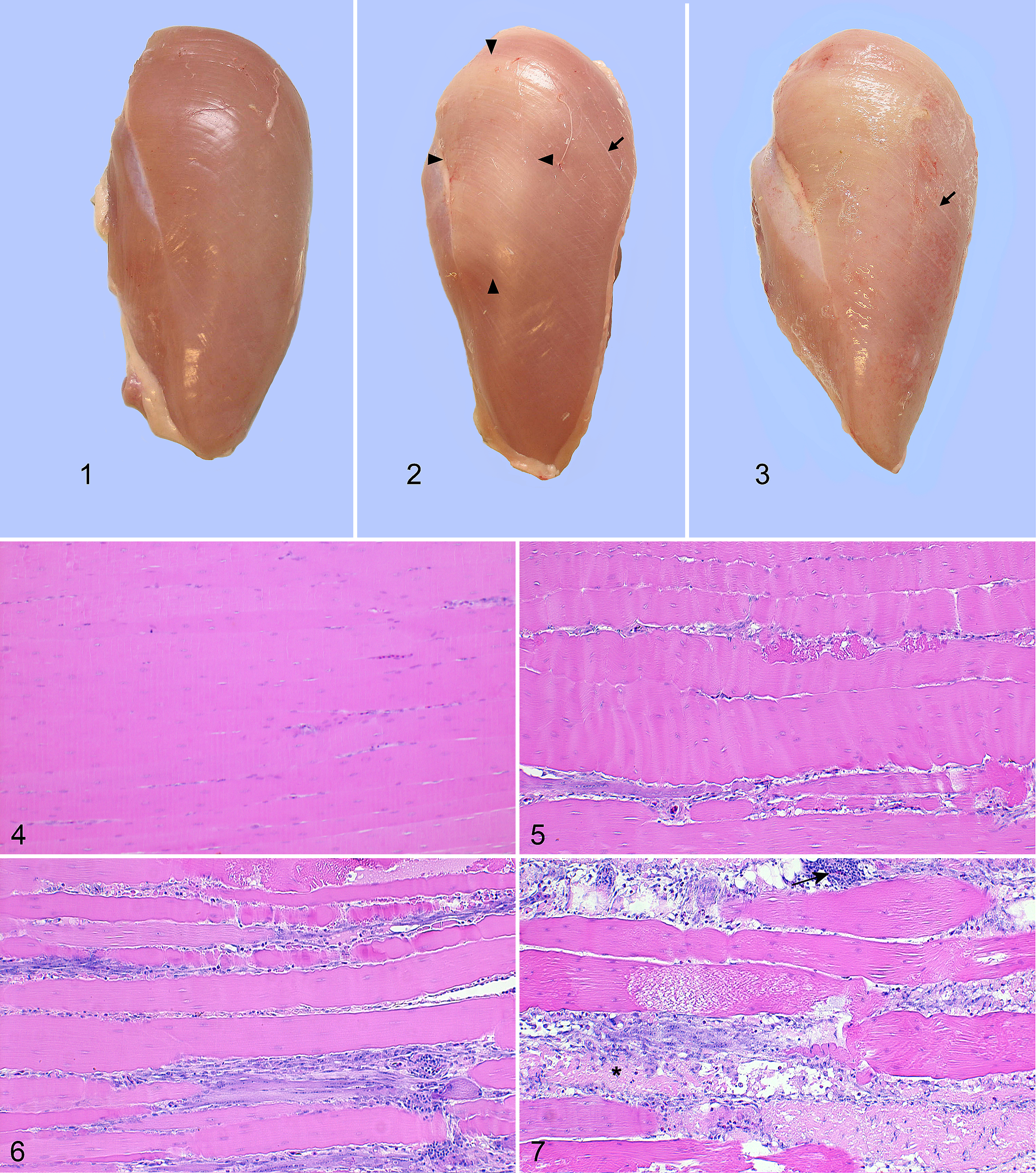
Birds were necropsied immediately after death. The necropsy and sampling crew were blind to the dietary treatment. After the evaluation of the skin, head, oral cavity, wings, legs, and footpads, the skin was bluntly dissected to reveal the pectoralis major muscles. Both pectoralis major muscles were visually evaluated for color change and white striping (present–absent) and palpated to evaluate the consistency of the muscle (normal, firm [mildly hardened] or markedly hardened [wood like]). The right pectoralis major muscle was then cut off and a second person graded its consistency by palpation as follows: grade WB0, normal consistency and macroscopically unaffected (Fig. 1), and grade WB1, hardened consistency and affected with WB (Figs. 2 and 3), including subcategories WB1mild, mildly hardened (firm) consistency, and WB1hard, markedly hardened consistency. Additionally, the distribution of the WB lesion was recorded as follows: focal (FOC) included cases with a single hardened lesion either caudally (Fcaud) or cranially (Fcran) within a muscle of otherwise normal consistency (Fig. 2); and diffuse (DIFF) included cases with diffuse or extensive lesions with the presence of very small areas of normal muscle consistency, usually in the middle lateral area (Fig. 3). When there was divergent scoring (n = 3, normal vs WB1mild), the 2 persons reevaluated the cases together to reach an agreement. The muscle was sampled for histology immediately after grading. The internal organs as well as muscles of the left thigh and large joints of the left limb were macroscopically evaluated (n = 226).
Histological Evaluation
Of the 226 macroscopically examined birds, 111 were evaluated histologically (WB0 n = 61; WB1 n = 50; Table 2). Our focus was to describe the early development of WB, so all cases at 18 and 24 days of age (the phase of early macroscopic lesions) were histologically evaluated. Additionally, at least 25% of other age-groups were randomly chosen for histological evaluation.
Histological Grades of Myodegeneration and Vasculitis by Age-Group in Broiler Chickens with Wooden Breast Myopathy.
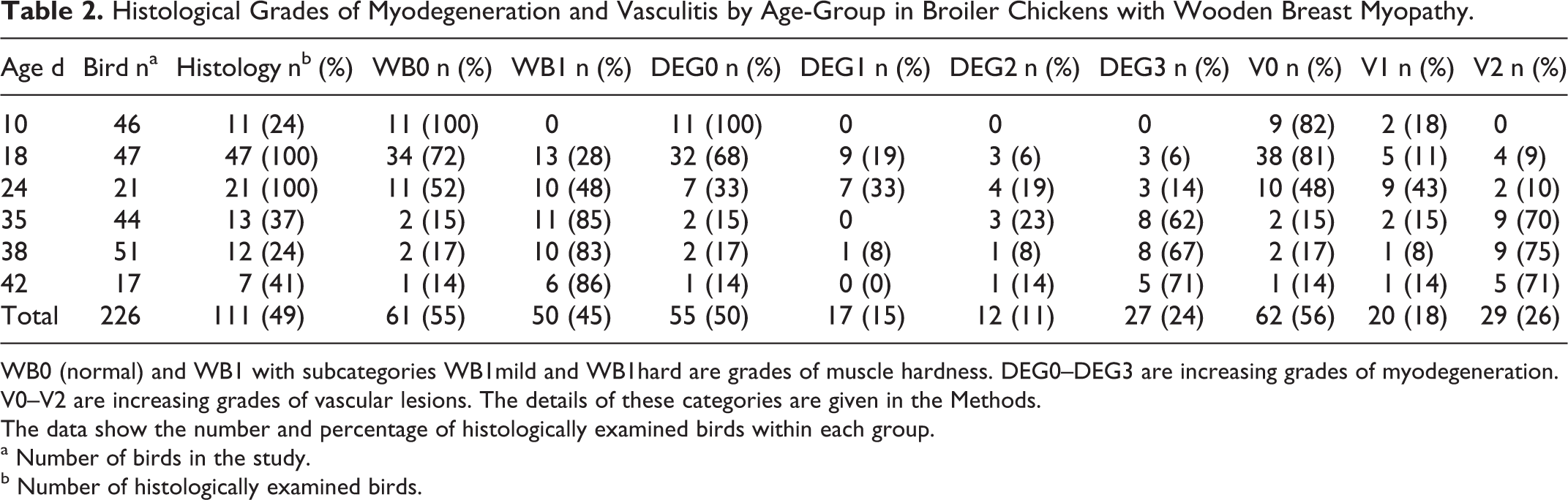
WB0 (normal) and WB1 with subcategories WB1mild and WB1hard are grades of muscle hardness. DEG0–DEG3 are increasing grades of myodegeneration. V0–V2 are increasing grades of vascular lesions. The details of these categories are given in the Methods.
The data show the number and percentage of histologically examined birds within each group.
a Number of birds in the study.
b Number of histologically examined birds.
For histology, a sample of 1 × 1 × 1 cm was collected immediately after the macroscopic grading from the right-side pectoralis major muscle. The sample was taken approximately 1 cm deep from the ventral surface of the muscle. The sampling sites were as follows: WB0 muscles were sampled from the middle region, the FOC cases from the focal lesion site, and diffusely affected muscles from the most severely affected region. Of FOC cases, a total of 15 cases at 18, 24, 35, and 38 days were also sampled from an area of normal consistency (non-lesion site) in addition to the lesion site, for the evaluation of intraindividual development of WB. The left biceps femoris muscle of 5 birds was sampled for histology: 2 WB0 and 2 WB1 cases with macroscopically normal biceps femoris muscles and 1 WB1 case at 38 days with a hardened biceps femoris muscle. Of the internal organs, the heart, spleen, liver, kidney, lung, gizzard, duodenum, and pancreas of 12 birds, 1 randomly selected WB0 and WB1 bird per each age-group, were sampled and evaluated histologically.
The tissue samples were fixed in formalin by immersion, embedded in paraffin, and sectioned at 4 μm thickness. Muscle samples were oriented transversally and longitudinally. Slides were stained with hematoxylin and eosin. CD3 antigen was detected by immunohistochemistry (polyclonal rabbit anti-human CD3; DakoCytomation, Glostrup, Denmark).
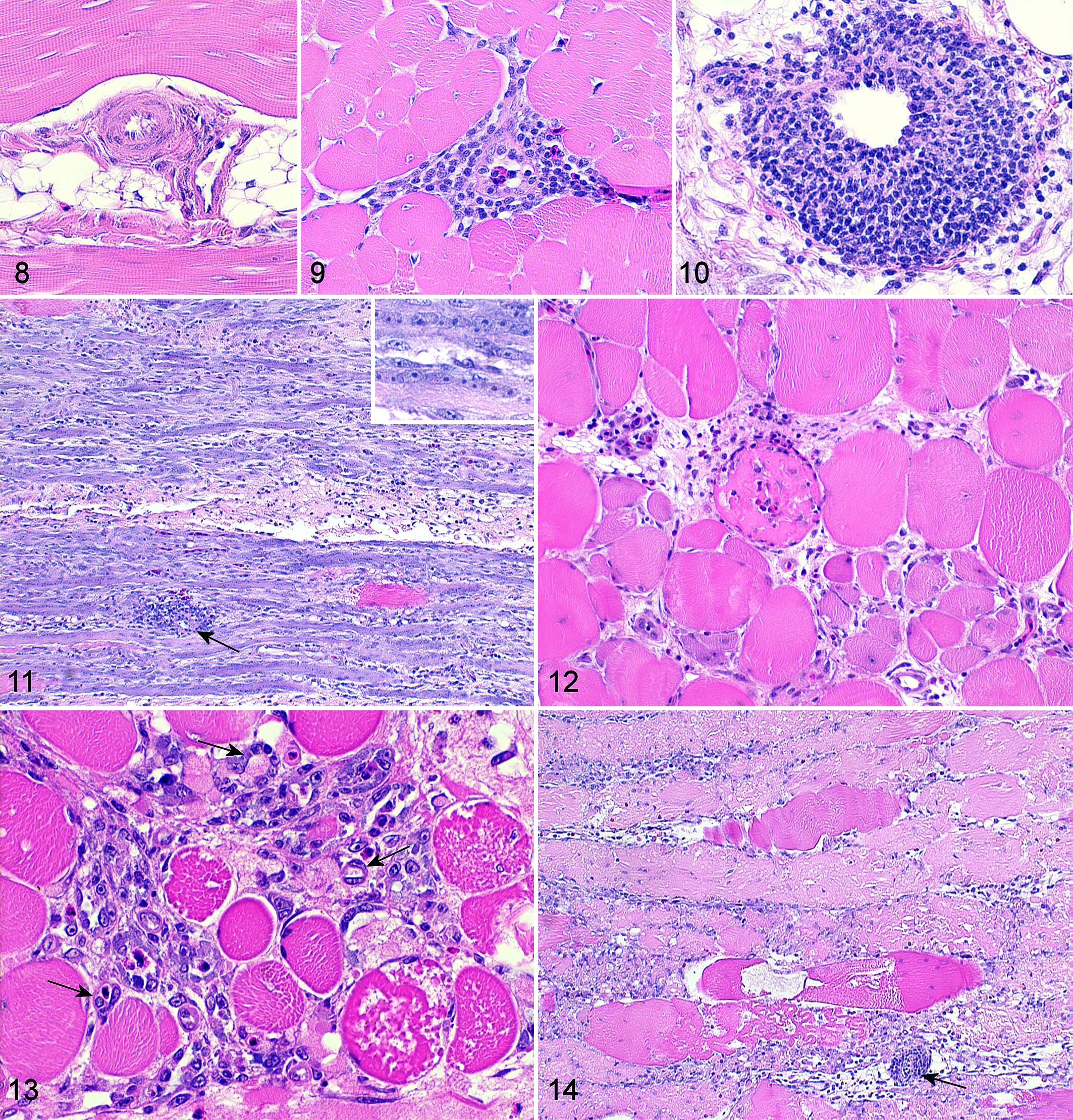
Histologically, the myodegenerative lesions were graded into 4 categories; DEG0 for absent or minimal, DEG1 for mild, DEG2 for moderate, and DEG3 for severe to excessive myodegeneration (Figs. 4–7). Myodegenerative fibers by definition included hyalinized fibers with the loss of cross striations as well as fragmented fibers that often were infiltrated or surrounded by macrophages or heterophilic granulocytes. Additionally, vasculitis and perivascular cell infiltration was graded into 3 categories according to the most severely affected vessel: V0 for normal vessel walls without surrounding cell infiltration, V1 for perivascular infiltrations of lymphocytes with or without mild intramural infiltration, and V2 for marked perivascular infiltration of lymphocytes with intramural infiltration sometimes obliterating the vascular wall (Figs. 8–10). The amount of perivascular adipose tissue was graded into 4 categories: A0 for absent, A1 for mild (thickness of the adipose tissue bed approximately similar to the largest diameter measure of the blood vessel), A2 for moderate (≤2× blood vessel diameter), and A3 for marked (≥3× blood vessel diameter) amount of adipose tissue. The presence of other histological features, such as regeneration, necrosis, and inflammatory cell infiltration, and the presence of loose connective tissue or fibrosis were also evaluated. The histological assessment was performed blinded to the macroscopic diagnosis and experimental dietary treatment. A board-certified veterinary pathologist (H.-K.S.) performed the necropsy and histological analyses.
Data Analysis
Descriptive analyses were performed for the variables. The association between the histological grades and WB or white striping was assessed twice with Fisher exact test; first by including only 18 and 24 days groups and then including all histologically evaluated birds. The relationships between WB severity and location or white striping were assessed with Fisher’s exact test. The association between weight and WB or dietary treatment was assessed by the analysis of variance. Logistic regression analysis was performed to analyze the effects of dietary treatment and age on WB prevalence; WB was the outcome variable. P values less than .05 were regarded as significant. Statistical analyses were performed using IBM SPSS Statistics version 23 (IBM Corp, NY).
Results
Of the 240 birds, 14 (5.8%; 7 for each dietary treatment) died during the experiment, exhibiting signs of sudden death syndrome (n = 7), ascites syndrome (n = 5) or other (n = 2) in the macroscopic postmortem examination. 14 These birds were excluded from the study due to the development of rigor and postmortem changes. In the remaining 226 birds, no significant clinical signs were detected except in 1 bird with difficulty walking but no gross lesions. WB was not suspected through clinical observation of any of the birds.
Macroscopic Findings
Palpatory hardness typical of WB was not exhibited by any of the excised pectoralis major muscles of the birds at 10 days of age. The first WB1 cases were seen at 18 days, with an age-group prevalence of 28% (Table 1). From 24 to 42 days, the WB1 prevalence remained close to or above 50% per group. When divided into the subcategories, the majority of the WB1 birds fell into WB1mild category at 18 days, but at 24, 35, and 38 days over 60% of the affected cases were of WB1hard. The last age-group, 42 days, slightly differed from this trend, with 18% in WB1hard but also had a markedly lower total number of cases. In addition to the hardened consistency, the WB lesion was characterized by pale color. Marked discrepancy between the right and left pectoralis major muscles in the presence or severity of the WB lesion was not noted.
The Fcaud location of the macroscopic WB lesion was detected only at 18 and 24 days; the Fcran and DIFF categories were more prevalent in the older age-groups (Table 1). The severity of the muscle hardness, WB1mild versus WB1hard, was associated with the location (P < .001); 2/13 cases (15%) of the Fcaud, 12/33 (36%) of the Fcran, and 38/47 (81%) of the DIFF were of the WB1hard category.
White striping and WB were significantly associated (P < .001; Supplemental Table S1). When white striping was present, it was usually seen throughout the pectoralis major and was not restricted to the WB area.
A macroscopic lesion similar to WB was detected in the biceps femoris muscle in the thigh of 1 bird at 38 days of age; the muscle focally exhibited mildly hardened consistency, whereas the bird had a diffuse WB1hard lesion in the pectoralis major muscle.
Histologic Findings
Myodegeneration was significantly associated with WB (P < .001; Table 3; WB0-1 vs DEG0-3). The myodegeneration grade for the 61 macroscopically unaffected (WB0) muscles was either DEG0, represented by all age-groups, or DEG1 that was seen only at ages 18 and 24 days. The majority of the WB1 cases exhibited DEG2 or DEG3, represented by all affected age-groups. DEG0 and DEG1 were more common in the younger age-groups of WB1 birds. In the WB1mild subcategory, all myodegeneration grades were rather equally represented, in contrast to the WB1hard, where 21/28 cases (75%) were of DEG3. Myodegeneration was segmental and accompanied by regeneration in all categories and age-groups (Figs. 5–7). In a few cases, areas of extensive regeneration were seen (Fig. 11). In cross section, the affected areas showed marked variation in shape and diameter of the fibers (Fig. 12). Some perivascular adipose tissue was present in all histologically evaluated cases. Within the 18- and 24-day-old birds, the amount of perivascular adipose tissue was not associated with WB, white striping, myodegeneration, or vasculitis grade (P > .05 for all variables). However, when all age-groups were included, the increased amount of perivascular adipose tissue was weakly associated with WB (P = .01) and with myodegeneration (P = .004; Table 3). Vasculitis and the amount of perivascular adipose tissue were not associated (P > .05). A few interstitial aggregations of adipose tissue without visible blood vessel bundles were observed in 3 cases (2 WB0 cases and 1 WB1mild case). Numerous spindle- to oval-shaped cells arranged in irregular vascular channel-like structures, with erythrocytes in the luminal area occasionally accompanying the myodegenerative changes (Fig. 13). The DEG2 and DEG3 grades were usually accompanied by moderate to marked interstitial edema and loose immature connective tissue as well as fibrosis in the older age-groups (35, 38, and 42 days). However, fibrosis was absent in the WB1 birds at 18 and 24 days of age, including the WB1hard cases (n = 8). Necrotic fibers were rare in general, but a few cases exhibited moderate to excessive numbers of necrotic fibers together with myodegeneration (Fig. 14). Features other than myodegeneration, perivascular inflammatory cell infiltration with vasculitis, and the amount of perivascular adipose tissue were not graded separately because they were either linked to myodegeneration or present in a minority of cases.
Association of the Macroscopic Wooden Breast (WB) Lesion With Histological Grades of Myodegeneration, Vasculitis, and Adipose Tissue.a
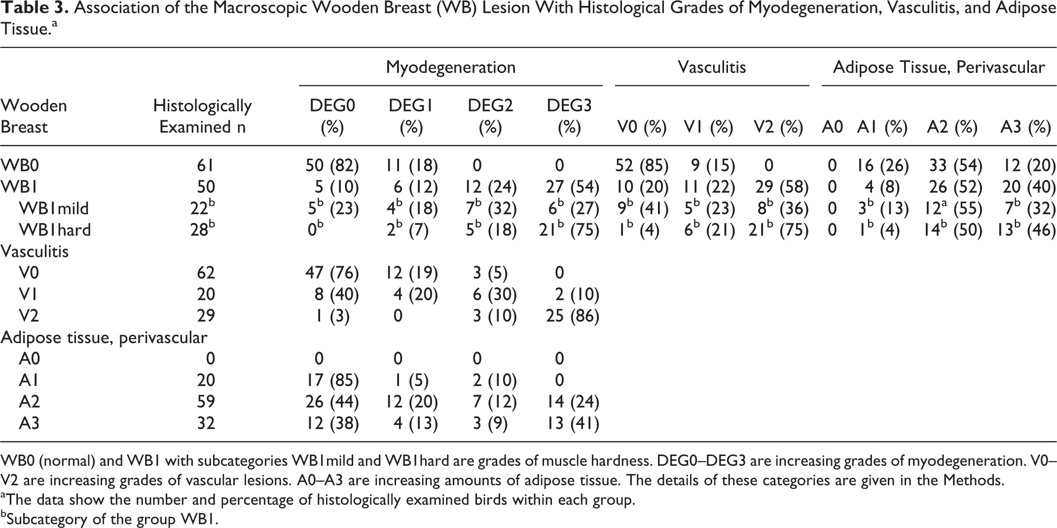
WB0 (normal) and WB1 with subcategories WB1mild and WB1hard are grades of muscle hardness. DEG0–DEG3 are increasing grades of myodegeneration. V0–V2 are increasing grades of vascular lesions. A0–A3 are increasing amounts of adipose tissue. The details of these categories are given in the Methods.
aThe data show the number and percentage of histologically examined birds within each group.
bSubcategory of the group WB1.
Vasculitis and WB were significantly associated (P < .001); none of the 61 WB0 cases, but 29/50 (58%) of the WB1 cases exhibited remarkable perivascular infiltration of lymphocytes with intramural infiltration, sometimes obliterating the vascular wall (V2; Table 3). Vasculitis was especially frequent in the WB1hard category; only 1 case did not show vasculitis or perivascular lymphocyte infiltration, whereas in 21/28 (75%) cases those lesions were extensive. The 10 WB1 cases that did not show any vascular lesions (V0) were either 18 or 24 days of age. Vasculitis scores V1 and V2 were exhibited by all affected age-groups. Nine WB0 cases that had perivascular infiltrations of lymphocytes (V1) were of the youngest age-groups: 10, 18, and 24 days.
Vasculitis and perivascular infiltrations were also significantly associated with higher myodegenerative scores (P < .001); 47/62 (76%) of the V0 cases were DEG0 and 25/29 (86%) of the V2 cases were of DEG3 (Table 3). Even though the majority of the V0 cases were associated with absent or minimal myodegeneration (DEG0), some of the DEG1 and DEG2 cases also lacked vascular changes; all these birds were either 18 or 24 days of age and WB0 or WB1mild macroscopically. All grade DEG3 cases had either perivascular infiltrations of lymphocytes (V1) or vasculitis (V2). Perivascular infiltration of lymphocytes (V1) was seen with all myodegeneration grades, but extensive infiltrations and vasculitis (V2) were associated mainly with grades DEG2 and DEG3. One DEG0 case also exhibited V2; this bird was 18 days of age and macroscopically was graded WB1mild.
Overall, vascular lesions and perivascular infiltrations were seen only in veins, typically venules or small veins; arteries did not exhibit significant changes even when the veins were severely affected. The cell infiltration around the veins formed dense cuffs and often was eccentric. Rare plasma cells or histiocytic cells were identified within the lymphocytic infiltration by morphology. Immunohistochemistry for the T-cell marker CD3 revealed a strong positive staining of the perivenular lymphocytes in 3 tested cases with V1 or V2, 10 or 18 days of age. Spleen tissue from the experimental birds was used as a control for the CD3 staining; their T-cell regions showed intense positive staining.
The nonlesion sites of the focally affected cases were sampled from 15 cases and exhibited histological myodegeneration scores of DEG0 (n = 1; lesion site was WB1hard DEG3), DEG1 (n = 7; lesion site WB1mild or WB1hard, DEG1-3), and DEG2 (n = 7; lesion site WB1hard, DEG3); none of them exhibited DEG3. Vasculitis and perivascular lymphocyte infiltrations were either absent or mild (V0 n = 8; V1 n = 6) in the majority of the nonlesion sites; only 1 nonlesion site exhibited V2. All nonlesion sites exhibited either A1 or A2 grades of perivascular adipose tissue.
Macroscopic white striping was significantly associated with myodegeneration (P < .001) when all birds were included. The association remained significant when it was tested with WB0 cases only; first only the birds of 18 and 24 days of age (P = .016) and then all age-groups (P = .011). Specific histological changes different from WB histology were not recognized for cases with macroscopic white striping.
In the internal organs, no significant pathological changes were seen. In the liver parenchyma and in the lamina propria of the duodenum, occasional aggregates of lymphoid cells were present. Perivascular lymphocytic infiltrations or vasculitis were not exhibited in any of the examined internal organs.
The biceps femoris muscle that had a macroscopic lesion similar to WB was histologically graded as DEG3, V2, and A1. The 4 histologically assessed but macroscopically normal biceps femoris muscles were of DEG0, V0, and A1.
Weight and Dietary Treatment
Mean live weights per age-group are displayed in Table 1. The weight between the WB0 and WB1 categories or between WB0, WB1mild, and WB1hard was not significantly different (P > .05). This was further analyzed by grouping together the birds near the conventional slaughter age (35, 38, and 42 days) but the differences were not significant (P > .05).
The dietary treatment SeLow did not have a significant effect on the occurrence of WB; the odds of having WB was 0.78 times lower for broilers that received SeNorm diet compared to SeLow diet (odds ratio 0.78; 95% confidence interval 0.42-1.46; P = 0.44; Supplemental Table S2). The weight of the birds did not significantly differ between the dietary treatments (P > .05; Supplemental Table S2).
Discussion
The morphology 27 as well as meat quality and processing technological properties 19,20 of the skeletal muscle affected with WB have been recently discussed, with focus on birds near slaughter age. In the present study, we describe the progression of WB myodegeneration in broilers over the breeding period until 42 days of age, and the associated morphological changes.
WB myopathy started to develop between 10 and 18 days of age. Some of the histological changes exhibited by the WB cases at 18 days, such as prominent regenerative fibers, take 1–2 days to appear, suggesting that the first birds succumb to histologically detectable WB at close to 15 days of age. 11 The severity of the palpatory hardness of the pectoralis major muscle, the macroscopic hallmark of the disease, was associated with both the age of the bird and the extent of the lesion. A milder and focal WB lesion was more common among the younger birds, whereas the birds close to the conventional slaughter age usually had a diffuse and more severe lesion. This indicates that the WB lesion begins focally and spreads as a diffuse form. This is further supported by the presence of myodegenerative lesions in the macroscopically unaffected (nonlesion) sites of the focally affected cases. The markedly hardened muscle consistency with lack of fibrosis histologically in several cases in the current study further corroborates previous findings that fibrosis is not the sole factor behind the hardening. 27
The WB lesion in the biceps femoris muscle of 1 bird in our study shows that the lesion is not strictly restricted into the pectoralis major muscles only. The ALD or other dorsal muscles were not evaluated in the present study and concurrent existence of a dorsal myopathy with WB cannot be excluded.
Histologically, WB presents as a polyphasic myodegeneration throughout the disease course, as the myodegeneration is accompanied with a variable amount of regenerative fibers in all affected age-groups. We consider the polyphasic myodegeneration, usually accompanied with lymphocytic phlebitis, as a constitutive microscopic lesion of WB. Other changes, such as connective tissue accumulation and changes suggestive of neovascularization, are likely to be secondary. Although the diagnosis of WB has until now relied almost entirely on the macroscopic findings, the diagnostic criteria should also include histological myodegeneration in the future, at least in studies of the pathomechanism. However, the microscopic changes alone are not specific for distinguishing WB from other myodegenerative diseases. For example, the presence of myofibers with an increased cross-sectional area (giant fibers), which are typically present in the pectoralis major of fast-growing broilers, 4 should not be used as a sole diagnostic criterion for WB.
Minimal myodegeneration is regarded as a frequent or even normal feature in the skeletal muscle of the broiler, and also in our study several cases exhibited minimal myodegeneration (DEG0) in the absence of WB. 10,25,28,30 On the other hand, the DEG1 myodegeneration exhibited by macroscopically unaffected birds at 18 and 24 days of age may represent cases in which the development of WB has started but is not yet macroscopically detectable. The 18-day prevalence of WB (28%) almost doubled by 24 days and remained rather steady and above 50% thereafter, indicating that many of the macroscopically unaffected cases at 18 days could have developed WB later on and should not be considered as a true unaffected control population.
Vasculitis and perivascular infiltrations of lymphocytes have been previously described in the WB lesions of slaughter-age broilers. 27 The present study confirms that similar CD3+ lymphocytic infiltration and vasculitis also affect the veins during the earlier phase of the WB disease. It remains unknown whether the phlebitis is primary or secondary in WB. In young birds, 18 and 24 days of age, DEG1 and DEG2 were seen without vasculitis, but extensive vasculitis without myodegeneration can also be seen. In general, conditions with lymphocytic vasculitis without arterial or systemic involvement are rare. Enterocolic lymphocytic phlebitis in humans is the best described. 22,26,26 It consists of a dense accumulation of lymphocytes around the veins and leads to thrombosis and ischemic lesions in the gut wall; the majority of the perivenular cells are CD3+ T lymphocytes. However, based on our study, thrombosis is not typical in WB and does not appear to be a primary cause of WB.
The potentially insufficient size or density of the capillary network to provide sufficient oxygen for the muscle, as well as remove the by-products, has been under discussion recently as both the growth rate and the diameter of individual myofibers in the broiler have increased in recent decades. 8,13,24 An increased expression of hypoxia-related genes in WB was also recently reported. 21 Intriguingly, we found tubular structures reminiscent of neovascularization accompanying the myodegenerative lesions of WB, possibly indicating hypoxia, which is a strong stimulus for the formation of new blood vessels. 6
The results on the condition known as white striping show that it is usually present when the muscle is affected with WB although it can occur without WB. In our study, different histological patterns could not be distinguished for WB and white striping, but white striping may result in the mild myodegenerative lesion in WB0 cases. The 2 conditions, WB and white striping, share very similar emergence temporally, both having become increasingly prevalent for at least half a decade now as well as many similar histological features. 3,17,25 These findings might suggest that white striping and WB are two macroscopic variations of one condition or share similar pathogenesis.
Although clinical signs could not be clearly related to WB by the routine daily observations of the birds, the presence of pain and welfare problems cannot be excluded and subtler studies are necessary. The mortality within the birds of our study was similar to what has been described in previous experiments with the same broiler hybrid, and the causes of death were usual for growing broilers. 14,18
Restriction of dietary selenium did not affect the occurrence of WB in the current study. The choice to use inorganic selenium for the lower-dose group and organic for the higher aimed at obtaining a higher bioavailability of the selenium for the higher group than the same concentration of inorganic selenium would have yielded. 5 We consider this as a preliminary study on the effect of selenium on the WB condition.
In conclusion, the present study describes that WB is a polyphasic myodegeneration and its morphological changes start to develop around 2 weeks of age in broilers. The usual macroscopic features are hardened consistency of the muscle, pale color, and white striping; and lymphocytic phlebitis accompanies the myodegeneration histologically. A mild and focal appearance is more common in the early phase, whereas birds close to the slaughter age usually exhibit a more severe and diffuse lesion. In the focally affected muscles, mild myodegeneration can be seen histologically also in the macroscopically unaffected areas of the muscle. Fibrosis is not a sole explanation for the hardened muscle consistency in WB but rather a secondary consequence of chronic myodegeneration, as markedly hardened muscle consistency without fibrosis is seen in the early phase of WB development. In the future, the diagnosis of WB should be based on both macroscopic and histologic findings, especially in birds younger than slaughter age.
Footnotes
Declaration of Conflicting Interests
The author(s) declared no potential conflicts of interest with respect to the research, authorship, and/or publication of this article.
Funding
The author(s) disclosed receipt of the following financial support for the research, authorship, and/or publication of this article: The study was supported by HKScan Finland Ltd and Atria Finland Ltd.
References
Supplementary Material
Please find the following supplemental material available below.
For Open Access articles published under a Creative Commons License, all supplemental material carries the same license as the article it is associated with.
For non-Open Access articles published, all supplemental material carries a non-exclusive license, and permission requests for re-use of supplemental material or any part of supplemental material shall be sent directly to the copyright owner as specified in the copyright notice associated with the article.