
Abstract
The assessment of tumor proliferation has been considered a determining prognostic factor in canine mammary tumors (CMTs). However, no studies have assessed the prognostic importance of proliferation in adjacent nonneoplastic mammary glands. We included 64 CMTs (21 benign and 43 malignant) and studied the proliferation index (PI) of Ki-67 and proliferating cell nuclear antigen (PCNA) together with several clinicopathological characteristics. A positive and statistically significant correlation between the PI of Ki-67 and PCNA in tumors and adjacent nonneoplastic mammary glands was observed in benign and malignant tumors. Tumor size, skin ulceration, histological type, mitotic index, nuclear grade, differentiation grade, histological grade of malignancy, lymph node metastasis, Ki-67, and PCNA expression in tumors and adjacent nonneoplastic mammary glands were statistically associated with overall survival by univariate analysis in malignant cases (n = 43). Histological grade of malignancy and high intratumoral PCNA retained their significance by multivariate analysis arising as independent predictors of overall survival. Interestingly, the PI of Ki-67 and PCNA of adjacent nontumoral mammary glands were associated with clinicopathological features of tumor aggressiveness and shorter overall survival, demonstrating the need to better explore this adjacent non-neoplastic tissue.
Keywords
The interest in studying the prognosis of canine mammary tumors (CMTs) has increased in recent decades. 21,33,43,46,54 The percentage of malignant tumors is high (≅50%), which alone would justify prognosis studies. The importance attributed by several authors to CMT as a biological model for the study of human breast tumors has contributed to the growing interest in research on this subject. 19,20,28,41,44,52
Several researchers have already studied the relationship between clinical stage, different criteria for morphological evaluation, and prognosis in malignant CMTs. 1,3,8,12,37 Even though among the context of these neoplasms, a wide range of clinicopathological parameters (animal’s age, tumor size, skin ulceration, clinical stage, and lymph node metastasis) have been identified with apparent prognostic value, 4,22,34,36 the number of clinical studies is still relatively limited, and solid criteria have not been established. Mammary cancer has variable biological behaviors, which hampers the estimates of individual clinical outcomes based solely on their histological and clinical characteristics. 26 Thus, it is evident that the interpretation of these tumors seems still far from complete. With the ceaseless pursuit to understand this phenomenon, the scientific community has focused its efforts on this theme in an attempt to uncover how the tumor process unfolds at a molecular level.
The identification of molecular markers, particularly with the use of immunohistochemistry, seems to be the key to identification of new therapeutic targets. 39,40 In recent years, new prognostic factors have been identified that have given essential aid, both to clinics and pathologists. Among them is a determination by immunohistochemical analysis of markers of cell proliferating, such as Ki-67 and proliferating cell nuclear antigen (PCNA). 33 Despite many studies showing the prognostic value of cell proliferation in CMTs, 7,29,33,46,55 the results are sometimes controversial, which justifies more studies in this area.
To our knowledge, no studies have focused on the role of the proliferative status of the adjacent nonneoplastic mammary tissue in the tumoral progression and prognosis of CMTs. Moreover, our objective was to investigate the proliferation index (PI) of Ki-67 and PCNA both of tumors and of the adjacent nonneoplastic mammary glands, together with several clinicopathological features, to clarify their potential value as predictors of a worse prognosis.
Materials and Methods
Animals and Clinical Procedures
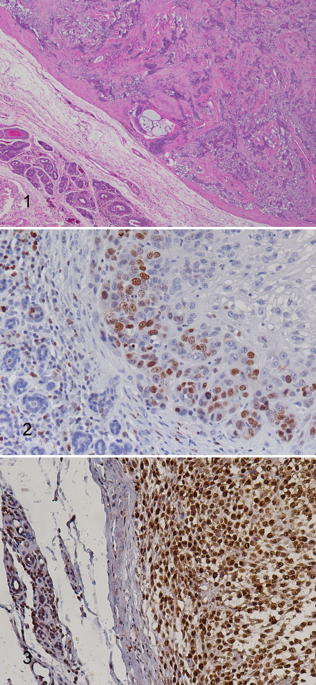
This work included 64 female dogs with mammary tumors (21 animals with only benign tumors and 43 with at least 1 malignant mammary tumor). For this study, 1 tumor was selected per animal. Therefore, in case of more than 1 benign tumor, the largest neoplasm was chosen. In the cases bearing more than 1 malignant tumor, the neoplasia with the most aggressive clinical (larger size, skin ulceration) and histological features (high histological grade of malignancy) was chosen, as described previously. 37 All tissues included in this study presented areas of adjacent nonneoplastic mammary glands without visible pathological changes such as hyperplasia and dysplasia (Fig. 1).
The animals ranged in age from 5 to 15 years, were of different breeds, and were received at the Hospital Veterinário do Porto, Portugal, for diagnosis and treatment. At the time of the surgery, all female dogs were spayed (n = 21) or in anestrus cycle confirmed by cytological examination of the vaginal smear. 37 At the first visit and through the physical examination, all the mammary glands and regional lymph nodes (axillary and inguinal) were evaluated. Animals with at least 1 malignant tumor were categorized by clinical stage using a modified TMN system. 31 Using this system, 3 clinical stages were established: local (without lymph node involvement), regional (metastasis at regional lymph nodes), and distant (presence of distant metastases). 37,38 In cases with regional lymph node enlargement, the involvement was investigated by fine-needle aspiration and confirmed by histological analysis after surgical removal. For each mammary tumor, the following clinical characteristics were evaluated: size (T1, <3 cm; T2, 3–5 cm; T3, >5 cm) 31,32 and skin ulceration. The absence of distant metastases was investigated with a thorax radiograph and an abdominal ultrasound. The study was approved by the University of Trás-os-Montes and Alto Douro, complying with the Portuguese legislation for animal protection (Law no. 92/1995).
Histopathological Evaluation
The samples for histopathology were fixed in 10% neutral formalin and paraffin embedded. Sections (4 μm) were cut and stained with hematoxylin and eosin (HE) for histological examination. Tumors were classified according to the World Health Organization (WHO) criteria for canine mammary neoplasms 26 and graded (mitotic index, 32 nuclear grade, differentiation grade, histological grade of malignancy [HGM]) in accordance with the method proposed by Peña and collaborators. 32 Additional clinicopathological characteristics evaluated were the presence of tumor necrosis and the regional lymph node involvement.
Immunohistochemical Analysis
In total, 3-μm sections were cut from each specimen and mounted on silane-coated slides. The detection of Ki-67 and PCNA was carried out by the streptavidin-biotin-peroxidase complex method, with a commercial detection system (Ultra Vision Detection System; Lab Vision, Fremont, California) following the manufacturer’s instructions. Both antigens were retrieved by microwave treatment for 3 × 5 minutes at 750 W in 0.01 M citrate buffer (pH 6.0), followed by cooling for 20 minutes at room temperature. For Ki-67, before microwave treatment, proteolytic treatment with trypsin (10 minutes at 37°C) was performed. After cooling at room temperature, the sections were immersed in 3% H2O2 for 30 minutes to block endogenous peroxidase activity. All slides were then incubated with a blocking serum (Lab Vision) for 10 minutes and then incubated with the specific antibody: Ki-67 (MIB-1; Immunotech, Monrovia, California, USA) for 24 hours at 4°C, with dilution 1:100 in phosphate-buffered saline (PBS), and PCNA (PC10; Dako, Glostrup, Denmark) for 24 hours at 4°C, diluted to 1:50 in PBS. After incubation, tissue sections were rinsed in PBS and then incubated with biotinylated secondary antibody, followed by streptavidin-conjugated peroxidase (Lab Vision). Subsequently, the color was developed with 3,3′-diaminobenzidine tetrahydrochloride (DAB) at 0.05% with 0.01% H2O2 (30%), and slides were counterstained with Gill’s hematoxylin, dehydrated, and mounted for evaluation by light microscopy. The primary antibody was replaced with PBS for negative controls, and epidermis was used as an internal positive control.
Immunoreactivity Evaluation
Ki-67 and PCNA immunoreactivity was evaluated using a quantitative method adapted from one study conducted by Peña and colleagues. 33 Immunoreactivity was considered when the staining occurred in the cell nucleus, regardless of the degree of intensity. The evaluation was carried out through observation of preparations in their entirety, and in each preparation, the fraction of positive nuclei in a total of 1000 cells (PI), after selecting the 2 areas with more intense and homogeneous nuclear positivity to count, was determined in percentage terms. The evaluation of the immunostaining was performed in tumors and adjacent nonneoplastic mammary glands. Therefore, all neoplastic cells in the tumor area were counted, independently of their origin, 33 whereas all epithelial cells (both acinar and ductal) were counted in the adjacent nonneoplastic mammary glands.
Follow-up Data
After surgical excision of the tumors, follow-up was carried out in the 43 animals with malignant mammary tumors, with a mean follow-up period of 16 months (minimum, 1 month; maximum, 26 months). Overall survival (OS) was defined as the period between surgery and natural death due to the tumor or euthanasia in advanced stages of the disease (confirmed with necropsy). None of the animals received any adjuvant chemotherapy or other antitumoral treatment after surgery. Only animals with malignant tumors at the local or regional clinical stage were included in the follow-up study.
Statistical Analysis
SPSS version 19.0 (SPSS, Inc, an IBM Company, Chicago, Illinois) was used for statistical analysis. Associations between Ki-67, PCNA, and categorical variables were studied with the application of analysis of variance (ANOVA) tests. The Pearson correlation test was performed to verify the presence of correlation between values of Ki-67 and PCNA in tumors and adjacent nonneoplastic mammary glands. Survival curves were generated by the Kaplan-Meier method, and the survival rates were compared using the log-rank test. In this work, there were no animals lost to follow-up or that died from causes other than neoplastic disease; therefore, censored observations correspond to the animals that remained alive at the end of the study period. 32 Cox proportional hazards model for multivariate analysis was used to study the effects of different variables on survival while taking other variables into account. Variables significantly associated with OS in univariate analysis were included in the multivariate study. The Ki-67 and PCNA cutoff points were calculated using the receiver operating characteristic (ROC) curve test. All values are expressed as means ± standard error. In all statistical comparisons, P < .05 was accepted as denoting significant differences.
Results
Clinicopathological Characterization of the Tumors
Benign tumors (n = 21) were classified as simple adenomas (n = 7), complex adenomas (n = 5), and benign mixed mammary tumors (n = 9). Malignant tumors (n = 43) were diagnosed as complex carcinomas (n = 16), solid carcinomas (n = 6), tubulopapillary carcinomas (n = 16), and carcinosarcomas (n = 5). Ulceration, only found in malignant tumors, was present in 10 cases (2 complex carcinomas, 2 solid carcinomas, 4 tubulopapillary carcinomas, and 2 carcinosarcomas). Tumor necrosis, absent in benign tumors, was observed in 20 of the malignant cases (9 complex carcinomas, 4 solid carcinomas, 5 tubulopapillary carcinomas, and 2 carcinosarcomas). Histological examination of regional lymph nodes, performed in all cases, showed regional lymph node metastases in 12 tumors, all of HGM III (1 complex carcinoma, 3 solid carcinomas, 5 tubulopapillary carcinomas, and 3 carcinosarcomas).
Ki-67 and PCNA Immunostaining
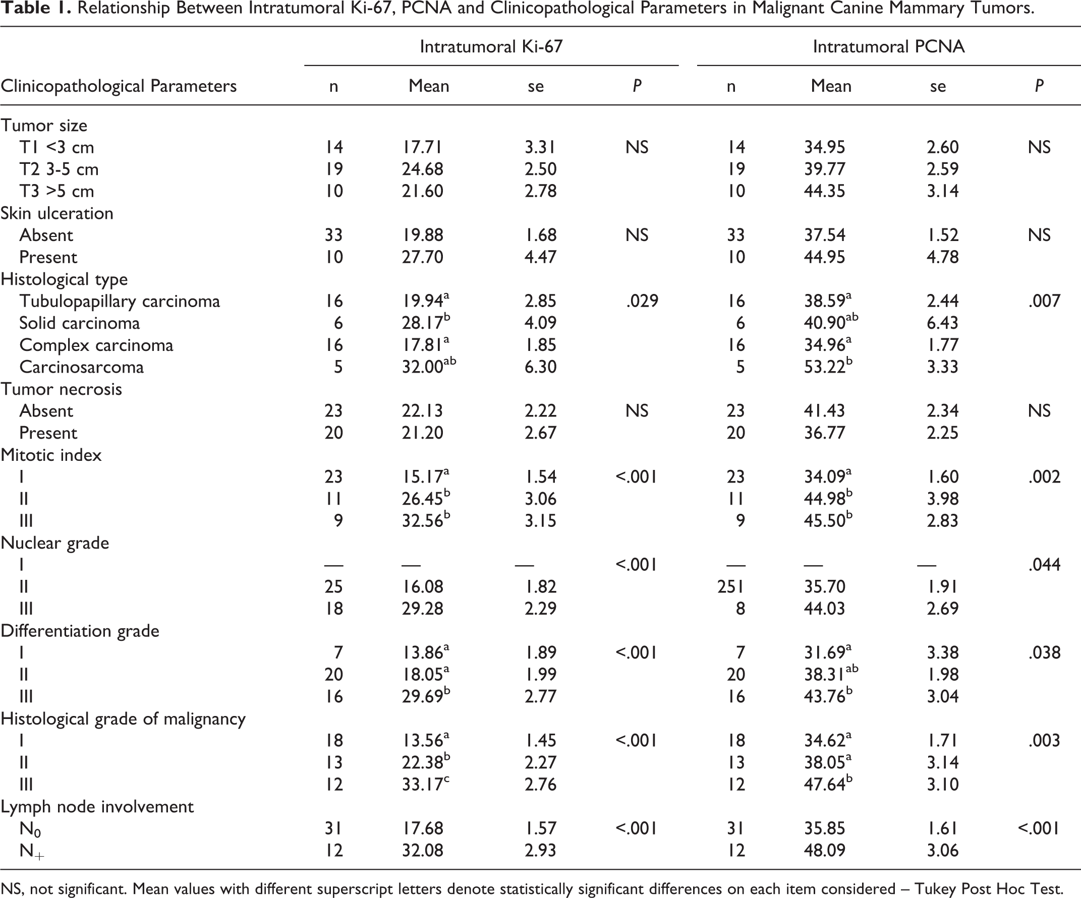
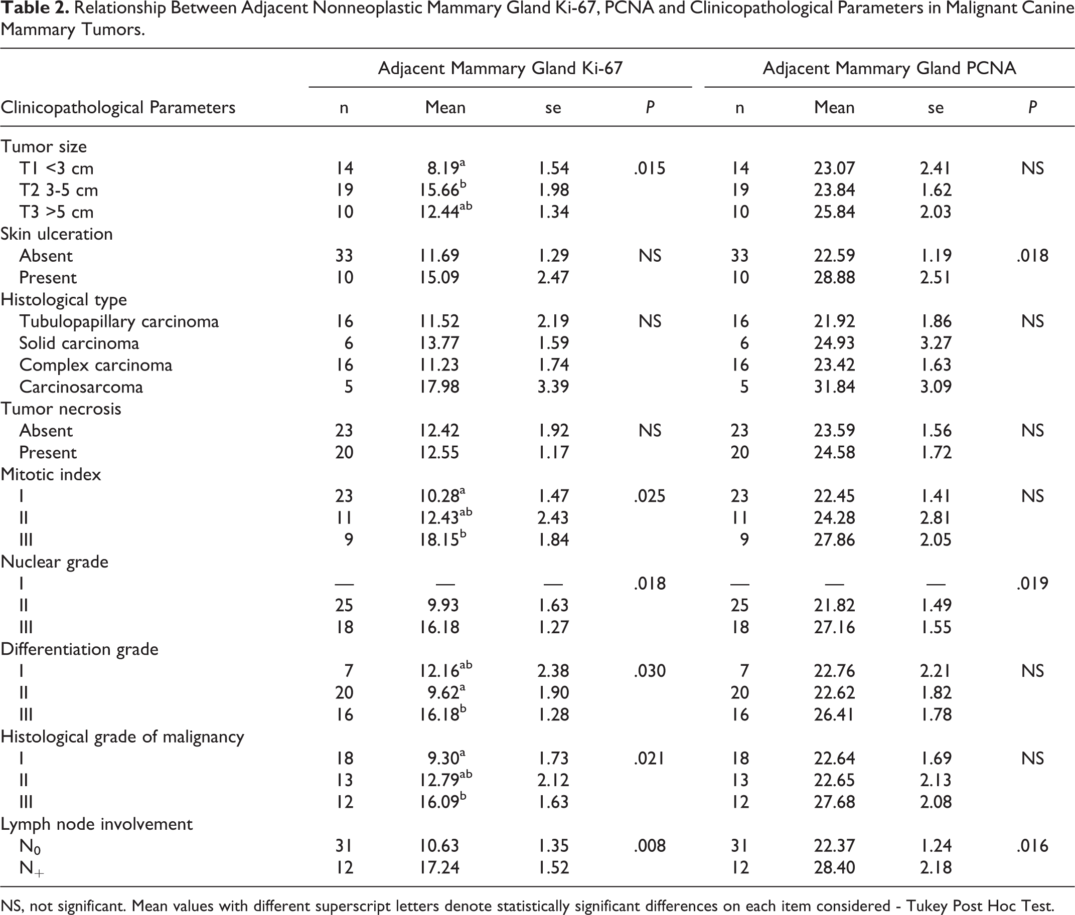
The immunoreactivity for Ki-67 and PCNA was observed always in the nucleus, appearing in a granular labeling pattern (Figs. 2, 3). In benign tumors, the PI of intratumoral Ki-67 and PCNA ranged between 3%–32% and 20%–49%, respectively. The PI in adjacent nonneoplastic mammary glands ranged from 0% to 24% for Ki-67 and 12% to 47% for PCNA. In malignant tumors, the PI of intratumoral Ki-67 and PCNA ranged between 3%–47% and 21%–69%, respectively. The PI in adjacent nonneoplastic mammary glands ranged from 1% to 29% for Ki-67 and 10% to 40% for PCNA.
Association Between Immunoreactivity of Ki-67, PCNA, and Clinicopathological Factors in Malignant Tumors
The PI of intratumoral Ki-67 was significantly associated with histological type (P = .029), mitotic index (P < .001), nuclear grade (P < .001), differentiation grade (P < .001), histological grade of malignancy (P < .001), and involvement of regional lymph nodes (P < .001). The PI of tumoral PCNA was significantly associated with histological type (P = .007), mitotic index (P = .002), nuclear grade (P = .044), differentiation grade (P = .038), histological grade of malignancy (P = .003), and involvement of regional lymph nodes (P < .001). All described associations are summarized in Table 1.
Relationship Between Intratumoral Ki-67, PCNA and Clinicopathological Parameters in Malignant Canine Mammary Tumors.
NS, not significant. Mean values with different superscript letters denote statistically significant differences on each item considered – Tukey Post Hoc Test.
The mean values of the Ki-67 PI in adjacent nonneoplastic mammary glands were statistically associated with tumor size (P = .015), mitotic index (P = .025), nuclear grade (P = .018), differentiation grade (P = .030), histological grade of malignancy (P = .021), and involvement of regional lymph nodes (P = .008). The PI of PCNA in adjacent nonneoplastic mammary gland, showed a statistical association with skin ulceration (P = .018), differentiation grade (P = .019), and presence of lymph node metastasis (P = .016). All described associations are summarized in Table 2.
Relationship Between Adjacent Nonneoplastic Mammary Gland Ki-67, PCNA and Clinicopathological Parameters in Malignant Canine Mammary Tumors.
NS, not significant. Mean values with different superscript letters denote statistically significant differences on each item considered - Tukey Post Hoc Test.
Correlation of Proliferative Indices Between Tumors and Adjacent Nonneoplastic Mammary Glands
The mean values of intratumoral Ki-67 PI were statistically significantly correlated with PI of Ki-67 in adjacent nonneoplastic mammary glands in benign and malignant tumors (r = .534, P = .013 and r = .577, P < .001, respectively). Present results also showed a positive and statistically significant correlation between the mean values of PI of intratumoral PCNA and PCNA in adjacent nonneoplastic mammary glands in benign and malignant tumors (r = .581, P = .006 and r = .689, P < .001, respectively).
Follow-up Study
To analyze the prognostic value of clinicopathological parameters and molecular markers, their relationship with OS was established (Table 3).
Association Between Variables and Overall Survival.
NS, not significant; *Multivariate Cox Proportional Hazard Analysis (Cox Regression) of overall survival (OS) in dogs with malignant mammary tumors in a prospective study with 2 years of follow-up.
With regard to clinical stage, 31 animals (72%) were in the local stage, 12 (28%) animals were in the regional stage, and no animal presented distant metastases at the time of diagnosis.
In this study, larger tumors and cases that presented skin ulceration were associated with shorter OS (P = .010 and P = .003, respectively). Animals with complex carcinomas presented longer survival times compared to the other tumor types. Carcinosarcomas were the most aggressive tumors and were related to shorter OS of animals (P = .006). Tumors with high mitotic index, high nuclear grade, high differentiation grade, and high HGM were associated with shorter OS (P < .001 for all parameters). Animals with regional lymph node metastases presented a decrease in OS time (P < .001). Molecular parameters significantly related to shorter OS included high intratumoral Ki-67 and PCNA (P < .001 and P = .001, respectively) and high Ki-67 and PCNA in adjacent nonneoplastic mammary glands (P < .001 and P = .006, respectively) (Figs. 4–7).
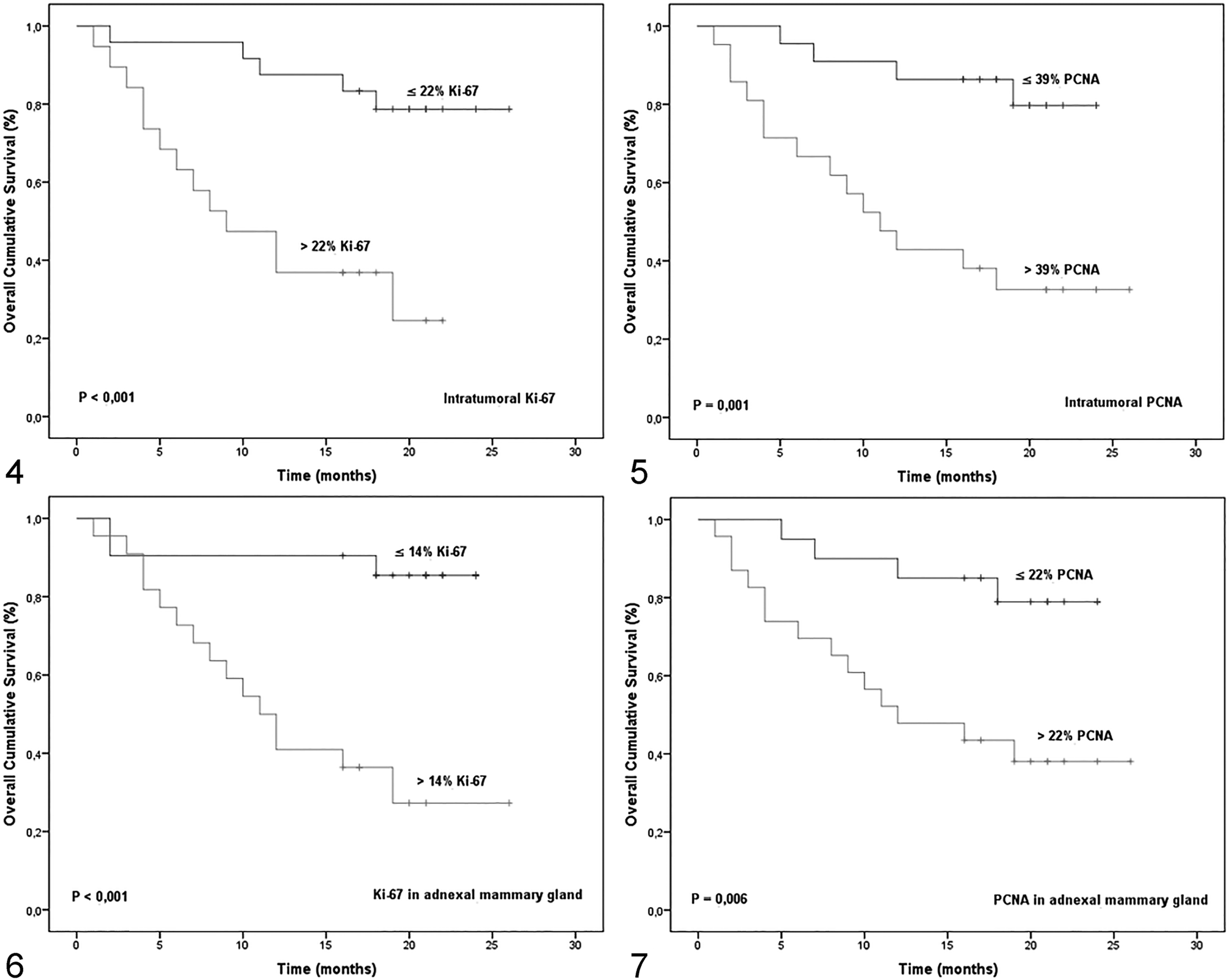
The multivariate analysis performed with the Cox proportional hazards model demonstrated that the high HGM and high intratumoral PCNA expression were independent predictors of OS (P < .001 and P = .016, respectively) (Table 3).
Discussion
Due to the heterogeneity of pathological types and clinical presentations of CMTs, histological evidence of malignancy does not necessarily imply a malignant clinical course of the disease. 30 Therefore, in veterinary medicine, the meticulous search and communication of reliable prognostic factors in a short period of time is important for the veterinary clinician.
Larger tumor size was associated with decreased survival. Tumor size is a clinical parameter classically associated with worse prognosis in dogs with malignant mammary tumors, and our findings are in agreement with other authors. 25,27,35,45 This study confirms that tumor size is a variable consistently related to prognosis, which clinicians should consider important. Our results highlight the need to excise the tumor as early as possible. The presence of skin ulceration, which has been suggested to be an indicator of higher tumor aggressiveness and poor survival, 6,15,35,37 was significantly related to OS in this study. Skin ulceration occurred exclusively in malignant tumors; thus, clinicians should pay special attention to ulceration in the clinical evaluation of these neoplasms. Some prognostic studies suggested that there is an increasing malignancy from complex carcinomas to simple carcinomas to sarcomas, 15,16,26,54 although this fact was not demonstrated in other publications. 6,20,36 In our study, carcinosarcoma was the most aggressive tumor type, related to a decrease in OS, while complex carcinoma was less aggressive. The present results are in accordance with recent studies. 45 However, these results should be prudently interpreted due to the small number of cases.
Corroborating previous findings, 33,35,45,47 our study showed that mitotic index, nuclear grade, differentiation grade, and HGM might be helpful to predict survival time. Interestingly, HGM retained the significance in multivariate survival analysis, arising as an independent predictor of prognosis. It must be remembered, however, that several grading methods 9,13 have been used to classify malignant CMTs, and the results of different studies may depend on them.
In our work, the presence of regional lymph node metastases was associated with decreased survival. The prognostic value of regional lymph node involvement at the time of surgery has been confirmed by several researchers. 15,17,27,35,45,54
Although in the context of CMTs, a range of clinicopathological factors with reliable prognostic value have already been identified, it is evident that the interpretation of tumor behavior still seems far from a complete understanding. To better understand this highly complex topic, the scientific community has been focusing their attention on an investigation at the molecular level. The identification of molecular markers allows, in addition, the identification of new therapeutic targets.
The antigen Ki-67 is a nuclear protein actively expressed in dividing cells but not after mitosis. 33 PCNA increases after the G1 phase of the cell cycle, reaching a maximum in the S phase and decreasing after G2, and then presents very low levels in the M phase and in quiescent cells. 50 The PI of intratumoral Ki-67 and PCNA was statistically associated, in the present study, with characteristics of higher aggressiveness, such as tumor histological grade, nuclear grade, and lymph node involvement. High intratumoral Ki-67 and PCNA were statistically associated with shorter survival, with high intratumoral PCNA maintaining its independent prognostic value in the multivariate analysis. Ki-67 and PCNA labeling has been found in a wide variety of tumors, in both humans 5,23,51 and dogs. 2,33,53 In humans, immunohistochemical studies with the antibodies Ki-67 and PCNA suggest that they are useful prognostic indicators for the relapse-free period in patients with breast cancer. 14,18,42,48 In dogs, previous studies also associated high Ki-67 and high PCNA with aggressive mammary tumor features 7,11,15,29 and decreased disease-free survival and OS, 33,46 strengthening our findings.
Present results revealed a positive and statistically significant correlation between tumoral Ki-67 and Ki-67 in adjacent nonneoplastic mammary glands, and between tumoral PCNA and PCNA in adjacent nonneoplastic mammary glands in benign and malignant tumors. Interestingly, in malignant tumors, the high Ki-67 and PCNA in the adjacent nonneoplastic mammary glands revealed a statistical association with clinicopathological features of tumor aggressiveness. To our knowledge, no studies in human or veterinary medicine have assessed the Ki-67 and PCNA in adjacent nonneoplastic mammary glands. Our results suggest that growth factors produced by the tumor might act on the adjacent nonneoplastic mammary gland in a paracrine manner, contributing to a high proliferative tumoral microenvironment.
PCNA is an antigen expressed in the nuclei of cells during the DNA synthesis phase of the cell division cycle. 10 The growth factors resulting from DNA damage induce the PCNA levels to remain high. 24 PCNA has a long cellular half-life and may be expressed in the phases of cellular repair since this antigen is a processivity factor for DNA polymerase δ involved in several specialized metabolic pathways for DNA damage repair, including nucleotide excision repair, base excision repair, mismatch repair, and double-strand break repair. 10,49 The participation of PCNA in DNA synthesis and DNA repair might explain the association between the high PCNA index in adjacent nonneoplastic mammary glands with the more aggressive tumor phenotypes found in our study.
Present results demonstrated a positive and significant correlation between tumor and adjacent nonneoplastic mammary gland proliferation, suggesting that tumoral growth factors might have a paracrine effect on adjacent normal tissues. Interestingly, the proliferation by Ki-67 and PCNA of adjacent nontumoral mammary glands was associated with aggressive tumor phenotypes, demonstrating the need to better explore this adjacent nonneoplastic tissue.
Footnotes
Declaration of Conflicting Interests
The author(s) declared no potential conflicts of interest with respect to the research, authorship, and/or publication of this article.
Funding
The author(s) disclosed receipt of the following financial support for the research, authorship, and/or publication of this article: The work was supported partially by a PhD scholarship SFRH/BD/78771/2011 financed by the Portuguese Foundation for Science and Technology (FCT). This work was also supported by European Investment Funds by FEDER/COMPETE/POCI– Operational Competitiveness and Internationalization Programme, under Project POCI-01-0145-FEDER-006958 and National Funds by the Portuguese Foundation for Science and Technology, under the project UID/AGR/04033/2013.