
Abstract
Tumor-associated macrophages (TAMs) are an important component of leukocyte infiltration in tumors. TAMs can be classified into M1 and M2 phenotypes. In the present study, the expression of CD204, an M2-polarized macrophage receptor, was investigated by immunohistochemistry in the area surrounding TAMs in 101 cases of canine mammary gland tumor (CMT). We examined the relationship between M2-polarized TAMs and malignancy, histological subtype, histological grade, molecular subtype, hormone receptor (HR) status, and clinical obesity indices. The mean number of CD204-positive macrophages was significantly higher in malignant CMTs than in benign CMTs (P = .000). The number of CD204-positive macrophages differed significantly between histological grades (P = .000) and were significantly higher in grade III than in grades I and II. Moreover, the mean number of CD204-positive macrophages was significantly higher in HR-negative malignant CMTs than in HR-positive malignant CMTs (P = .035) and in malignant CMTs with lymphatic invasion compared to malignant CMTs without lymphatic invasion (P = .000). These findings suggest that CD204-positive macrophages might affect the development and behavior of CMTs and highlight the potential of CD204 as a prognostic factor.
The inflammatory microenvironment is recognized as an important component in carcinogenesis. 16,46 Many leukocytes, including macrophages, penetrate tumor tissues and form a tumor microenvironment along with fibroblasts and endothelial cells. 23 These macrophages are also a major leukocyte in all tumors. 41
Among the various types of macrophages, tumor-associated macrophages (TAMs) mediate common inflammatory pathways. 33 TAMs derived from circulating monocytes exhibit dual actions toward tumor cells. Previously, activated TAMs were suspected to have cytotoxic activity toward tumor cells, but protumoral functions of TAMs have also been reported. 15,24,45 Thus, TAMs can kill tumor cells as well as promote tumor growth and angiogenesis. 2
TAMs can be classified into M1 and M2 phenotypes. 35 –37 Higher numbers of total TAMs or M2-polarized TAMs are linked to increased tumor cell proliferation and histological malignancy in some kinds of tumors. 24,25 A higher density of M2-polarized TAMs is associated with worse clinical course in some tumor types and with poor prognosis in others. 17
TAM infiltration is linked with poor prognosis in breast carcinoma in humans, 9 and these TAMs have been reported to have more M2-like characteristics. 3 M2-polarized macrophages show high CD204 (also called scavenger receptor A) expression. 26 A high number of CD204-positive TAMs are associated with worse prognosis in various tumors. 18,42 In addition, many recent studies have discussed the relationship between obesity and macrophage polarization. 31,59 Therefore, it is rational to propose that CD204-positive TAMs are important in the biological behavior of the still poorly understood neoplasm, canine mammary gland tumors (CMTs). With this background, the objectives of the present study were (1) to compare the expression of CD204-positive TAMs between benign and malignant tumors; (2) to compare the expression of CD204-positive TAMs between tumors with different clinicopathological characteristics such as histological subtype, histological grade, mitotic count, nuclear grade, tubule formation, tumor necrosis, lymphatic invasion, molecular subtype, and expression of hormone receptor; and (3) to determine the relationship between CD204-positive TAMs and clinical obesity index.
Materials and Methods
Tissue Samples and Group Assignment
Canine mammary tumor specimens from the histopathological database of the Department of Veterinary Pathology, Konkuk University, Animal Teaching Hospital (Seoul, Korea) from 2014 to 2016 were analyzed. Among a total of 921 CMT samples, 71 malignant and 30 benign CMT cases that had all the necessary information recorded, including the breed, age, neuter status, body condition score (BCS) using a 5-point scale, and body weight (BW), were selected. The specimens were then divided into 2 groups based on the BCS and BW as follows: group 1, both BCS = 2/5 (underweight) or 3/5 (ideal) and BW <15% of the maximal ideal body weight (lean or optimal body weight); group 2, both BCS = 4/5 (overweight) or 5/5 (obese) and BW >15% of the maximal ideal body weight (overweight or obese). 54
Histopathology
The histological classification system proposed by Goldschmidt et al 14 was used to determine the histological subtype. The tumor grade was assessed based on the criteria described by Clemente et al. 10 The clinicopathological characteristics evaluated in each sample were histologic grade, mitotic count, nuclear grade, tubule formation, presence of tumor necrosis, and presence of lymphatic invasion. The evaluation of histopathological parameters was performed by 3 authors (B.J-S., J.I-S., and H.W-K.), and the results were rechecked by 1 author (J.H-S.). All the samples were fixed in 10% neutral buffered formalin and embedded in paraffin wax, and 4-μm sections were used for hematoxylin and eosin staining.
Immunohistochemistry
All the CMT samples were immunostained to examine the expression of CD204 (scavenger receptor A), estrogen receptor (ER), progesterone receptor (PR), and human epidermal growth factor receptor 2 (HER2). To determine the expression of M2-polarized TAMs, immunohistochemistry (IHC) was performed using a specific primary CD204 antibody (M2 phenotype marker) that has been shown to specifically bind to canine macrophages. 20
IHC analysis was performed using primary antibodies specific for each factor on formalin-fixed, paraffin-embedded tissue sections. Each slide was deparaffinized in xylene and rehydrated in graded ethanol prior to staining. After 3 washes with phosphate-buffered saline (PBS; pH 7.4, 137 mM NaCl, 2.7 mM KCl, 10 mM Na2HPO4, and 2 mM KH2PO4), endogenous peroxidase activity was blocked by incubation in 3% H2O2 for 20 minutes at room temperature. The sections were then again washed thrice in PBS. Heat-induced antigen retrieval was performed using Tris-EDTA (pH 9.0) and citric acid (pH 6.0) for all the primary antibodies in a microwave oven at high power. After antigen retrieval, the sections were cooled in cold water and washed in PBS thrice. After washing, the sections were blocked using 5% normal goat serum in PBS and then incubated with primary antibodies according to each protocol (Table 1). After incubation, the sections were washed thrice in PBS for removing unbound antibodies, and primary antibody binding was detected using secondary antibodies (REAL Envision kit; DAKO, Glostrup, Denmark) for 40 minutes at room temperature. Horseradish peroxidase and 3,3′-diaminobenzidine were used for labeling, and the reaction was stopped by washing in distilled water. The sections were then counterstained with Gill’s hematoxylin, dehydrated with graded ethanol, and covered with coverslips.
Primary Antibodies and Protocols Used for Immunohistochemical Analysis.
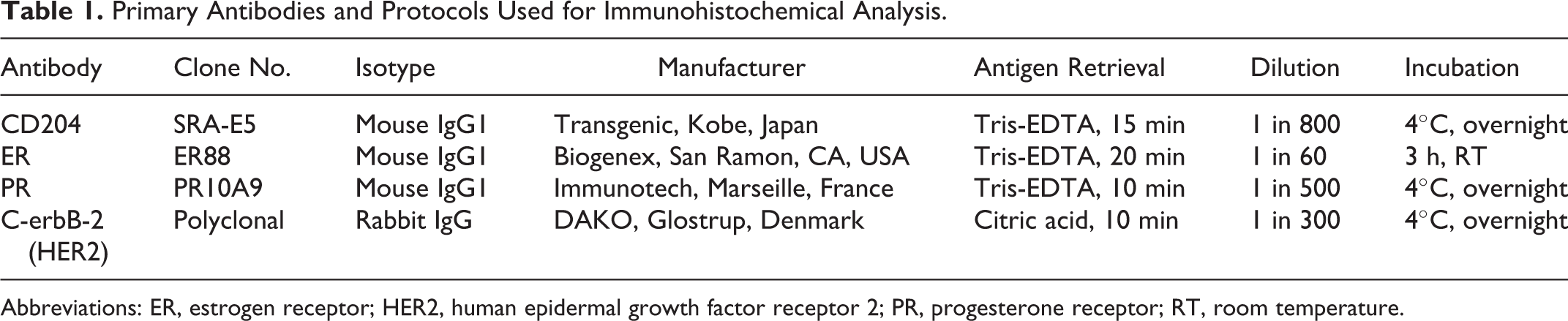
Abbreviations: ER, estrogen receptor; HER2, human epidermal growth factor receptor 2; PR, progesterone receptor; RT, room temperature.
Normal mammary glands and normal to hyperplastic lesions adjacent to tumors were used as positive controls for the ER and PR antibodies. Canine mammary carcinomas with HER2-overexpression were used as positive controls for the HER2 antibody by referring to our previous study. 22 A normal dog spleen sample was used as the positive control for the CD204 antibody. To ensure the reactivity of the primary antibodies, isotype-matched immunoglobulins were used as the negative control.
Evaluation of Immunohistochemical Expression
To count CD204-positive macrophages, sections were first observed at a low magnification under a microscope to identify the 3 areas with the greatest numbers of macrophages, the so-called “hotspots,” by 1 author (B.J-S.). Images were acquired at 400× magnification. 40 Only the tumor cell and stroma areas were selected for hotspots. Necrotic and bleeding areas were excluded from the evaluation. Digital images were acquired using an Olympus BX51 microscope (Olympus, Tokyo, Japan) and Image transfer software (Olympus). The number of CD204-positive macrophages was analyzed using automated image analysis software (Image Pro Plus 5.1; Media Cybernetics, Silver Spring, MD). Average values were used for statistical analysis.
To evaluate ER and PR expression, the percentage of neoplastic epithelial cells with nuclear immunoreactivity was determined. Tissue samples with more than 10% clear nuclear staining of neoplastic epithelial cells were considered positive. 19 The single ER- or PR-positive CMTs and both receptor-positive CMTs were all regarded as hormone receptor (HR) positive. Evaluation of anti-HER2 staining was based on recent guidelines 44 recommended by the American Society of Clinical Oncology/College of American Pathologists (ASCO/CAP). 60 Only 3+ tumors (with more than 30% complete membrane labeling of epithelial neoplastic cells) were considered HER2 positive.
According to IHC results, malignant CMTs were categorized into 4 molecular subtypes: luminal A (ER- and/or PR-positive and HER2-negative), luminal B (ER- and/or PR-positive and HER2-positive), HER2-overexpressing (ER- and PR-negative and HER2-positive), and triple-negative (ER-, PR-, and HER2-negative) subtype.
Statistical Analyses
The correlations between the number of CD204-positive macrophages and histological subtype, histological tumor grade, mitotic count, nuclear grade, tubule formation, and molecular subtype were determined using the Kruskal-Wallis test. The correlation between each pair of histological grades was determined using the Bonferroni corrected post hoc Mann-Whitney test (P < .017 was considered to indicate significance). The correlation between the number of CD204-positive macrophages and clinicopathological parameters (malignancy and obesity status) was determined using the Student t tests (P < .05). The correlations between the number of CD204-positive macrophages and other parameters (tumor necrosis, lymphatic invasion, and hormone receptor status) were determined using the Mann-Whitney test (P < .05). All statistical analyses were performed using Statistical Package for the Social Sciences for Windows, version 22.0 (SPSS, Inc, an IBM Company, Chicago, IL).
Results
Clinical and Histopathological Characteristics
We studied tissues from 52 intact and 49 neutered female dogs. The breeds included Maltese (n = 45), Shih-Tzu (n = 14), Poodle (n = 10), Yorkshire Terrier (n = 7), Miniature Schnauzer (n = 6), Cocker Spaniel (n = 5), Miniature Pinscher (n = 4), English Cocker Spaniel (n = 2), Jindo (n = 2), Pekingese (n = 2), Pomeranian (n = 2), Beagle (n = 1), and Dachshund (n = 1). The age range of the dogs was 3 to 19 years (mean ± standard deviation [SD]: 10.5 ± 3.0 years). The CMTs were classified as benign (n = 30) and malignant (n = 71). The histological subtype of malignant CMTs was classified as complex carcinoma (n = 10), simple tubular carcinoma (n = 21), simple tubulopapillary carcinoma (n = 7), intraductal papillary carcinoma (n = 6), carcinoma and malignant myoepithelioma (n = 3), comedocarcinoma (n = 5), solid carcinoma (n = 5), anaplastic carcinoma (n = 5), carcinoma arising in benign mixed tumor (n = 7), adenosquamous carcinoma (n = 1), and carcinosarcoma (n = 1). Malignant tumor grades included grade I (n = 37), grade II (n = 15), and grade III (n = 19). Of all samples, 27 cases of malignant CMTs exhibited tumor necrosis, and 9 cases of malignant CMTs exhibited histological evidence of lymphatic invasion. Lean dogs or those of optimal body weight were assigned to group 1 (n = 60), and overweight or obese dogs were assigned to group 2 (n = 41).
Expression of CD204, ER, PR, and HER2
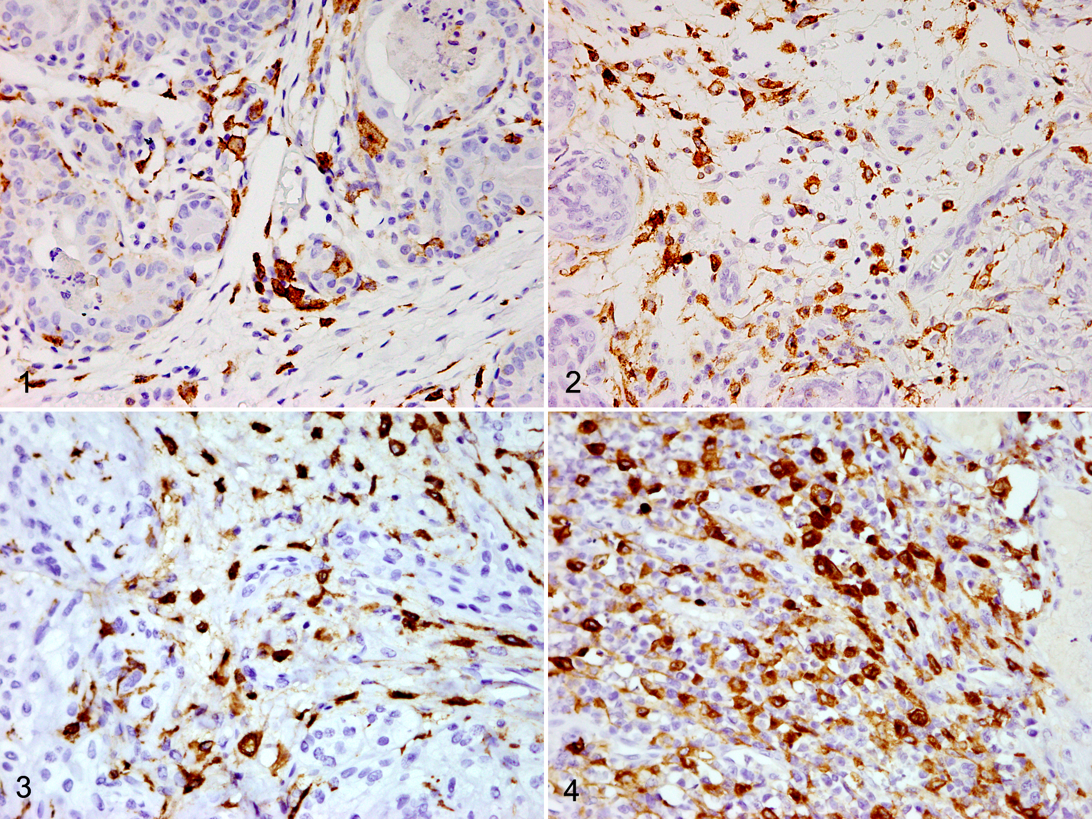
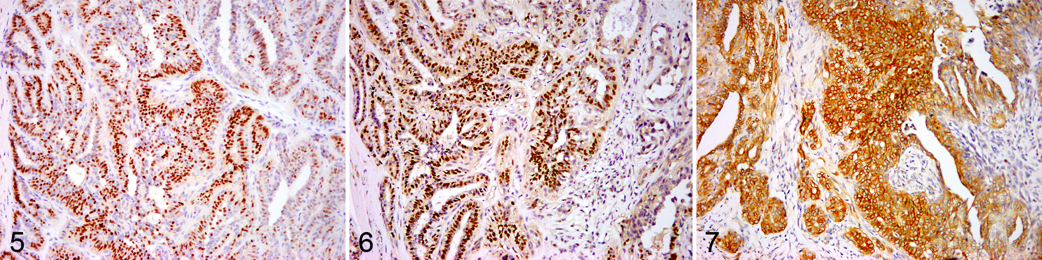
The CD204 antigen was expressed in macrophages infiltrating within and around tumors with cytoplasmic and membranous staining pattern (Figs. 1–4). Malignant CMTs were classified as luminal A (n = 33), luminal B (n = 18), HER2 overexpressing (n = 3), and triple negative (n = 17) based on the IHC results (Figs. 5–7). A total of 51 cases of malignant CMTs were HR (ER and/or PR) positive, and 21 cases of malignant CMTs were HER2 positive.
Association Between IHC Results and Clinicopathological Parameters
Associations between the immunohistochemical results and clinicohistopathological features, including malignancy, histological subtype, histological grade, mitotic count, nuclear grade, tubule formation, tumor necrosis, presence of lymphatic invasion, molecular subtype, HR status, and obesity status, are summarized in Tables 2 and 3. The number of CD204-positive macrophages was significantly higher in malignant CMTs (mean ± SD, 104.0 ± 38.1) than in benign CMTs (Fig. 1) (72.1 ± 24.7) (P = .000) (Fig. 8). There were statistically significant differences in number of CD204-positive TAMs among histological subtypes (P = .002), the 3 histological grades (P = .000), mitotic count (P = .016), nuclear grade (P = .000), and tubule formation (P = .000), based on Kruskal-Wallis tests. In addition, significant differences between the 3 histological grades were observed based on the Bonferroni-corrected post hoc Mann-Whitney test (grade I [Fig. 2] vs grade III [Fig. 4], P = .000; grade II [Fig. 3] vs grade III [Fig. 4], P = .010) (Fig. 9). The number of CD204-positive TAMs was significantly higher in malignant CMTs with tumor necrosis (114.8 ± 34.6) than in those without tumor necrosis (97.4 ± 39.0) (P = .020). The number of CD204-positive TAMs was also significantly higher in malignant CMTs with lymphatic invasion (144.2 ± 42.4) than in those without lymphatic invasion (98.2 ± 34.0) (P = .002). The number of CD204-positive TAMs was also significantly higher in HR-negative malignant CMTs (118.4 ± 43.8) than in HR-positive malignant CMTs (98.4 ± 34.5) (P = .035). No associations were found between the molecular subtype of the CD204-positive macrophages and the obesity status of dogs.
Comparisons of CD204-Positive Macrophages According to Obesity Status in Canine Mammary Tumors.
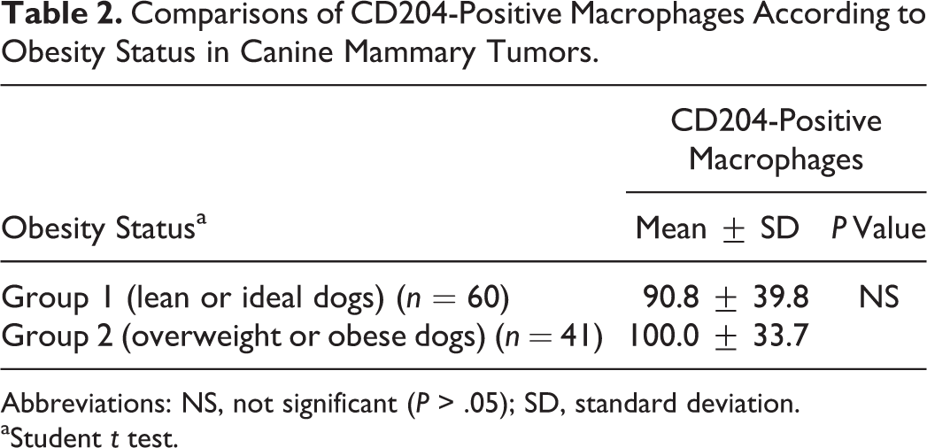
Abbreviations: NS, not significant (P > .05); SD, standard deviation.
aStudent t test.
Comparison of the Number of CD204-Positive Macrophages with Pathological Parameters in Malignant Canine Mammary Gland Tumors.
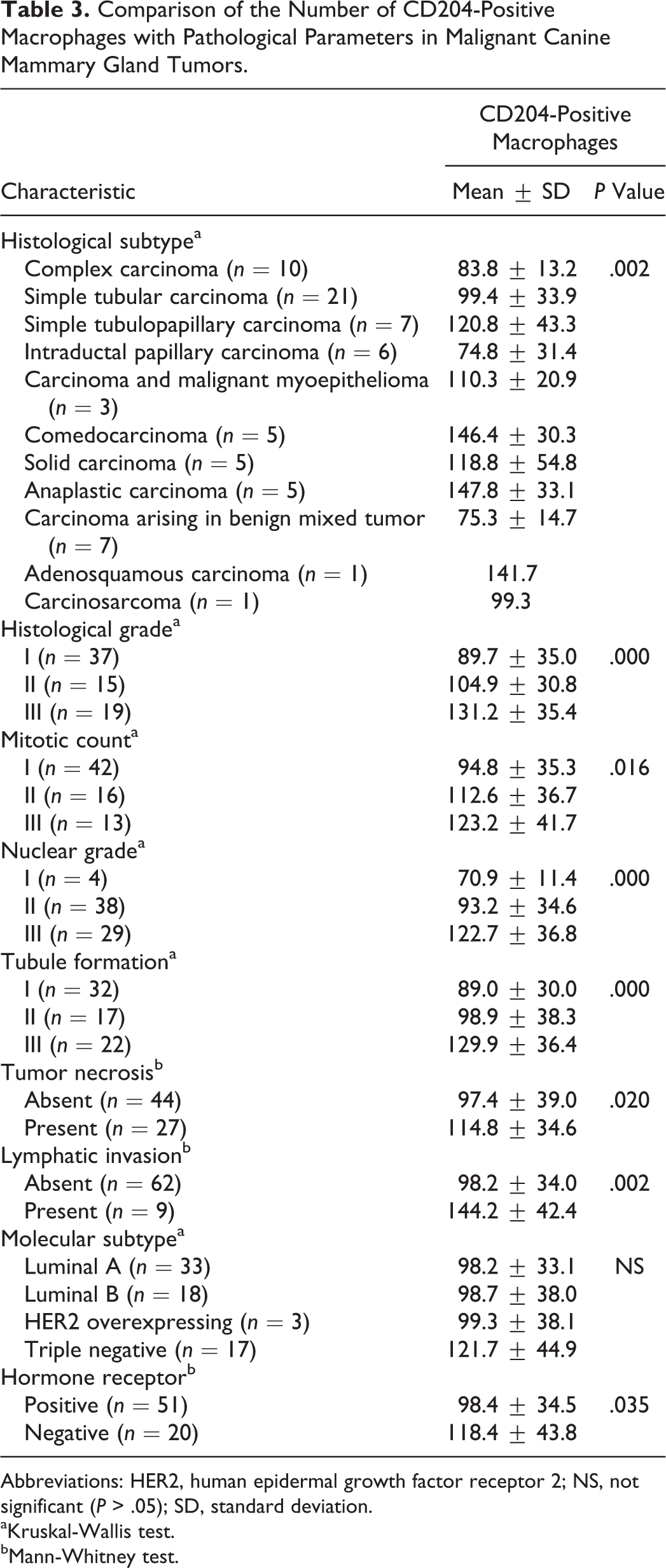
Abbreviations: HER2, human epidermal growth factor receptor 2; NS, not significant (P > .05); SD, standard deviation.
aKruskal-Wallis test.
bMann-Whitney test.
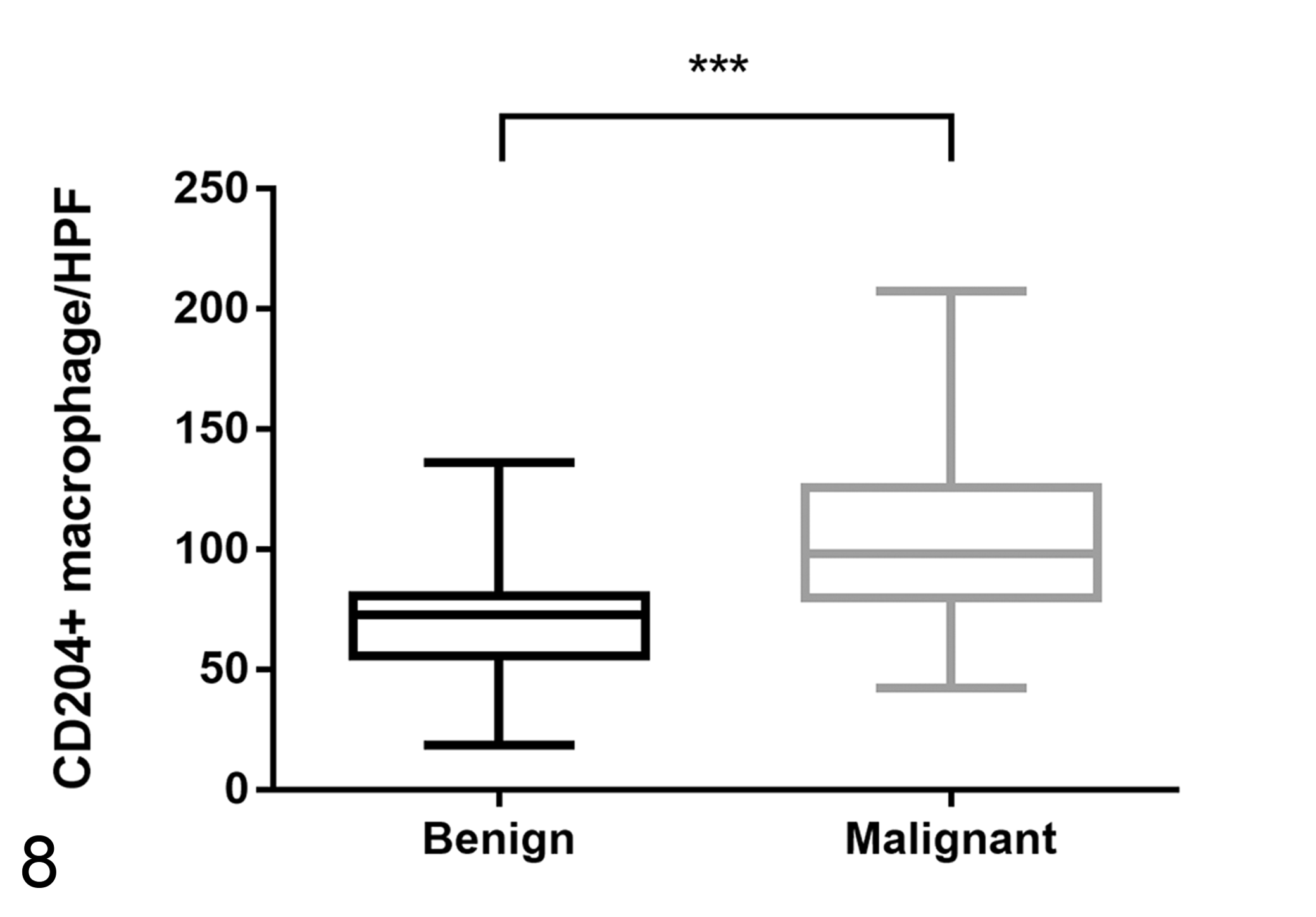
Comparison of the number of CD204-positive macrophages in benign and malignant canine mammary gland tumors (CMTs). Overall, the number of CD204-positive macrophages is significantly higher in malignant CMTs than in benign CMTs. HPF, high-power field. ***P < .001.
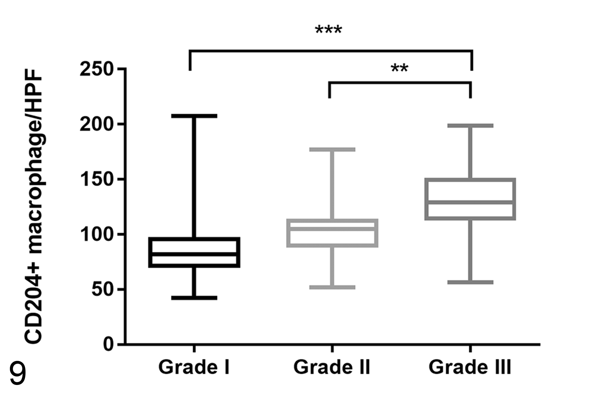
Comparison of the number of CD204-positive macrophages in grade I, grade II, and grade III malignant canine mammary gland tumors (CMTs). Overall, the number of CD204-positive macrophages is significantly higher in grade III malignant CMTs than in grade I and grade II malignant CMTs. HPF, high-power field. **P < .01. ***P < .001.
Discussion
In humans, macrophage infiltration in tumors has already been described, 34 and numerous studies have indicated that TAMs are correlated with the progress of breast cancer in humans. 28,32,51 High density of TAMs is associated with poor prognosis in a variety of tumors, including breast cancer in humans. 9,12,30,51 This observation has already been described in our previous study and in other articles. 7,29,47
In this study, we confirmed that the number of CD204-positive TAMs (M2-polarized TAMs) was higher in malignant CMTs than in benign CMTs. We also found that the number of CD204-positive TAMs was correlated with histological subtype, consistent with previous evidence that there are fewer CD204-positive TAMs in subtypes known to have a better prognosis, 49 such as complex type and carcinoma arising in benign mixed tumor type. We also found that an increased number of CD204-positive TAMs were correlated with a higher histological tumor grade, higher mitotic count, higher nuclear grade, higher tubule formation, and the presence of tumor necrosis and lymphatic invasion. To our knowledge, this is the first study to examine M2-polarized macrophage infiltration in benign and malignant CMT. Previous studies on CMT focused on the total TAMs rather than on polarized TAMs. 6,27,29,47,48 Some of these studies used myeloid/histiocyte antigen (MAC387), which is a general marker for myelomonocytic cells 29,47 that reacts with macrophages as well as granulocytes (neutrophils, basophils, and eosinophils). 4 In comparison, CD204 (scavenger receptor A) is preferentially expressed on macrophages, 21 and the antibody has been shown to have specific reactivity to canine macrophages. 20 Consistent with this, we confirmed that the CD204 antibody specifically reacted to tissue macrophages in our study. Studies focused on M2-polarized TAMs are important because these are considered to play a protumoral role. 53 M2-polarized TAMs have been shown to be associated with aggressive behavior in human breast cancer, 51 which suggests that CD204-positive TAMs have protumoral properties such as enhancing tumor progression and metastasis. Since infiltration of CD204-positive macrophages has been found to be highly correlated with an aggressive tumor behavior index in humans, our results suggest that CD204-positive macrophages might affect the development and behavior of CMTs. Furthermore, the findings indicate the possibility of using CD204-positive macrophages as a novel prognostic biomarker in CMT cases.
In humans, breast cancers have been molecularly classified as luminal A (ER/PR positive and HER2 negative), luminal B (ER/PR and HER2 positive), HER2 overexpressing (ER/PR negative and HER2 positive), and triple negative (all negative). 55 Based on the above criteria, the tissue samples in our study were divided into molecular subtype and HR-positive or negative groups. No significant association between the CD204-positive macrophages and molecular subtype was found (P = .163). However, the number of CD204-positive macrophages was higher in the HR-negative group (HER2-overexpressing and triple-negative subtype) than in the HR-positive (luminal A and luminal B subtype) group (P = .035). In humans, the intensity of macrophage infiltration is strongly correlated with ER-negative and PR-negative status in breast cancer, 58 and the correlation between TAM index and HR negativity has also been confirmed. 1,5,52 Consistent with this, our findings also showed that HR-negative tumors, which are known to be associated with poor prognosis, correlated with an increased number of CD204-positive macrophages in CMTs. In humans, targeted therapies according to molecular subtypes are well established in breast cancer, and various agents such as tamoxifen and trastuzumab are used for targeted therapy. 43,56 In CMTs, the molecular subtype is associated with clinicohistopathological parameters such as histological subtype and histological grade. 22,50 However, the individual prognostic values of ER and PR in CMTs are still a matter of debate; malignant CMTs with ER and PR expression had a better prognosis in some studies 8,39 but not in another study, 11 and the overexpression of HER2 in CMTs is still unclear. 13 Additional large-scale studies are needed to determine the prognostic value of molecular subtypes in malignant CMTs, and the mechanism explaining how TAMs are associated with HR status needs to be answered in both humans 57 and dogs in future studies.
Another purpose of our present study was to examine the association between M2-polarized TAMs and obesity indices. In a previous study, total TAM infiltration was found to be higher in obese dogs. 29 In the present study, the number of CD204-positive TAMs seemed to be slightly elevated in group 2, but no statistically significant associated with obesity indices (BCS and BW) was found (P = .227). Since the accumulation of excessive adipose tissue is associated with adipose tissue macrophage polarization, 53 there is a limit to the accuracy of the prediction of the amount of fat accumulation in the body based on clinical obesity indices such as BW and BCS. For an accurate analysis of TAM polarization, accurate measurement of body fat mass and determination of its correlation with TAM are required.
There is growing evidence indicating that CD204-positive macrophages are deeply associated with tumor progression in a specific tumor microenvironment. TAMs are recruited to tumors owing to various inflammatory responses and chemokines. 45 A recent study shows that tumor progression and metastasis were to be inhibited in CD204 knockout mice in vivo, and treatment of tumor-bearing mice with 4F, a small ligand of CD204, was found to suppress tumor progression and metastasis. 38 These results suggest that CD204-positive macrophages and tumor cells cooperate with each other and participate in aggressive tumor behavior. It has been hypothesized that M2-polarized TAMs can be used as a target therapy to prevent tumor metastasis as well as a novel prognostic biomarker in humans. 23 In veterinary medicine, further investigations are warranted to determine the mechanism underlying the association between TAMs and carcinogenesis to explore the potential of TAMs as a therapeutic target.
Footnotes
Acknowledgements
We thank Ms. E.-M. Yu for her excellent technical assistance and the participating private veterinary clinics for providing access to tissue samples of canine mammary tumors. This report represents a part of the PhD thesis by Byung-Joon Seung.
Declaration of Conflicting Interests
The author(s) declare that there are no potential conflicts of interest with respect to the research, authorship, and/or publication of this article.
Funding
The author(s) disclose the receipt of the following financial support for the research, authorship, and/or publication of this article: This research was supported by the Bio & Medical Technology Development Program of the National Research Foundation (NRF) funded by the Korean government (MSIT) (2016M3A9B6903437).