
Abstract
Ki-67 is a nuclear protein and a proliferation marker frequently used in establishing the prognosis for breast cancer patients. To investigate the prognostic value of the Ki-67 proliferation index in female cats with mammary carcinoma, a prospective study was conducted with 96 animals. The Ki-67 index of primary tumors (n = 96) was initially determined, and whenever possible, the Ki-67 index of regional lymph node metastasis (n = 38) and distant metastasis (n = 16) was also estimated. The optimal cutoff value for the Ki-67 index was determined by univariate and multivariate analysis. Ki-67 indices ≥14% were detected in 72.9% (70 of 96) of the tumors. Tumors with a Ki-67 index ≥14% were significantly associated with large size (P = .022), poor differentiation (P = .009), presence of necrotic areas (P = .008), estrogen receptor-negative status (P < .0001), fHER2-negative status (P = .003), and shorter overall survival (P = .012). Moreover, Ki-67 expression in the primary tumor was strongly and positively correlated with both regional metastasis (P < .0001; r = 0.83) and distant metastasis (P < .0001; r = 0.83), and was significantly higher in distant metastases when compared with the primary tumor (P = .0009). A similar correlation was also observed between regional and distant metastasis (P < .0001; r = 0.75). On the basis of the above results, the authors propose the adoption of the 14% value as the optimal cutoff for Ki-67 to identify tumors with high risk of disease progression.
Feline mammary carcinomas (FMCs) are among the most prevalent tumors in cats, with an incidence that can reach 40% of all tumor cases in this species. 23,39,42 Contrary to those of humans and dogs, 85% to 93% of feline mammary tumors are considered malignant, and the most common histotypes are the tubular, papillary, solid, and cribriform carcinomas. 39 Feline mammary tumors may metastasize to the regional lymph nodes, lungs, and liver, as well as other organs. Although some studies correlate age, tumor size, presence of regional metastasis, and malignancy grade with overall survival (OS), 19,37,43 still scarce information is available regarding the proliferation index of FMC. Based on this scenario, additional prognostic and predictive factors may provide useful insights into tumor biology to achieve a more efficient therapy and a better follow-up of the affected female cats.
Ki-67 is a protein found only in growing, dividing cells. It is expressed in all cell cycle phases, except in the resting phase (G0). With an intranuclear localization, the expression levels of Ki-67 are low during the G1 and S phases, rapidly increase during the G2 phase, and reach a peak in mitosis. This well-defined expression pattern makes the Ki-67 antigen a good proliferation marker, very useful and reliable in the prognosis of human breast cancer. 9,45 Several studies of women demonstrated that high Ki-67 expression is a poor prognostic indicator of 5-year recurrence-free survival in breast cancer patients, 6,7,9,18,27,41 being also used for chemotherapy response prediction. 6,9,18 However, different Ki-67 cutoff values and tumor heterogeneity require an adaptation of the scoring system, with cutoff values varying from 3.5% to 34%. 7,9,14,29 To overcome this problem, a large study recently recommended the standardization of a Ki-67 cutoff point at 14% in breast cancer to improve prognosis. 5
In veterinary medicine, although the Ki-67 index has been investigated as a prognostic factor in FMC, few data are available on how the Ki-67 index correlates with lower survival. 23,32,37 Furthermore, the evaluation of cell proliferation by Ki-67 immunostaining has not been subject to standardization of protocols. The lack of clear indication on a cutoff level explains why no values have been validated for FMC. 4,30,32,37
The main goal of the present prospective study is to validate the prognostic value of the Ki-67 index in FMC. First, the Ki-67 index of 96 primary mammary tumors was determined and compared with corresponding lymph node and distant metastasis. Second, the ideal cutoff value was calculated by evaluating a set of threshold values based on univariate and multivariate survival analysis. Finally, the association between the Ki-67 index and 19 other clinicopathologic features of cats with mammary carcinoma was evaluated.
Materials and Methods
Animal Population
The studied population consisted of 96 female cats with mammary tumors admitted to the Small Animal Hospital of the Faculty of Veterinary Medicine of the University of Lisbon from June 2011 to December 2013. Animals were followed for at least a month to be included in survival analysis, and the last follow-up was in May 2014. For each animal, the following clinical data were recorded: age, breed, reproductive status (intact vs spayed), administration of progestogens, number and location of tumors, tumor stage (TNM system), 39 treatment prescribed (none, mastectomy, mastectomy combined with chemotherapy), and OS. Additionally, the presence of microscopic regional lymph node metastasis was evaluated in a majority of cases (86 cats). A full postmortem examination was performed in 18 cats, and metastatic disease was confirmed histologically in 16 of those animals.
All mammary and respective metastatic lesions excised during surgery from 89 cats or during necropsy (7 cats) were fixed in 10% buffered formalin for 24 to 48 hours and embedded in paraffin. The histologic specimens were evaluated following the World Health Organization classification system. 24 The degree of malignancy was determined by the assessment of 3 morphologic features (tubule formation, nuclear pleomorphism, and number of mitosis) 10 and scored from I (well-differentiated tumor) to III (poorly differentiated tumor), according to the Elston and Ellis grading system. Detailed information was also collected about tumor size, histopathologic classification, malignancy grade, presence of necrotic areas, lymphatic invasion by neoplastic cells, lymphocytic infiltration, and skin ulceration.
Immunohistochemistry
The Ki-67 proliferation index, the status of estrogen receptor (ER) and progesterone receptor (PR), and the expression of feline epidermal growth factor receptor type 2 (fHER2) and cytokeratin 5/6 (CK 5/6) were evaluated in all mammary carcinomas (n = 213), lymph node metastasis (n = 59), and distant metastasis (n = 69) collected during surgery or necropsy. A representative area of each lesion (diameter, 0.6 cm) was selected and used to prepare 5 serial 3-µm sections, which were later attached to SuperFrost Plus microscope slides (Thermo Scientific, Rockford, IL, USA). Tissue sections were then dried at 60°C for 1 hour and deparaffinized. The antigen retrieval was performed by boiling samples for 2 minute in a sodium citrate buffer solution (0.01M NaCH3COO, pH 6.0) using a pressure cooker (2 atm). 3,38 For PR immunodetection, antigen retrieval was achieved with the same sodium citrate buffer in a water bath for 60 minutes at 95°C. For CK 5/6, samples were microwaved at 900 W for 15 minutes in Tris-EDTA buffer (pH 9.0). 2
Immunohistochemical analysis was performed with the primary antibodies summarized in Supplemental Table 1, and staining was achieved with a modified streptavidin–peroxidase conjugate method (Novolink MaxPolymer Detection System, Leica Biosystems, Wetzlar, Germany). Finally, tissue sections were counterstained with Mayer’s hematoxylin (Merck, NJ, USA).
Univariate and Multivariate Analysis of Ki-67 Proliferation Index.
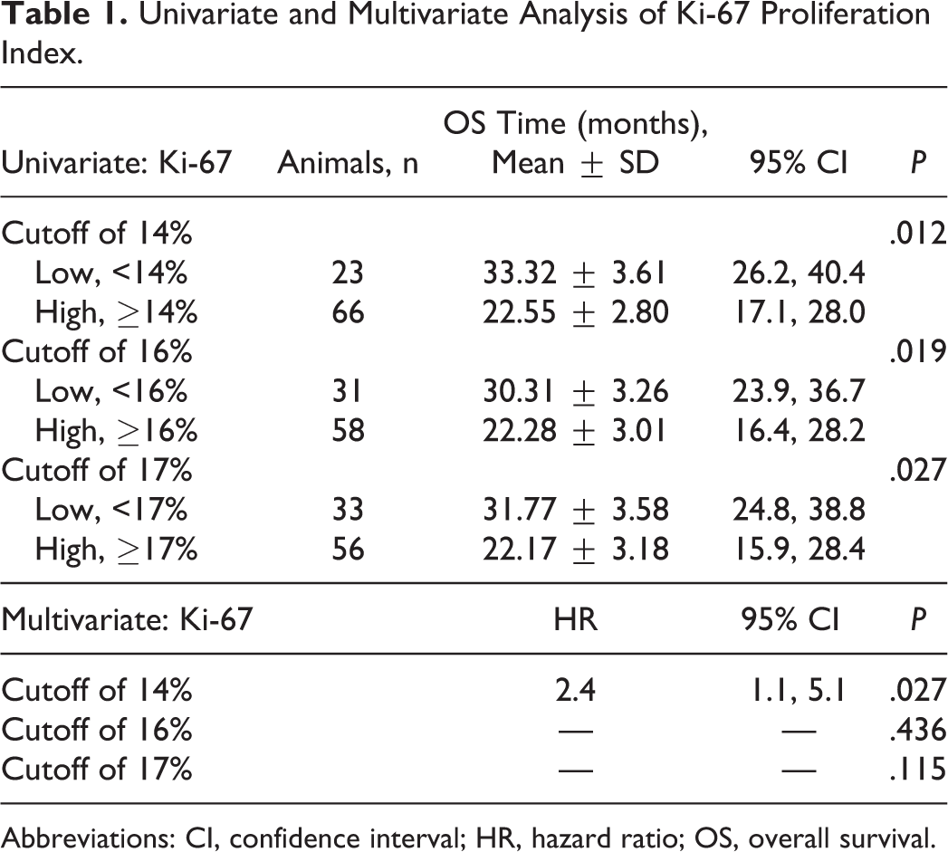
Abbreviations: CI, confidence interval; HR, hazard ratio; OS, overall survival.
Feline tonsil was used as positive control for Ki-67 index, while normal skin was used as a positive control for CK5/6 expression. Feline mammary samples with known positive and negative ER, PR, and fHER2 status were also employed as positive controls.
Immunohistochemistry staining was scored by 2 independent observers, and discordant interpretations were settled using a multiviewer microscope. Tumor cells were considered Ki-67 positive in the presence of brown nuclear staining of granular or diffuse type. 5,9,30,45 The Ki-67 proliferation index was determined by assessing the percentage of positively staining tumor cell nuclei in 1000 tumor cells. For each lesion, 5 to 6 images were randomly taken with an Olympus DP25 camera (Pennsylvania, USA) on an Olympus BX51 light microscope and analyzed using Image J (Open Source Software, version 1.46r; National Institutes of Health, Bethesda, MD, USA).
ER and PR expression was evaluated in tumor tissues using the Allred score system 15,25 (Suppl. Table 2), where a score ≥3 is considered positive. 13 fHER2 staining intensity was evaluated by using the Food and Drug Administration–approved scoring system, 13,44 in which tumors with immunohistochemistry scores of 3+ (uniform and intense membrane staining of at least 10% of tumor cells) or 2+ (complete membrane staining, not uniform or weak in intensity but with obvious circumferential membrane distribution in at least 10% of cells) were considered HER2 positive. 2,3,21,35,38 Finally, tumors were considered CK 5/6 positive when >1% of cells were immunoreactive. 1,2,16
Determination of the Optimal Cutoff Value for Ki-67 Index
Whenever a cat exhibited multiple mammary tumors, the lesion with higher risk of malignancy was selected for further statistical analysis based on tumor size and malignancy grade, which both have been associated with worse prognosis in FMC. 19,37,43
After testing for normality, the paired t test was applied to make a comparison between the Ki-67 index of primary tumors and regional metastasis, between the Ki-67 index of primary tumors and distant metastasis, and between the Ki-67 index of regional and distant metastasis. Correlation analyses were performed by using the Pearson correlation test. When multiple metastases were present (regional and/or distant), the lesions with higher Ki-67 expression were chosen for statistical analysis.
To determine the optimal cutoff value for Ki-67 in FMC, an univariate survival analysis was performed testing the following range of values, also used in human breast cancer patients: 5%, 10%, 12%, 13%, 14%, 15%, 16%, 17%, 18%, 19%, 20%, 25%, 30%, and 35%. 7,9,29 Regarding the survival analysis, OS was defined as the time, in months, between the initial diagnosis and death or the date of the last follow-up for surviving cats (censored observations). Only deaths attributed to mammary carcinoma progression were considered. In this study, 2 deaths were caused by other diseases (censored observations), both belonging to the group of cats with high Ki-67 indexes: one was caused by renal failure, and the other animal was euthanized because owners could not afford the proposed surgery. Among the total of the cats in the study (n = 96), 7 were excluded for survival analysis, once they were followed for less than a month. OS curves were estimated using the Kaplan-Meier method and compared with the log-rank test.
The cutoff values that showed significance were subjected to multivariate analysis using the Cox proportional hazard model. The hazard ratio was calculated with a 95% confidence interval (95% CI).
After the establishment of the Ki-67 cutoff, the positive predictive value (PPV) and negative predictive value (NPV) were calculated for predicting death and survival after 1 and 2 years after the surgery. For this analysis, only the animals subjected to surgery were considered, with those that also received chemotherapy being excluded.
The Fisher exact test was used to assess the association between Ki-67 expression and 19 clinicopathologic features: age, breed, reproductive status, previous administration of progestogens, tumor number, location and size, stage of disease, histopathologic classification, malignancy grade, presence of necrotic areas, lymphatic invasion, lymphocytic infiltration, cutaneous ulceration, regional lymph node metastasis, receptor status (ER, PR, fHER2), and CK 5/6 expression. For each significant association, odds ratio (OR) was calculated with a 95% CI.
The association between Ki-67 labeling index and tumor size was analyzed after dividing the population into 3 subgroups, according to the tumor size, using the levels established in the TNM classification system (<2 , 2–3 , and >3 cm). To evaluate the effect of age, the population was split into 3 subgroups: <8, 8–12, and >12 years old.
Quantitative data were processed and analyzed with SPSS 21.0 (IBM, New York, NY, USA), and a 2-tailed P < .05 was considered statistically significant.
Results
Animal Population Data
Ninety-six female cats with mammary carcinoma were followed up, and their clinicopathologic features are summarized in Supplemental Table 3. The mean ± SD age of the animals was 11.49 ± 2.85 years (range, 5–19 years). The majority of the cats were subjected to surgery (n = 89), including unilateral mastectomy in 71 (79.8%), regional mastectomy in 11 (12.4%), and bilateral mastectomy in 7 (7.8%). Eight mastectomized cats received anthracycline-based chemotherapy after surgery (doxorubicin, 25 mg/m2, intravenously, every 3 weeks for 5 cycles). 39
In this study, 213 mammary carcinomas were collected from the 96 queens. In the 60 animals that showed >1 tumor, the most malignant lesion was chosen (see Materials and Methods). The mean size of the tumors was 2.71 ± 1.5 cm (range, 0.5–7 cm).
By the end of the study, 48 animals had died or were euthanized due to metastatic progression of the disease, and for those animals, the median OS was 10.5 months (mean, 12.2 months).
Ki-67 Index of the Primary Tumor Is Strongly and Positively Correlated With the Ki-67 Index of Regional and Distant Metastasis
The immunohistochemical analysis of the Ki-67 expression revealed a moderate to strong nuclear staining of tumor cells (Fig. 1a), especially in distant metastases (Fig. 1b). The mean Ki-67 index was 26% ± 1.8% in primary tumors (range, 0.2%–85.8%; median, 21.4%), rising to 30.1% ± 3.1% in regional lymph node metastases (range, 3.5%–78.8%; median, 26.4%, n = 38). Distant metastases (n = 16) showed a mean Ki-67 index of 48.2% ± 5.1% (range, 13.3%–92.1%; median, 44.6%).
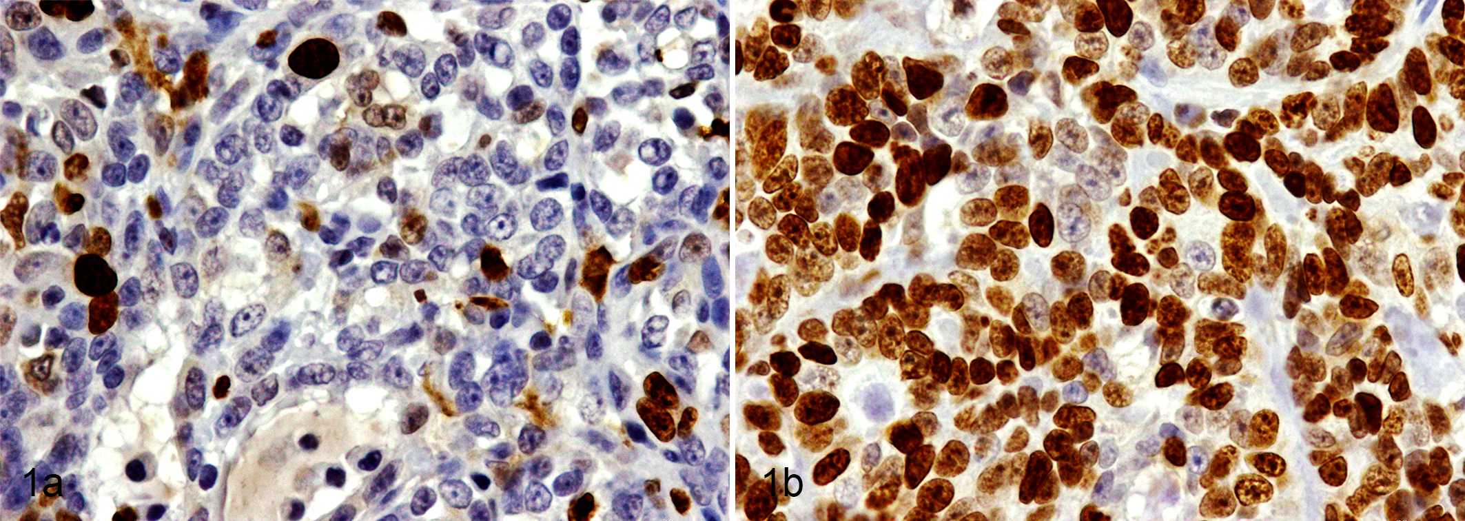
Cribriform carcinoma, mammary gland, cat. Nuclear Ki-67 staining. (a) Primary tumor with a Ki-67 index of 13.7%; (b) lung metastasis with a Ki-67 index of 88.6%. Immunohistochemistry for Ki-67.
A strong and positive correlation was found between the Ki-67 index of the primary tumor and regional metastasis (P < .0001; r = 0.83, n = 38), between the Ki67 index of the primary tumor and distant metastasis (P < .0001; r = 0.83, n = 16), and between the Ki-67 index of regional and distant metastasis (P = .0052; r = 0.75, n = 16).
In addition, the Ki-67 index of primary tumors was significantly lower when compared to the Ki-67 index of distant metastasis (P = .0009, n = 16). Differences between regional and distant metastasis approached significance (P = .08, n = 16), with distant lesions showing higher Ki-67 scores. Finally, primary tumors and regional metastasis presented distinct Ki-67 scores, which were not statistically significant (P = .186, n = 38).
The Optimal Cutoff Value of Ki-67 Index Is 14% in FMC
To determine the optimal cutoff point for the Ki-67 index, a univariate survival analysis was performed using different cutoff values (5%, 10%, 12%, 13%, 14%, 15%, 16%, 17%, 18%, 19%, 20%, 25%, 30%, and 35%). Statistically significant differences in OS curves were observed only for the cutoff of 14% (P = .012), 16% (P = .019), and 17% (P = .027; Table 1, Fig. 2). Further multivariate analysis showed that a 14% cutoff value could better distinguish cats with highly malignant mammary carcinomas from those with low malignant potential. In addition, only a cutoff value of 14% was identified to be a Ki-67 independent prognostic indicator of OS (P = .027), showing a hazard ratio of 2.4 (Table 1).
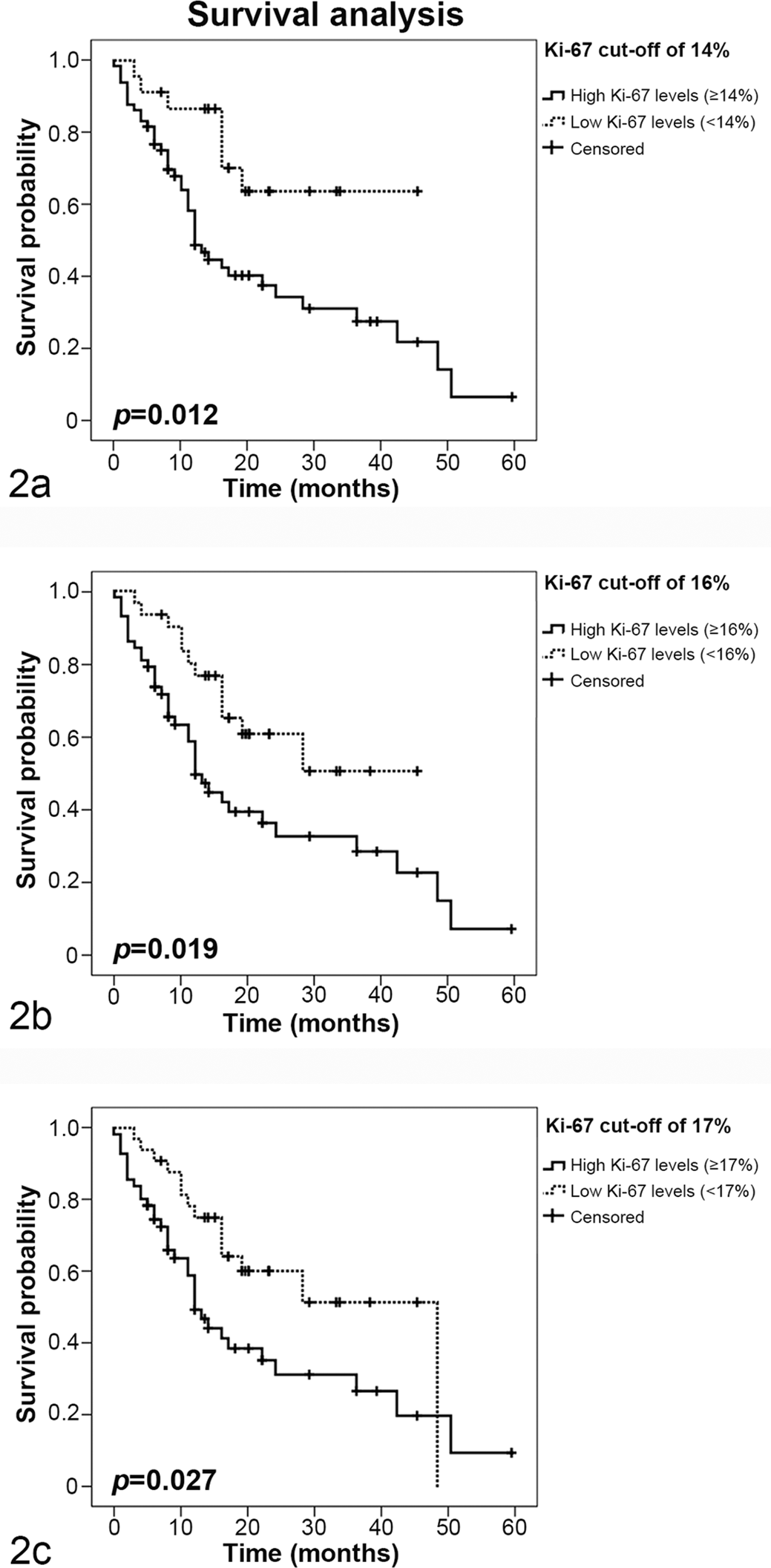
Kaplan-Meier overall survival curves using different cutoff values of Ki-67. Overall survival curves with Ki-67 cutoff values of 14% (a), 16% (b), and 17% (c). In all scenarios, cats with feline mammary carcinoma showing a Ki-67 index below the cutoff value survived significantly longer than those with higher Ki-67 indexes.
The majority of the animals that presented low Ki-67 values were alive by the end of the follow-up period (69.6%, 16 of 23), while only 37.9% (25 of 66) of cats with higher Ki-67 values (≥15%) were alive. Moreover, most of the cats that died had a primary tumor with a Ki-67 index ≥14% (85.4%, 41 of 48), and only 7 presented low Ki-67 values (14.6%, 7 of 48).
Animals With High Ki-67 Index (>14%) Have Higher Probability of Dying Within the 2 Years After Surgery
With the 14% as a threshold value, the PPV and NPV were calculated for predicting death or survival at 1 and 2 years after surgery. For 1 year after surgery, the PPV for predicting death was 50%, and the NPV was 81.8%. For 2 years of survival after surgery, the PPV was 80%, and the NPV was 27.3%.
Ki-67 Index ≥14% Is Significantly Associated With Unfavorable Features
Of the 96 cats with mammary carcinoma, 70 showed mammary tumors with a Ki-67 index ≥14% (72.9%), while 26 cats showed lower Ki-67 indices (27.1%). The majority of the cats with mammary carcinoma presenting with regional lymph node metastasis had a Ki-67 index ≥14% (78.9%, 30 of 38). Of 16 metastatic disease cases, 15 (93.8%) also had distant metastasis with a Ki-67 index ≥14%.
Moreover, primary mammary carcinomas that showed a Ki-67 index ≥14% were significantly associated with large size (P = .022; OR, 4.4; 95% CI: 1.12, 21.65), poor differentiation (grade III, P = .009), presence of necrotic areas (P = .008; OR, 3.7; 95% CI: 1.25, 11.13), negative ER status (P < .0001; OR, 7.12; 95% CI: 2.38, 22.66), and low fHER2 expression (P = .003; OR, 4.31; 95% CI: 1.47, 12.98; Supplemental Table 4). In contrast, no significant statistical association was found between Ki-67 index ≥14% and age, breed, reproductive status, previous administration of progestogens, tumor location, multiple mammary tumors, lymph node metastasis, disease stage, histopathologic classification, lymphatic invasion by neoplastic cells, lymphocytic infiltration, skin ulceration, and PR or CK5/6 status of the primary tumor (Suppl. Table 4).
Discussion
Until now, the World Health Organization histopathologic classification has been extensively used to establish the prognosis in cats and dogs with mammary tumors. 10,24,39 Although tumor cell proliferation has emerged as an important diagnostic and prognostic tool in human breast cancer patients, 5,9 –11,17,28,31,36 no standard cutoff point for the Ki-67 index is known in FMCs. 37 Recently, some studies assessed Ki-67 expression in feline mammary tumors and showed that malignant lesions exhibited higher Ki-67 levels that benign lesions. 3,4,23,32,33,37 In the present study, we demonstrated that the Ki-67 labeling index in the primary tumor reveals a strong and positive correlation with the Ki-67 index of local and distant metastasis. Moreover, and besides the small sample (n = 16), the metastatic lesions had significantly higher Ki-67 indices than the primary lesions (means of 48.2% vs 26%), corroborating the association between Ki-67 overexpression in the primary tumor and aggressive tumor behavior.
Employing the methodology used in human clinical studies, 27,29 the optimal cutoff value for Ki-67 was determined. The Ki-67 cutoff of 14% was considered as the optimal value to predict clinical outcome of FMC. Indeed, cats whose primary tumors showed a Ki-67 index ≥14% displayed a 2.4-fold increased risk of tumor-related death, in comparison with cats having mammary tumors that exhibited a lower Ki-67 index (Table 1). Most primary tumors (72.9%) and metastatic lesions (93.8%) showed a Ki-67 index ≥14%, with the corresponding cats presenting significantly shorter survival. In fact, the majority of cats that died from mammary cancer disease during the follow-up showed a primary tumor with a Ki-67 index ≥14% (41 of 48, 85.4%).
The Ki-67 index also demonstrated a strong predictive value, with a high proportion of cats with low levels of Ki-67 alive after 1 year of the surgery (NPV = 81.8%). Moreover, high Ki-67 index levels also demonstrated an important predictive value, as the probability of the cat dying within the 2 years after surgery was 80% (ie, PPV = 80%).
In parallel, we also observed a strong association between Ki-67 levels and several clinicopathologic features associated with shorter OS, such as size of the tumor, 19,43 malignancy grade, 37 presence of necrotic areas, 30,37 and low ER expression, 22 which reinforces the relevance of this marker for the clinical follow-up of FMC.
In human breast cancer, a positive ER status is associated with low cancer mortality. 8,13 Our results corroborate this scenario in FMC. Regarding HER2 tumor status, its overexpression is associated with worse prognosis in breast cancer 20,44 ; however, its role in FMC is still controversial. It was reported that overexpression of fHER2 is associated with short OS periods. 21 However, a recent report claimed that fHER2 overexpression is associated with feline tumors that show nonmalignant features. 35 Our findings support these results, since mammary tumors with Ki-67 levels ≥14% were significantly associated with a negative fHER2 status, indicating that fHER2 overexpression could be associated with a better clinical outcome.
Recent studies also showed that Ki-67 immunohistochemistry measurements can significantly predict breast cancer outcome in women treated with anthracycline-based chemotherapy. 26,34,40 Anthracyclines are a class of antitumor drugs (eg, doxorubicin) that inhibit a family of enzymes essential for cell division (topoisomerases type II), making these drugs a primary choice in the treatment of tumors with high growth rates, being also used to treat a variety of malignancies in cats, including mammary carcinoma. 39 To the best of our knowledge, there are no studies showing that chemotherapy improves survival in cats with mammary carcinomas showing a high Ki-67 index. Unfortunately, in our study, only 8 cats (8.3%) with mammary carcinoma were treated with doxorubicin after surgery, not allowing us to assess the value of Ki-67 in predicting the benefit of anthracycline-based chemotherapy.
In conclusion, our study demonstrated that the Ki-67 index can be used as a prognostic biomarker in cats with mammary carcinoma—specifically, it showed that Ki-67 values ≥14% are associated with lower OS and with other aggressive clinicopathologic features. All results presented above suggest that a Ki-67 cutoff of 14% can be regarded as a useful tool to identify animal patients with worse prognosis.
Footnotes
Acknowledgements
We thank João Matos and José Cabeçadas, MD, from Instituto Português de Oncologia de Lisboa; Dr Manuel Mestre, DVM, Ana Mota, DVM, MSc, and Tiago Rafael, DVM, MSc, from Clínica Veterinária Zoomédica; Mafalda Lage, DVM, MSc, from Clínica Veterinária Villa Animal; Rafaela Lalanda, DVM, MSc, and Miguel Caninhas, DVM, from Clínica Veterinária Mvet; Verónica Azevedo, DVM, MSc, from Hospital Sul do Tejo; António Ferreira, DVM, PhD, Ana Murta, DVM, MSc, and Rodrigo Bom, DVM, from the Small Animal Hospital from the Faculty of Veterinary Medicine of the University of Lisbon, for the clinical follow-up.
Declaration of Conflicting Interests
The author(s) declared no potential conflicts of interest with respect to the research, authorship, and/or publication of this article.
Funding
The author(s) disclosed receipt of the following financial support for the research, authorship, and/or publication of this article: This research was supported by “Fundação para a Ciência e Tecnologia” (FCT) project grant UID/CVT/00276/2013 and PhD fellowship SFRH/BD/70720/2010.