
Abstract
The application of medical knowledge to the purpose of law is the foundation of forensic pathology. A forensic postmortem examination often involves the expertise of multiple scientific disciplines to reconstruct the full story surrounding the death of an animal. Wildlife poses additional challenges in forensic investigations due to little or no associated history, and the disruptive effects of decomposition. To illustrate the multidisciplinary nature of wildlife forensic medicine, the authors outline a case of secondary pentobarbital/phenytoin toxicosis in a bald eagle (Haliaeetus leucocephalus). The eagle was the single fatality in a group of 8 birds that fed on euthanized domestic cat remains that had been improperly disposed of in a landfill. Cooperation between responding law enforcement officers, pathologists, and other forensic scientists led to the successful diagnosis and resolution of the case.
The practice of diagnostic pathology—the assessments used to discover and delineate the disease or condition that caused an animal’s death—is a multimodal exercise. Infrequently is a definitive cause-of-death judgment based on gross or microscopic examination alone. Gross findings must be married with microscopic, toxicologic, microbiologic, and/or clinicopathologic findings to build the full and complete story about the death of that animal. The case is similar in the practice of forensic pathology. To fully discover the “who, what, when, where, and how” of a veterinary forensic pathology story, multiple aspects of the forensic sciences are often utilized. Sciences that may contribute to a full forensic analysis—filling in the outline provided by a forensic pathology examination—include genetics, morphology, toxicology, and criminalistics.
Domestic animals, by definition, live in close proximity to humans. Their health and well-being depend on the presence and assistance of people. Investigation of cases of death due to neglect, therefore, involves suspects and crime scenes not too far from the victim. In contrast, wild animals are independent, often avoiding civilization and places that humans frequent. Because of this separation, linking a suspect to a wildlife victim can be problematic. Adding to this challenge, and inherent to the separation between wildlife and humans, is an often extended time between death and discovery of the remains. Autolysis and the effects of scavenging often obscure or remove changes in the body that could tell the story of an animal’s death.
To illustrate the multidisciplinary nature of wildlife forensic pathology and the challenges that may arise, we present a case report of fatal pentobarbital and phenytoin toxicosis in a bald eagle (Haliaeetus leucocephalus) that was examined at the US Fish and Wildlife Service’s National Fish and Wildlife Forensics Laboratory (NFWFL). Eight adult and subadult bald eagles were found in and near a municipal landfill site. At the time of discovery, the eagles exhibited signs of neurologic depression, regurgitation, and/or convulsions. Several eagles were depressed to the point of appearing to be dead to onlookers. Staff from a wildlife rehabilitation program collected 7 of the 8 eagles; 1 eagle flew away during the capture attempts. The 7 captured birds received supportive treatment and showed improvement over the next several days. Samples of regurgitated material were frozen for potential analysis.
Examination of the landfill site around where the eagles were found revealed the partially scavenged remains of 2 mammals appearing to be domestic cats (Fig. 1). The remains were found uncontained and exposed to the environment.
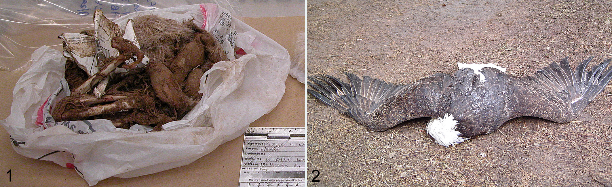
Later the same day, a dead eagle was found on a snowmobile trail located 25 miles from the municipal landfill site (Fig. 2). The eagle appeared to have regurgitated prior to death. The body was prone and the wings of the eagle were spread indicating a relaxed state prior to death. Law enforcement personnel responding to both scenes suspected that the eagle that flew away during capture attempts at the landfill site was the same eagle found on the snowmobile trail. The dead eagle and regurgitated material were submitted with the remains of the mammal by the responding law enforcement officer to NFWFL for cause of death determination.
The eagle underwent a forensic postmortem examination which included radiographs, complete skinning of the carcass, and removal of the viscera. The ventriculus contents were collected in aluminum foil, officially sealed, and submitted for chemical analysis. Per standard forensic analysis guidelines, photographs were taken to document both normal and abnormal gross findings.
The eagle weighed 4.5 kg just prior to necropsy and had a good postmortem preservation status. Musculature over the shoulders and chest was moderately depleted, but fat stores were present in adequate amounts. Ventriculus contents consisted of only 2 ml of dark green, mucoid ingesta. Small amounts of digesta were present in the intestinal tract. Aspirated ingesta was present in the tracheal lumen. The lungs, liver, heart, brain, and kidneys were unremarkable. There was no gross evidence of trauma, gunshot wounds, electrocution, or systemic disease.
Samples for the forensic chemistry examination consisted of regurgitated material from an eagle that survived, and the ventriculus contents from the bald eagle submitted for necropsy. These samples, as well as a control sample, were subjected to extraction with methylene chloride, and then a portion of each extract was concentrated to dryness. Triphenyl phosphate was added as an internal standard and the samples were analyzed employing an Agilent 6890 Gas Chromatograph coupled with an Agilent 5973 Mass Spectrometer.
Results of the chemical analyses of the eagle’s postmortem ventriculus contents and antemortem regurgitated material indicated that the barbiturate pentobarbital and the anticonvulsant phenytoin were present in both samples.
Forensic genetic analysis was conducted to determine the species of origin of the contents of the regurgitated material . DNA was extracted from a subsample of the regurgitated material and a segment of the mitochondrial 16 S rDNA gene was amplified by PCR (Short-16S-F: gAAATTgACCTTCCCgTgAAgAgg; Short-16S-R: CgCTgTTATCCCTAGggTAACT) and subjected to Sanger sequencing by capillary electrophoresis. The resulting 212 base pair 16 S rDNA sequence was compared to the NFWFL mammal reference sequence database.
A 212 base pair segment of the 16 S rDNA locus of mitochondrial DNA was amplified from DNA extracted from the regurgitated material. This sequence was compared to those in the NFWFL mammal reference sequence database and found to be identical to that of domestic cat (Felis catus) at 212/212 bases. The sequence was compared to and found to be unlike those of other species in the genus Felis, as well as other North American wild cats including cougar, bobcat, and lynx.
Forensic morphology examination was used to identify mammalian remains found at the landfill site and considered a probable source of any toxic material. The remains consisted of mangled limbs and partial cranium covered in white fur. Standard morphological protocols were used to determine the taxonomic identification of these remains. Analysis consisted of extracting those anatomical elements that offer diagnostic details at the species level, which in this case included dentition and claws.
Dental observations of the mammalian remains revealed lateral grooves on the canine teeth, as well as retractile claws on the feet. Both of these features are diagnostic for felids. Size and fur color completed the morphological determination of domestic cat as the species represented by the remains.
Based on the combined forensic analyses, it was determined that the eagle submitted to the NFWFL for necropsy had died of pentobarbital and phenytoin toxicosis through ingestion of a euthanized cat at a municipal landfill site. The regurgitated material found with the dead eagle, and the eagle’s ventriculus contents both contained pentobarbital and phenytoin. Though the eagle’s body was found 25 miles from the landfill site, the eagle could be placed at the site through genetic analysis of the eagle regurgitated material and morphological analysis of the mammalian remains at the landfill, both of which were determined to be of domestic cat origin. Histology was determined to be unnecessary in the determination of the eagle’s cause of death and was not performed. The 7 eagles that were initially captured and treated fully recovered within 2 months and were released into the wild.
The NFWFL does not conduct genetic matching (individual identity) analysis of domestic species such as cats, as this analysis falls outside the mission scope of the laboratory. Genetic matching of many domestic species is available at other institutions. 1 In the case presented here, the circumstantial evidence of domestic cat tissue and pentobarbital in the dead eagle and regurgitated material, taken in conjunction with events and findings at the municipal landfill crime scene was considered to be adequate for successful prosecution of the case.
Incidents of pentobarbital toxicosis of wildlife have been rarely documented in the literature. 4,5 A retrospective survey of cases at the US Geologic Survey’s National Wildlife Health Center in Madison, Wisconsin, showed that 4.3% of bald eagle poisonings and 2.6% of golden eagle (Aquila chrysaetos) poisonings were due to barbiturate intoxication. 5 From this study population of 2980 bald eagles and 1427 golden eagles that were submitted for necropsy, 33 bald eagles and 3 golden eagles succumbed to barbiturate poisoning. 5 Langelier reported an incident involving 29 bald eagles that fed on a euthanized domestic cow. 4 Of this group, 5 were found dead or died within an hour of presentation, and 1 was found 21 km from the cow carcass. As exemplified by Langelier and the bald eagle in this case report, the clinical course of pentobarbital toxicosis may be such that birds can fly or soar great distances from the point source of the toxin. Removal from the scene of the crime eliminates environmental and proximity clues that could assist an investigation. Diligent and thorough analysis of these cases is required to provide an accurate diagnosis.
A review of case records dating from 1992 to 2015 at the NFWFL revealed 62 animals with suspected or definitive mortality due to secondary barbiturate intoxication in a total of 41 separate events (Supplemental Table 1). Animals diagnosed with fatal barbiturate intoxication included bald eagles (40), golden eagles (11), 2 red-shouldered hawks (Buteo lineatus), 1 Cooper’s hawk (Accipiter cooperii), 1 red-tailed hawk (Buteo jamaicensis), 1 black-billed magpie (Pica hudsonia), 1 common raven (Corvus corax), 1 coot (Fulica americana), 1 common crow (Corvus brachyrhynchos), 1 bobcat (Lynx rufus), and 1 coyote (Canis latrans). Species that were presumptively euthanized and eaten by the affected animals included horse, mule, cat, sheep, goat, cow/calf, dog, pig, and llama or alpaca. Chemical analysis of liver or stomach contents of the affected animals showed both pentobarbital and phenytoin in 11 of the 62 animals; pentobarbital alone was detected in 50 animals, and the bobcat had ingested phenobarbital. The combination of pentobarbital and phenytoin is present in several veterinary formulations for domestic animal euthanasia, including Beuthanasia-D Special (manufactured by Intervet/Merck Animal Health) and Euthasol Euthanasia Solution (manufactured by Virbac).
Each state within the United States creates and administers laws regarding the disposition of euthanized animals. Disposal practices outlined in the laws are designed to prevent negative impacts to wildlife and the environment by making the animals and tissue drug residues unavailable for ingestion or contamination of the environment. It is the responsibility of the veterinarian licensed to administer euthanasia solutions to either dispose of a euthanized animal appropriately, or advise staff at a holding entity, such as an animal shelter, of correct disposal practices.
The successful resolution of a forensic pathology case is rarely an individual effort. As in this case example in which several scientific disciplines added pieces of data to determine the cause of the eagle’s death, reconstructing the events surrounding the death of an animal often requires the efforts of more than 1 analyst.
As in clinical medicine and diagnostic pathology, a good investigation starts with a good history. Unfortunately, in many wildlife cases, a clinical history is unavailable as animals may be found dead in the environment. A thorough investigation of the scene by the responding law enforcement personnel may glean information or evidence that can help the investigation. Communication of these findings, as well as the location of the body (eg, under a power pole or near a baited carcass) can help the veterinary pathologist direct his/her analysis. In the case presented here, the law enforcement officer relayed to the veterinary pathologist information about the possible link between the eagle found dead at the remote snowmobile trail and the clinically affected eagles at the landfill site. This information allowed the veterinary pathologist to refine a list of possible causes of death and prioritize tests that would support or disprove the primary hypothesis.
Other challenges that wildlife forensic pathologists frequently face are the effects of decomposition and scavenging. Wild animals rarely die at the doorstep of a veterinary clinic or rehabilitation center where their remains can be either appropriately preserved or expeditiously examined. Wild animals tend to die in remote or hidden venues, exposed to the factors of weather, climate, and opportunistic feeders. These agents effect changes on the body—a process termed “taphonomy”—that may alter or obscure evidence that points toward a cause of the animal’s death.
Taphonomic processes may render a carcass unidentifiable to the untrained eye. Invertebrate and vertebrate scavengers may deflesh bones and scatter identifying characteristics. Even with highly altered remains, depending on the extent of degradation, certain modalities may be used to (1) identify the species and (2) determine cause of death. In the case report herein, genetic and morphologic examination of heavily scavenged remains revealed domestic cat as the origin of the toxin that affected the eagles. Muscle remnants, regurgitated material, teeth, and claws were all that were needed for genetic and morphologic species assignment.
Pathologic processes may remain evident even in highly degraded remains, but must be differentiated from changes induced by aggressive scavengers, such as rodents and canids. 2,3 While bullets often cause perforating or excoriating lesions in bone, so too can scavenging activity. Use of an x-ray florescence analyzer may reveal minute particles of lead that are neither grossly visible nor evident on standard radiographs of heavily degraded remains. The presence of lead particles around a fracture strongly supports a diagnosis of gunshot injury. Analysis of equivocal fractures by a chemist or ballistic analyst trained in elemental analysis can help to differentiate postmortem taphonomic changes from antemortem lesions.
Similarly, the degraded and scavenged bones of animals that died of pentobarbital toxicosis/euthanasia may still contain residues of the drug in the skeletal remains. Watterson et al 6 were able to detect pentobarbital in the bones of a pig 2 years after euthanasia. While variable drug distribution patterns and the effects of environmental exposure invalidate quantitative results, the presence of the drug as determined by a toxicologist still provides a valuable data point in a forensic pathology investigation.
Investigation of a veterinary forensic case often requires the skill sets and knowledge of multiple individuals, including law enforcement personnel, veterinarians, other scientists, and workers in the legal profession. Wildlife forensics presents unique challenges with regards to history, crime scene investigation, and postmortem degradation/scavenging. As exemplified by this case of pentobarbital toxicosis in a bald eagle, the cooperation between these different groups can overcome some challenges and lead to the successful resolution of a veterinary forensic pathology investigation.
Footnotes
Acknowledgements
We wish to thank US Fish and Wildlife Service Special Agent S. Stoinski for investigation of the pentobarbital case and submission of case materials, and Colleen Wilson for pathology technical assistance. The findings and conclusions in this article are those of the authors and do not necessarily represent the views of the US Fish and Wildlife Service.
Declaration of Conflicting Interests
The author(s) declared no potential conflicts of interest with respect to the research, authorship, and/or publication of this article.
Funding
The author(s) received no financial support for the research, authorship, and/or publication of this article.
References
Supplementary Material
Please find the following supplemental material available below.
For Open Access articles published under a Creative Commons License, all supplemental material carries the same license as the article it is associated with.
For non-Open Access articles published, all supplemental material carries a non-exclusive license, and permission requests for re-use of supplemental material or any part of supplemental material shall be sent directly to the copyright owner as specified in the copyright notice associated with the article.