
Abstract
The expanding presence of red foxes (Vulpes vulpes) in urban and suburban regions could potentially lead to increased instances of human aggression towards this species. We studied 10 deceased red foxes that were submitted by law enforcement agencies in the metropolitan area of Madrid in 2014–2022 because of suspected abuse. Forensic autopsies were performed to establish the cause and manner of death. In 4 of the 10 cases, the cause of death was deemed unnatural, involving blunt-force trauma (n = 2), asphyxia resulting from hanging (n = 1), and firearm injury (n = 1). Among the remaining cases, most had succumbed to natural causes (n = 4), often marked by severe emaciation and a high burden of parasites, primarily Sarcoptes scabiei. In 2 cases, death was undetermined given the poor preservation of the carcass. The growing prevalence of wildlife species in urban areas, particularly red foxes, may require forensic veterinary investigation of deaths potentially related to abuse.
The rapid growth of major population centers, combined with habitat loss and urbanization, has led to the displacement of native species from their natural habitats. Some displaced wildlife species have successfully adapted to coexist with humans, such as the wild boar (Sus scrofa) and the red fox (Vulpes vulpes).1,14 The red fox is one of the most abundant carnivores worldwide, and its colonization of urban and suburban areas has increased.10,14 The expansion of wildlife habitat can lead to the transmission of infectious and parasitic diseases to humans and companion animals. The role of the red fox as a reservoir of rabies (Lyssavirus spp.), tuberculosis (Mycobacterium tuberculosis complex), sarcoptic mange (Sarcoptes scabiei), echinococcosis (Echinococcus multilocularis), and tularemia (Francisella tularensis) is of public health concern.7,10,14,16
Red foxes, like many wildlife species, have historically been regarded as detrimental to human activities in rural areas, mainly livestock rearing, exacerbating human–wildlife conflicts.9,12 However, red foxes have been shown to be irrelevant as a livestock predator species. 18 This unjustified fear has sometimes resulted in the legal or illegal killing of these animals, sometimes having a counterproductive effect.9,17 Aggression toward wildlife has been successfully managed by local agencies in most cases, whereas in others it has led to human–wildlife conflict. 9 Reports on cases of red fox abuse are scarce in the literature. Furthermore, studies on the contribution of veterinary pathology to the investigation of wildlife crime are limited in Spain. Here, we present the postmortem findings of a group of deceased red foxes found with signs of potential abuse in the metropolitan area of Madrid, Spain.
Ten red foxes (Vulpes vulpes) that were found dead in the metropolitan area of Madrid were referred to the VISAVET Health Surveillance Centre (Centro de Vigilancia Sanitaria Veterinaria, Complutense University of Madrid, Madrid, Spain) by law enforcement agencies between 2014 and 2022. The discovery scene and witnesses led the authorities to suspect that the deaths were related to abuse. All available information was recorded in the submission reports including nonspecific information regarding locations where the bodies were found (e.g., bodies found near roadways). During the autopsy, age, sex, carcass preservation, and postmortem interval (PMI) were recorded (Table 1). For age assessment, tooth eruption and tooth wear were used as a reference, classifying animals as juveniles or adults. 2 PMI was established by assessing postmortem changes and using forensic entomology, and was divided into 5 categories: fresh, bloated, active decay, advanced decay, and dry remains. 15 Body condition score (BCS) was assessed by adapting the World Small Animal Veterinary Association body condition score for dogs. 19 Fractures were assessed by visual examination and palpation of individual bones.
Date of submission, age, sex, carcass conservation, and submission report detailed for each case.
F = female; M = male.
For rabies testing, half of the specimen’s brain (when available) was placed in glycerol buffer (50%) in PBS. Tests were performed at the National Center for Microbiology (Carlos III Health Institute, Madrid, Spain). Rabies tests included lyssavirus nucleic acid detection by PCR assay, simultaneous detection of rabies virus (RABV; Rhabdoviridae, Lyssavirus rabies) and European bat 1 lyssavirus (EBLV1; Rhabdoviridae, Lyssavirus hamburg) using a multiplex real-time PCR assay, and RABV antigen detection by indirect immunofluorescence. Parasite identification was carried out based on morphology and employing a stereoscopic microscope. Samples of all tissues were fixed in 10% neutral-buffered formalin, processed routinely, and sections stained with H&E. Samples for subsequent toxicologic studies were collected and frozen, and processed if requested by the Court (judge or prosecutor).
Cause and manner of death were established according to previous publications. 15 Briefly, cause and manner of death were interpreted based on the gross findings, histopathology, and ancillary testing. Cause of death was the main lesion leading to death; manner of death was sub-classified as natural, non-natural, or undetermined, according to a forensic approach adapted to veterinary medicine. 15 We interpreted the manner of death as non-natural in 4 of 10 cases: blunt force trauma (n = 2), asphyxiation by hanging (n = 1), and gunshot injuries (n = 1; Table 1).
In the first blunt force trauma case (case 1), the BCS was adequate (5 of 9). Externally, mucous membranes were pale, and there were focally extensive hemorrhages in the subcutaneous tissue and muscle in the cranial, thoracic, and abdominal areas. In the thoracic cavity, there was ~60 mL of free and clotted blood, the lungs were atelectatic, emphysematous, and edematous. Hemoperitoneum was also present. Catarrhal enteritis with intralesional nematodes (Toxocara canis) was observed.
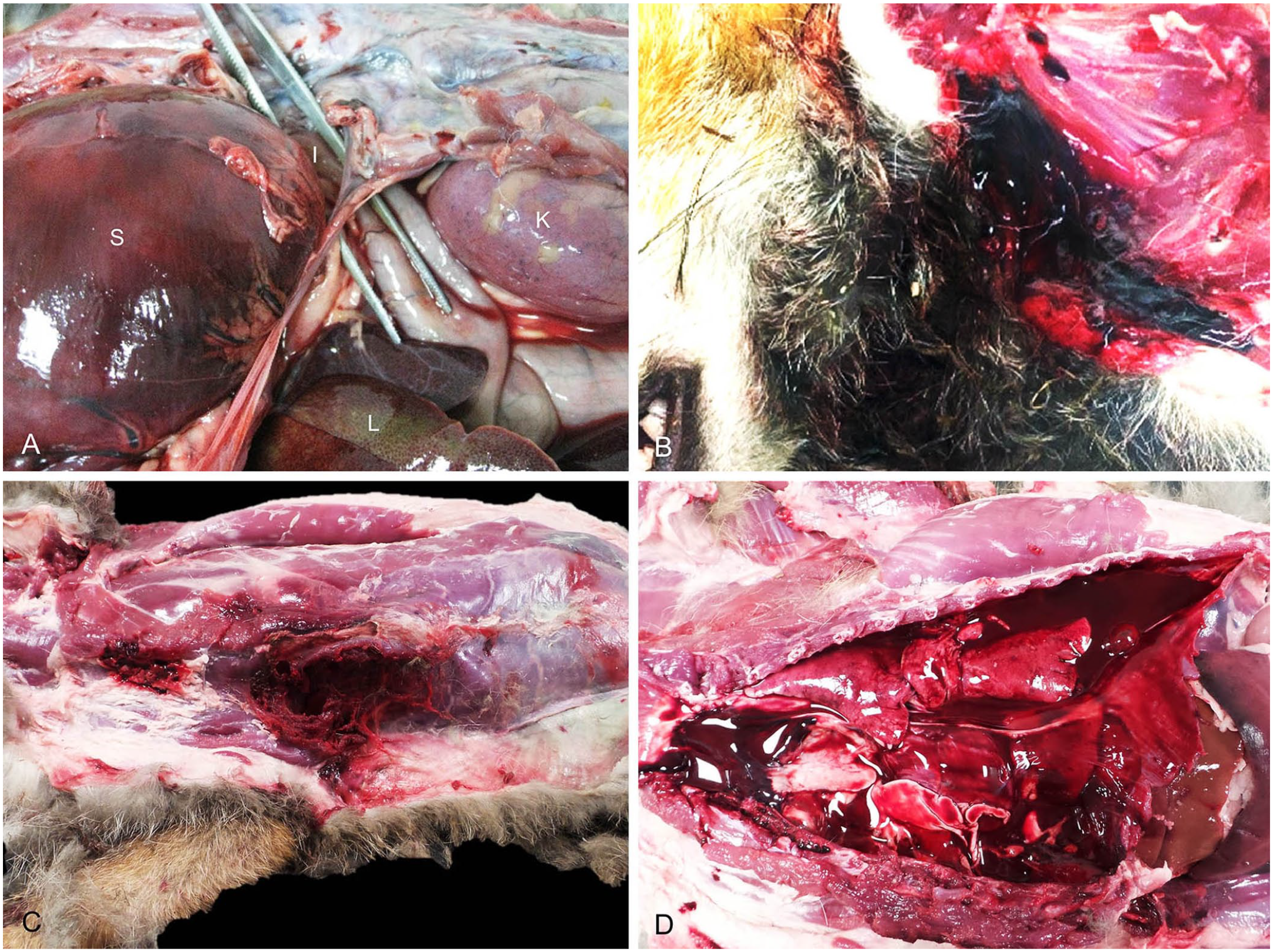
In the second case of blunt-force trauma (case 2), the fox had an adequate BCS (5 of 9), pale mucous membranes, and extensive focal hemorrhages throughout the subcutaneous tissue and muscle in the cranial, thoracic, and abdominal areas. There was a 10-cm rupture in the diaphragm, with displacement of the spleen, stomach, and small intestine into the thoracic cavity (Fig. 1A). Hydropericardium (5 mL) and dilation of the right ventricle were also observed, as was hemoperitoneum. The stomach was markedly dilated, with congestion of the gastric blood vessels and necrosis of the gastric wall.
Forensic cases of suspected abuse of red foxes (Vulpes vulpes) in the metropolitan area of Madrid, Spain.
One red fox (case 4) was found hanged. This animal had a low BCS (1 of 9, emaciated). Externally, the hair on the neck was stained with blood, and a focal laceration was observed in the ventral area of the neck (Fig. 1B). The subcutaneous tissue 8-cm cranial to the focal laceration had a linear pale subcutaneous area (ligature mark), subcutaneous edema, and focally extensive hemorrhage, extending caudally to the left scapular area. Cranial thoracic lymph nodes had marked dilation of subcapsular sinuses by abundant RBCs. Multifocal pulmonary emphysema was severe, and the right ventricle appeared dilated. In the abdominal cavity, the spleen was contracted, and the stomach was filled with plastic material.
The fox that had died of a gunshot wound (case 8) had an adequate BCS (5 of 9). The mucous membranes were pale, and externally there was blood around the nares and mouth. There was postmortem loss of the left forelimb distal to the carpus associated with bone fractures. A penetrating wound involving skin, subcutaneous tissue, and muscle was observed on the right side of the thorax (Fig. 1C). After dissection, subcutaneous and muscle hemorrhages with intralesional ammunition were found in the tissue adjacent to the penetrating wound. In addition, hemorrhages in the subcutaneous tissue of the head were observed macroscopically and histologically. Marked hemothorax (Fig. 1D) and cardiac hemorrhages were present.
Cases 5–7 and 9 were determined to be natural deaths. The foxes were markedly emaciated (BCSs of 1 of 9), and external examination revealed pale mucous membranes and focally extensive areas of cutaneous alopecia and lichenification (head, dorsum, hindlimbs, tail base) with focal cutaneous ulceration in case 7. Histologically, the parakeratotic hyperkeratotic epidermis contained parasites consistent with S. scabiei (adult forms) within serocellular crusts, associated with mild eosinophilic infiltrates. Also present were ascites (case 7), mild splenomegaly (case 9), hepatic congestion (case 9), distension of the gall bladder (case 5), mild chronic interstitial nephritis (cases 5–7, 9), urolithiasis (case 7), nodular cortical hyperplasia of the adrenal glands (cases 5, 7), chronic hyperplastic gastritis (case 9), and moderate hemorrhagic enteritis with intralesional Dipylidium caninum (case 9). In the thoracic cavity, complete loss of pericardial fat (case. 7), dilation of the right ventricle (cases 5–7, 9), pulmonary atelectasis (case 5), and discrete pulmonary hemorrhages (cases 6, 7) were observed. Autopsy of cases 3 and 10 did not allow a conclusive diagnosis because of poor preservation of the carcass (undetermined death). Test results for RABV and EBLV1 were negative.
The annual distribution of the cases we received suggests that red foxes are most prevalent in the metropolitan areas of Madrid in the autumn and winter months (October–January). Indeed, it has been reported that red fox movements into urban areas were highest during the breeding season, which coincides with the coldest months of the year. 14 In our study, epidemiologic data on sex and age may indicate a higher proportion of males and juveniles, which may be associated with movement toward urban environments in search of resources and territory expansion.
We aimed to distinguish natural deaths from abuse-related deaths. Four of 10 red foxes presented because of suspected abuse died of non-natural causes; the cause of death was not determined in 2 cases because of autolysis. Blunt-force trauma was seen in 2 cases with severe subcutaneous and muscle hemorrhages, hemothorax, and hemoperitoneum, and a diaphragmatic hernia in one case. Blunt-force trauma can be attributed to several causes, and it is difficult to determine definitively the event inducing these injuries. Blunt-force trauma due to dog attack or human beating of red foxes has been reported in 2 red fox cases.5,12 The distribution and severity of injuries observed in our 2 cases suggest that the most likely cause was a collision with a vehicle, 6 which is reported as a common cause of death in urban and peri-urban wildlife.4,12 Of the 10 submitted cases of suspected animal abuse, only 2 were found to likely be cases of abuse, namely one of hanging and one of gunshot injury.
Witness testimony in one case stated that the red fox was found hanged in a tree. The cause of death was determined to be asphyxia caused by hanging, and the main postmortem findings were the ligature mark, and subcutaneous and muscle hemorrhages, which are frequent findings in canine and feline cases.11,15 We retrieved no reports of hanging in foxes in a search of Google, PubMed, CAB Direct, Web of Science, and Scopus using search terms “red fox abuse,” “red fox forensic necropsy,” “red fox hanging,” and “red fox asphyxia,” suggesting that this condition has not been reported previously in red foxes.
Firearm injuries are a common cause of death in wildlife species and have been reported sporadically as the cause of death in red foxes. 3 Although red fox hunting is allowed in Spain under certain conditions, the use of firearms in cities is forbidden. The case reported here had a gunshot injury in the right side of the thorax, which caused extensive hemorrhages in the subcutaneous and muscle tissues, and hemothorax. Other authors have described a case in which abdomen and upper thorax were the main region affected in shot foxes. 3
The cases classified as natural deaths had alopecia and lichenification affecting thorax, hindlimbs, and head, and were confirmed as sarcoptic mange. The hypersensitivity form of sarcoptic mange is characterized by alopecia, lichenification, and fissuring, 13 and has been reported as causing high mortality in red foxes. 5 Severe emaciation and death in such cases have been attributed to heavy parasitism. 13 Additionally, we observed nodular adrenocortical hyperplasia and chronic interstitial nephritis, reported previously in red foxes. 8 Our limited resources did not allow further investigation of nutritional, autoimmune, infectious (Leptospira spp.), or parasitic diseases (Leishmania spp.) as potential causes of disease in foxes. 8 Lastly, although some authors have described a significant proportion of red foxes with cardiovascular lesions, 8 we attributed the right ventricular dilation that we observed to the postmortem accumulation of blood or autolysis.
Endangered animals and wildlife suspected of violent death should be subjected to autopsy to elucidate whether injuries are consistent with animal abuse. Postmortem examination is of great value in monitoring wildlife health and disease and assessing zoonotic risks to humans. We identified no significant zoonotic agents previously reported in red foxes, except for S. scabiei.
Footnotes
Acknowledgements
We thank Gabriela Torre (VISAVET Health Surveillance Centre) for her technical support.
Declaration of conflicting interests
The authors declared no potential conflicts of interest with respect to the research, authorship, and/or publication of this paper.
Funding
Agustín Rebollada-Merino was the recipient of a Spanish Government–funded PhD contract for Research Staff Training (FPI) granted by the Spanish Ministry of Science and Innovation and the Spanish Ministry of Universities (RTI2018-098658-B-C22; PRE2019-087439).