
Abstract
Decades after the problem was first identified, power line electrocution continues to be a cause of avian mortality. Currently, several federal laws protect eagles and other migratory birds, meaning that utility companies may be liable for electrocution-related deaths. Veterinarians and veterinary pathologists called upon to diagnose and treat electrocuted birds should keep this in mind when conducting clinical and postmortem examinations. This review details necropsy findings and methods used to diagnose electrocution. A combination of gross, subgross, and radiographic examinations can aid in identification of subtle injury. Diagnosis is made based on the presence of skin and/or feather burns. Other necropsy findings may include skin lacerations, subcutaneous burns, bruising, limb avulsion, hemopericardium, and vascular rupture. At the US Fish and Wildlife Service’s National Forensics Laboratory, from 2000 to 2015, 417 raptor deaths were determined to have been caused by electrocution. Bald eagles and golden eagles were the most commonly submitted species. In a retrospective review of 377 cases, for which whole bodies were submitted, 18% of the electrocuted birds had only a single, small (less than 3 cm in diameter) external burn. Small, isolated burns tended to occur on the undersides of the wings at and distal to the elbow and on the lower legs and feet. These areas should be most carefully examined in cases where electrocution injury is not immediately apparent.
Keywords
Electrocution of birds from overhead power lines has been a recognized cause of mortality in the United States since the early 1970s. 10,13 Today, power lines are still a significant cause of death, fatally electrocuting an estimated 0.9 to 11.6 million birds each year. 15 Overall, the incidence of electrocution does not appear to have decreased despite over 3 decades of research and mitigation procedures. 14 Although not the single most common cause of anthropogenic avian mortality, it is of particular concern as a hazard to species with low reproductive rates and declining populations. 1,15 Power line electrocutions are also costly for the utility company. Because the cost of wildlife-related outages can be in the millions of dollars (a burden that may be transferred to the consumer), it is in everyone’s best interest to limit the potential for electrocutions. 1
Three federal laws protecting birds may be invoked in electrocution cases. 13 The Migratory Bird Treaty Act (MBTA) of 1918 protects most native migratory birds in the United States. The MBTA does not require proof of intent, so any “take” may be a violation. The Bald and Golden Eagle Protection Act (1940) and the Endangered Species Act (1973) provide additional, species-specific protections. Under these laws, utilities must work with the US Fish and Wildlife Service or state agencies to identify and acquire permits and procedures for all avian interactions. Utility companies may be liable for power line mortalities; therefore, it is important to approach all avian electrocutions as forensic cases.
Mechanisms of Injury
Birds are electrocuted when they make contact with 2 pieces of electrical equipment or electricity and a grounded object. 10 On an overhead power line, this may mean contact between 2 wires, or between a wire and a noninsulated pole or pole equipment (such as transformers). 14 Narrow horizontal or vertical wire separation predisposes wing-to-wing and head-to-body contact points. Minimal horizontal and vertical separations of 60 inches (152.4 cm) and 40 inches (101.6 cm), respectively, are recommended for eagles, although these distances likely underestimate the optimal spans. 1,8,9 It should also be noted that high-voltage electrocutions do not always require direct contact as electrical arcing may occur with proximity. 5
Avian electrocutions reportedly occur most often at distribution lines, which are considered to have a moderate voltage of 2.4 to 60 kilovolts (kV). 10,13 Distribution lines send electricity from the distribution station to the residence or business and are those lines typically seen along roads. In comparison, transmission lines carry high voltages, equal to or greater than 60 kV. Low voltages (600 V or less) are used by the service lines that deliver power from the pole transformer directly to a house as well as electrical sources within a house. Low voltages are most commonly implicated in human electrocutions but do not appear to be a factor in avian mortalities. 1,14,15 The range of voltages associated with distribution lines includes what is, from a medical perspective, both low and high voltage. 5 For the purposes of this article, the term high voltage will refer to currents greater than 600 V.
Body size, habitat, young age, power line configuration, and wet weather are predisposing risk factors for electrocution. 1,10 Eagles, Buteo hawks, and large owls appear to be the most commonly electrocuted groups of birds, with golden eagles (Aquila chrysaetos) in particular being identified as the species most often affected. 1,10,14 The most important factor in establishing 2 contact points is skin-to-skin contact. Dry feathers provide substantial resistance, but wet feathers have 10 to 15 times less resistance. 1,14 Feathers are unlikely to protect against the extremely high voltages carried by transmission lines.
When a bird completes an electrical circuit, damage is sustained over the pathway taken by the current. The path of least resistance in high-voltage situations is the shortest distance between the entry and exit points. Although the precise mechanism of death in high-voltage electrocutions is still not completely understood, it is generally agreed that death results from the passage of current through the cardiac and/or respiratory centers of the brain or directly through the heart. Depending on variables such as contact points, cardiac or pulmonary arrest may be caused by brainstem damage, paralysis, muscle spasm, and/or direct injury to the heart. 2,5,17 Ventricular arrest is not preceded by fibrillation as occurs in cases of low-voltage electrocution. 5 It is important to keep in mind that cardiopulmonary failure may not be associated with a substantial visible burn, meaning that severe gross injuries are not required to arrive at a diagnosis of electrocution.
At a cellular level, there are 2 main types of electrothermal damage: thermal and electroporation. The electric current generates heat within the tissues, while the electric field causes pores to form in cell membranes. Cells with larger surface areas, such as neurons and myocytes, appear to be more severely affected by electroporation and may become fatally damaged in the apparent absence of thermal injury. Although there is overlap between the 2 types of injury, electroporation is likely to occur directly along the path, while thermal injury is visible in areas of higher resistance (not necessarily directly along the current path). 3
Examining Electrocution Injuries
Electrocution should not be assumed from the history. A necropsy is necessary to make a diagnosis. Birds may be found dead under power lines for any number of reasons, including collision and gunshot. Alternatively, electrocution injury may be subtle and missed if only a cursory evaluation is performed. 14 Because bird-friendly modifications on power poles are not foolproof, electrocution should be considered for any bird found under or near a power line regardless of pole configuration or the presence of retrofitted perches or bird guards.
Birds may survive the initial injury and recover or die later from complications. Harris’s hawks (Parabuteo unicinctus), for example, have been found surviving in the wild with healed leg amputations suggestive of prior electrocution injury. 9 Bilateral cataracts presumed secondary to electrocution were reported in a great horned owl (Bubo virginianus). 4 Thorough documentation of electrocution burns is recommended once the patient is stabilized as evidence of burns will eventually be lost during the healing process.
The cause of death is considered to be electrocution for cases where birds are euthanized or die later from secondary infections, if in the pathologist’s opinion the inciting event was electrocution. In humans, for the purposes of death certification, this underlying cause of death is referred to as the proximate cause of death. The proximate cause of death is the original event or disease that triggered the chain of events leading to the demise. In contrast, the immediate cause of death is the final event (such as euthanasia or a secondary infection). Although this terminology is not often used in veterinary medicine, it should be understood by the veterinarian because the US court system uses terminology established by human forensic medicine. 7
External Injury
Feather and skin burns may be present in one or a few, small to large areas. Small burns in close proximity to each other are suggestive of arcing injury. In humans, high-voltage electrocution reportedly always results in visible surface burns, often severe and coalescing. 5 In contrast, although raptor electrocutions almost always involve high voltages, visible burns are not necessarily severe and may in fact be difficult to locate. This could reflect the highly resistant nature of feathers compared to human skin and clothing. Burns may be focal, small, and/or hidden under overlapping feathers (Fig. 1). Skin and beak burns can be hard to differentiate grossly from dirt or blood staining (Fig. 2). Rictal bristles should not be neglected as, particularly in owls, they may be the only feathers visibly burned.
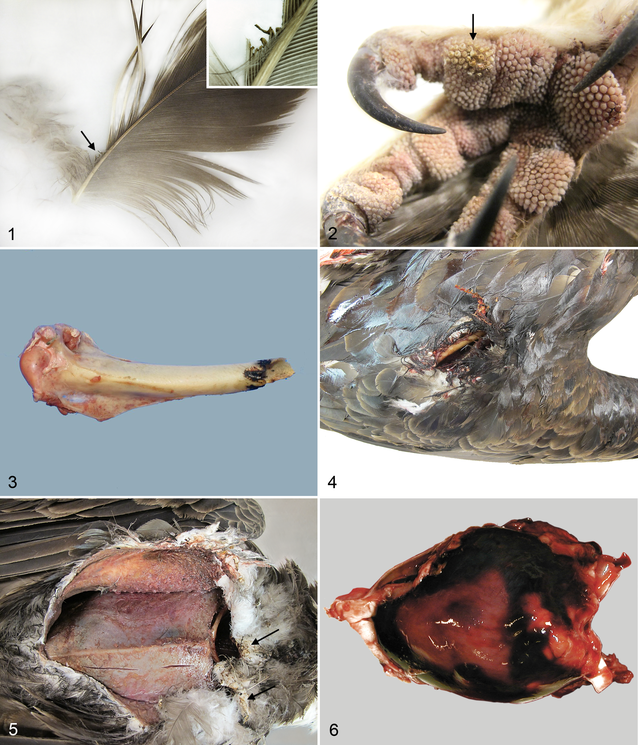
Electrocution-related injuries.
Electrocution can cause limb fractures, presumably through muscular contractions initiated by the current. 5 In birds, some of the more striking injuries associated with electrocution are fractures resulting in traumatic amputation of one or more wings, legs, or digits. Ends of amputated bones and skin are often charred (Fig. 3). Traumatic amputation as a direct result of electrocution has not, to the author’s knowledge, been reported in humans, and the mechanism in birds is uncertain. Although muscular contraction could play a role, there may be a thermal component as well.
Lacerations at contact sites can be quite large, sometimes resulting in partial extrusion of viscera (Fig. 4). Lacerations could be mistaken for sharp-force trauma. Findings to indicate electrocution include a dry, coagulated appearance to the underlying tissue and feather burns around the lacerated edges of skin. Burned feathers may be difficult to identify if the area is dirty or bloody.
Internal Injury
With high voltages there can be severe internal organ injury, including ruptures of viscera and thermal damage. 5 Fully skinning the body is recommended as it may reveal larger burns in the underlying tissue and help to pinpoint the location of small contact points. There often is substantial internal damage compared to the exterior burn. 2 Pocket-like areas of dermal-muscular separation may be large and coalescing. The separated portions of deep dermis and muscle have a tan, dry, slightly coagulated appearance (Fig. 5).
Evidence of blunt-force (impact) trauma is common as birds fall from the pole after being electrocuted. Liver fractures, pectoral girdle fractures, rib fractures, and/or contusions may be present and should not preclude a further search for burn injury. Vascular tears causing hemocoelom, hemorrhage around the base of the neck, and/or hemopericardium may occur (Fig. 6). In the absence of other signs of blunt-force trauma, it is suspected that these ruptures are caused by muscular contractions from the electrical current.
Histopathology
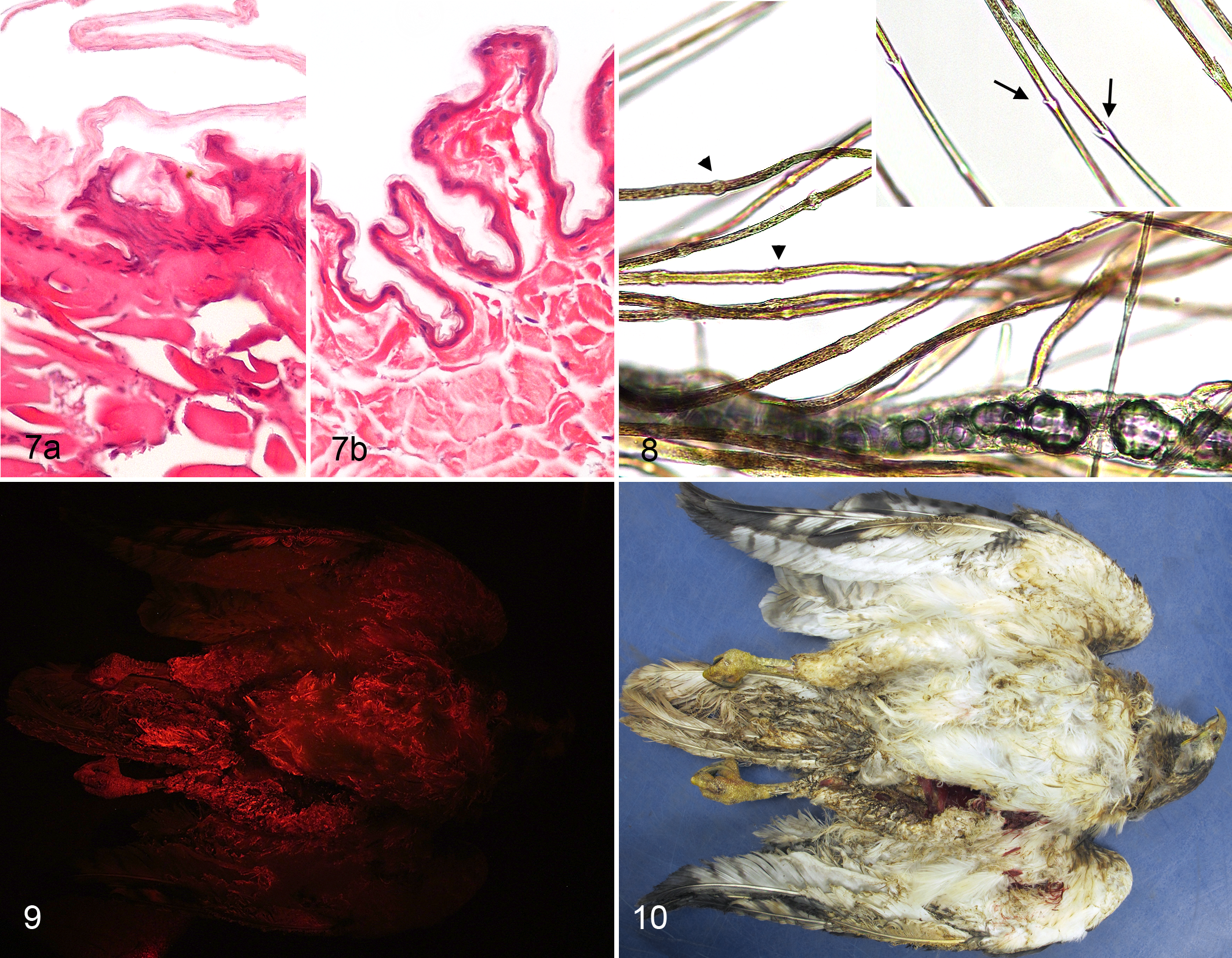
In humans, low-voltage electrical exposure results in intraepidermal and subepidermal separation, epidermal coagulation necrosis, epidermal nuclear elongation, dermal (collagen) homogenization, and dark staining of epidermal nuclei (Fig. 7). Although intraepidermal separation, coagulation necrosis, and nuclear elongation are statistically most common in electrocution, none of these features are pathognomonic. Similar changes can be found secondary to flame burns, abrasions, or freezing. 18 Histopathologic examination of skin can be challenging in raptors due to the thin epidermis.
An important histologic differential diagnosis for lesions of electrocution is flame burns. While the history is usually sufficient to differentiate the two, there also are differences in lesion distribution. Electrocution injury, as opposed to flame burns, may be multifocal. This is thought to be due to variations in resistance and electrolyte content among tissues. 18 The presence of more than one external wound (suggesting entrance and exit points) and the depth of the damage can also support a diagnosis of electrocution. In addition, electrical injury can cause deep burns and muscle damage with little apparent surface injury, whereas, excepting incineration, thermal burns from fire are relatively superficial or cover large surface areas. 6
Necrosis of the vascular tunica media is reported in humans surviving the initial electrocution event. 6 Necrosis resulting in delayed rupture of damaged vessels and/or thrombosis can cause further injury and death. This is one of the factors contributing to “progressive” damage described in humans. 2
Other Diagnostic Tools
For the most part, electrocution burns are fairly obvious and should not present a diagnostic challenge; however, sometimes burns can be very small or ambiguous in appearance. In these instances, additional techniques may be helpful in determining the cause of death.
Using a dissecting microscope to examine feathers and skin can help differentiate between burned and dirty or otherwise damaged tissue. Burned feathers have broken barbs and/or vanes with melting and charring of the truncated ends. Burned skin on the legs and feet typically has a slightly tan to ash-gray tinge and consolidation (melting) of the papillae. A wet mount will show melting of the barbs and barbules, whereas grossly they may appear only slightly discolored (Fig. 8).
Small foci of metal deposition in tissues have been identified through histochemical analysis and scanning electron microscopy. 12,17 Studies of low-voltage electrocution deaths, with electrical wire as the source, have demonstrated copper deposition in electrocuted skin. 11 This metallization of tissues occurs at contact points when metallic ions from the electrical source meet tissue anions. Metal deposition may also occur as a result of arcing. 17 In the United States, power lines are made of aluminum or, less commonly, copper. It seems likely that metal in birds could be identified histologically from high-voltage electrical encounters where skin makes direct contact with the wire; however, feathers may act as a barrier similar to clothing in humans. 16
Metallization of skin can sometimes be seen on radiographs but is too uncommon to be relied upon. Radiology is a more useful tool to help rule out other causes of death or complicating factors, such as gunshot and lead ingestion. Fractures from impact trauma can also be more easily documented.
Examination with an alternate light source at 530 to 570 nm under a red filter can highlight charred skin, feathers, and beak keratin (Figs. 9, 10). 19 If the equipment is available, this is an extremely useful tool for identifying and documenting subtle burns. Charred tissues will photoluminesce bright red, whereas feathers, skin, beak, and talons that are unaffected or merely dirty will photoluminesce poorly or not at all. Questionable areas can be isolated for confirmation with a dissecting microscope. It should be noted that photoluminescence may be lost if the burnt areas have fallen off. This may occur if the body is wet or has been exposed to the environment for a long period of time.
Retrospective Review of Cases
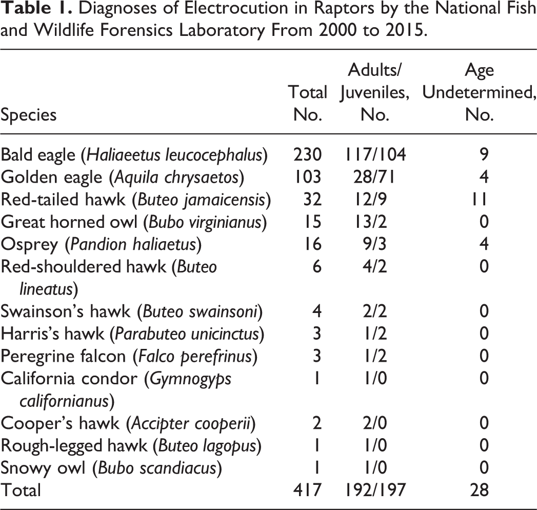
To further examine external burn distribution and severity, raptor electrocution cases submitted to the US Fish and Wildlife Service’s National Forensics Laboratory (NFWFL) from 2000 to 2015 were reviewed (Table 1). A total of 2810 raptors were examined. Of these, the cause of death for 417 (14.8%) was electrocution. Injury distribution was further analyzed in a subset of 377 birds for which whole carcasses had been submitted. Bodies lacking internal organs due to decomposition or scavenging and nonintact carcasses (bodies with postmortem removal of limbs) were excluded. Definitive diagnosis of electrocution in all cases relied on the finding of burned feathers or skin. The presence of poorly distinct burns was confirmed with a dissecting microscope and/or alternate light source illumination. Based on location on the body, electrocution injuries were assigned zones (Table 2). Externally visible electrocution burns were also assigned a level of severity designated as grades 1 to 4 (grade 1, least severe to grade 4, most severe) (a complete description of the grades is provided in the Table 2 caption). The total numbers of birds with burns of a particular grade in each zone are recorded in Table 2.
Diagnoses of Electrocution in Raptors by the National Fish and Wildlife Forensics Laboratory From 2000 to 2015.
Distribution and Severity of External Burns on Raptors.a
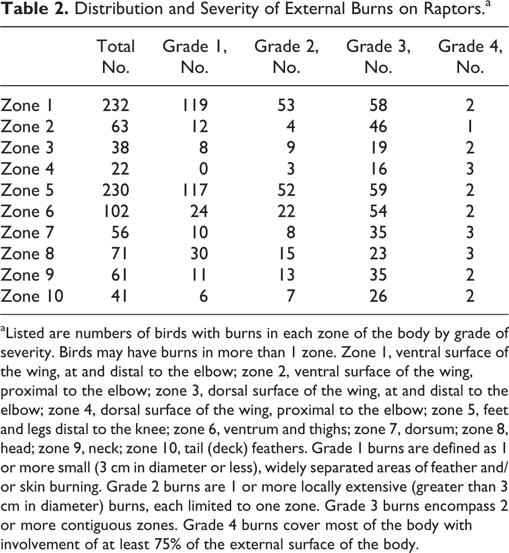
aListed are numbers of birds with burns in each zone of the body by grade of severity. Birds may have burns in more than 1 zone. Zone 1, ventral surface of the wing, at and distal to the elbow; zone 2, ventral surface of the wing, proximal to the elbow; zone 3, dorsal surface of the wing, at and distal to the elbow; zone 4, dorsal surface of the wing, proximal to the elbow; zone 5, feet and legs distal to the knee; zone 6, ventrum and thighs; zone 7, dorsum; zone 8, head; zone 9, neck; zone 10, tail (deck) feathers. Grade 1 burns are defined as 1 or more small (3 cm in diameter or less), widely separated areas of feather and/or skin burning. Grade 2 burns are 1 or more locally extensive (greater than 3 cm in diameter) burns, each limited to one zone. Grade 3 burns encompass 2 or more contiguous zones. Grade 4 burns cover most of the body with involvement of at least 75% of the external surface of the body.
Bald eagles (Haliaeetus leucocephalus) were the most commonly submitted raptor (230/417 birds; 55%). This is not substantially different from the percentage of bald eagles submitted for all causes of death (1269/2810; 45%). Their large dimensions make them more vulnerable to electrocution; however, their size, celebrity, and tendency to live in closer proximity to humans also make them more likely to be found and turned in as law enforcement cases. The second most commonly submitted bird was the golden eagle. The higher proportion of bald eagles within NFWFL cases differs somewhat from the literature, which reports golden eagles to be the more commonly electrocuted raptor. The argument has been made that previous studies focused on areas of golden eagle habitation and that young bald eagles may have been improperly identified. 10 In either case, there may be a selection bias.
Juveniles (subadult or younger) did not consistently present in higher numbers than adults (total of 192 adults, 197 juveniles, and 28 undetermined age), except for golden eagles, for which there were 28 adults, 71 juveniles, and 4 of undetermined age. Smaller raptors, which can decompose much more rapidly, could be falsely underrepresented. Smaller body size may also mean that these birds are comparatively safer on power poles.
In summary, 85 of 377 (22.5%) birds had a single burn, with 68 (18%) of those being isolated, small burns (grade 1 injury). When there was only 1 grade 1 burn, it was most likely to be found on the ventral surface of the wing at and distal to the elbow (ie, zone 1; P = .00668, 2-tailed Fisher exact test) or on the leg or feet distal to the knee (ie, zone 5; P = .00656). The pathologist should be especially thorough in examining these areas in suspected electrocution cases because they are most often affected by subtle electrocution burns.
Conclusions
The problem of raptor and other avian electrocutions from manmade electrical structures has been a persistent one that, unfortunately, is not likely to be resolved any time soon. As a cog in the wheel of justice, the wildlife pathologist or clinician should know how to diagnose and document electrical injuries following best practices for forensic investigation. The hope is that, with accountability, responsible parties will act to mitigate damage and be inspired to prevent future deaths.
Footnotes
Author Note
The findings and opinions contained herein are those of the author and do not necessarily reflect the views of the US Fish and Wildlife Service.
Acknowledgements
The author would like to acknowledge the contributions of the NFWFL and US Fish and Wildlife Service Office of Law Enforcement staff, particularly Darby Morrell for formatting the photographs, Colleen Wilson for histologic preparation, and Tabitha Viner for manuscript review.
Declaration of Conflicting Interests
The author(s) declared no potential conflicts of interest with respect to the research, authorship, and/or publication of this article.
Funding
The author(s) received no financial support for the research, authorship, and/or publication of this article.