
Abstract
The aim of this study was to evaluate gross and histologic lesions and epidemiologic factors of foot lesions in farmed mink. The feet of 1159 mink from 4 Danish farms were examined and lesions described. Swabs from the lesions were taken from 27 mink for microbiology, and tissue samples from a representative spectrum of feet with and without lesions (n = 22) were examined histologically. Feet were grouped according to gross inspection: no lesions (55.1%), hair loss (7.1%), hyperkeratosis (35.8%), and crusting (5.3%). Lesions were predominantly located in plantar metatarsal skin (98.1%). Staphylococci were the most prevalent microorganisms cultured from the lesions. There was a significant association between presence of lesions and sex (P < .0001), age (P < .0001), and color type (P = .023). Lesion size was significantly different between hair loss and crusts and between hyperkeratosis and crusts (P < .0001). Histologically, lesions included varying degrees of orthokeratotic to parakeratotic hyperkeratosis and granulomatous to pyogranulomatous dermatitis with trichogranulomas as a dominant feature in all mink. The gross and microscopic lesions were comparable to physically induced changes in other species that develop as a response to repetitive friction or pressure. The condition may have an impact on animal welfare in mink production.
In recent years, the occurrence of outbreaks of foot lesions in farmed mink in several countries has increased the awareness of skin diseases affecting the feet of mink. The lesions have been described under the term pododermatitis or simply as ulcerative lesions, for which a cause is being sought. 3,10,14 The occurrence of callus on the feet of mink has been mentioned only briefly in the literature 3 ; however, during routine diagnostics at our department, callus-like lesions in the plantar metatarsal skin area have been observed in a large proportion of mink submitted from certain farms. Skin callus has been reported in dogs, rabbits, and other species, and it develops as a localized hyperplasia of the stratum corneum in response to continuous friction or pressure, typically affecting skin covering pressure points or bony prominences. 5,7,12,21 –23 The impact of callus on animal welfare is unknown; however, in humans, callosities of the feet can be highly uncomfortable. 28 Furthermore, in other species, calluses may be vulnerable to ulceration through the formation of cracks in the hyperkeratotic epithelium. 12
The aims of the present study were to describe the gross pathology and histopathology of foot lesions in Danish farmed mink, as well as to look into some factors epidemiologically associated with the lesions and affected animals.
Materials and Methods
Animals and Tissue Collection
A cross-sectional study was conducted in November 2012 on 4 Danish mink farms (A–D) located on the Zealand and Funen islands. The farms were included because of a history of foot lesions, and each farm was visited once during time of pelting. Carbon monoxide–euthanized mink were placed on cooling shelves and the feet examined immediately prior to pelting. The mink had, prior to euthanasia, been fed commercial mink diets and kept in standard cages elevated from the ground with galvanized or stainless metal wire mesh floors (1 × 1.5 or 1 × 1 in). The nest boxes were made of wood, had solid floors, and were fitted with removable plastic-coated wire mesh (1 × 0.5 in) covering the floor and sides. Wire thickness varied from 2 to 2.5 mm.
In total, 1159 mink were included in the study (farm A: n = 296; farm B: n = 262; farm C: n = 264; farm D: n = 337). For all animals, sex, color, and age (juvenile or adult) was recorded with foot lesions. Weight was obtained for 80 randomly sampled clinically normal mink and 77 randomly sampled mink with lesions. All feet, including the carpus and tarsus, were examined by inspection and palpation. Lesions observed on the feet were described macroscopically, and the size (length and width) and location (on which foot and what position) were recorded. Lesion areas were calculated according to whether a lesion was circular (A = r2 × π) or square (A = length × width).
For histologic characterization, 22 tissue samples were obtained from a representative spectrum of feet with (n = 16) and without (n = 6) lesions. The lesions were categorized as hair loss (n = 4), hyperkeratosis (n = 5), or crusts (n = 7) and were representative of the pattern of changes seen in this study as well as in mink submitted to our department across a number of years for other research purposes. Skin samples of approximately 2 × 2.5 cm were excised with a scalpel, dissected as deep as possible to ensure adequate sampling of all affected tissues and rim areas.
Histopathology and Microbiology
Skin specimens were processed routinely after preservation in 10% neutral buffered formalin for up to 1 week. Paraffin-embedded tissues were sectioned at 4 to 5 µm, stained with hematoxylin and eosin, and examined by light microscopy. All specimens were viewed in polarized light for visualization of birefringent material. Von Kossa, 4 Levaditis, 1 and periodic acid–Schiff stains 18 were used for identification of mineral, spirochetes, and fungi, respectively.
Skin swabs (sterile BBL CultureSwab) for bacteriologic and fungal analysis were taken from foot lesions from 27 mink (hyperkeratosis: n = 15; crusts: n = 12). Swabs were taken between the toes and around the central foot pad. They were cultured on blood agar for detection of bacteria and Sabouraud agar for detection of fungi and incubated aerobically at 37°C for 48 hours. Subcultures of each morphologic bacterial colony type were examined by matrix-assisted laser desorption ionization time-of-flight mass spectrometry (bioMérieux, Marcy l’Etoile, France) as described elsewhere. 9
Statistical Analysis
Since no major differences in lesion distribution were discovered among the farms, data from the 4 farms were pooled. Statistical analyses were performed with SPSS (IBM, Chicago, IL) and SAS 9.4.2014 (SAS Institute Inc, Gary, NC). Color types were grouped as either light (light and gray) or dark (brown and black). Testing for association between lesion type and any of the variables of sex, age, fur color, and location of lesions was conducted with χ2 analysis and described by relative risk (RR). Analysis of variance and independent samples t test was performed for testing differences between weight of mink with different lesion types and size of different lesion types. Finally, a multivariate logistic analysis was carried out for the factors of sex, age, and color type via the likelihood ratio test for model selection. P < .05 was considered statistically significant.
Results
Gross Pathology, Histopathology, and Microbiology
Mink had either no gross skin lesions (n = 639, 55.1%) or lesions that could be characterized by hair loss, hyperkeratosis, or crusting (n = 520, 44.9%; Table 1).
Data From 1159 Mink Examined for Foot Lesions Defined as No Lesions, Hair Loss, Hyperkeratosis, or Crusting.
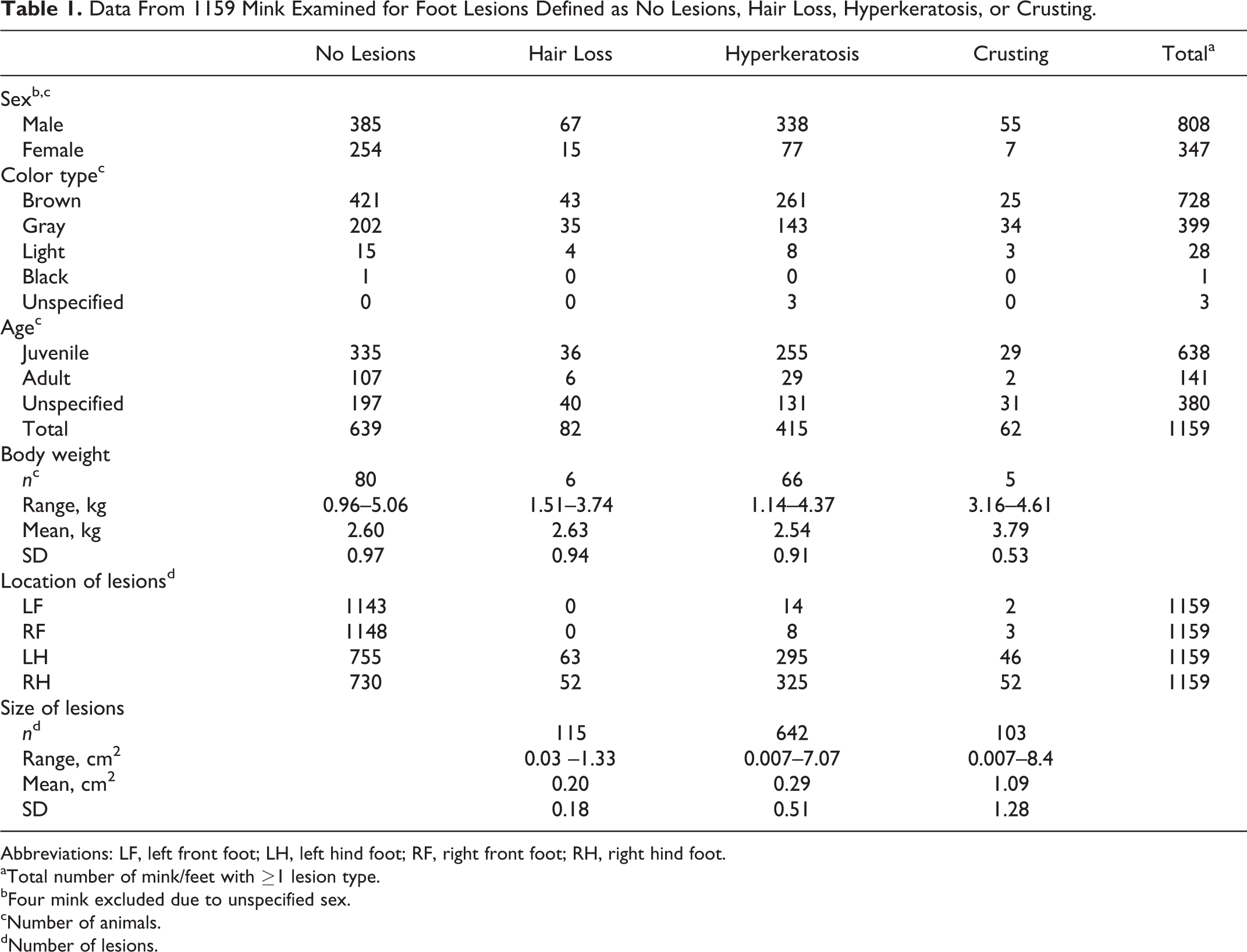
Abbreviations: LF, left front foot; LH, left hind foot; RF, right front foot; RH, right hind foot.
aTotal number of mink/feet with ≥1 lesion type.
bFour mink excluded due to unspecified sex.
cNumber of animals.
dNumber of lesions.
Feet without gross lesions had a dense, glossy fur, which was fixed to the skin. The skin was of normal thickness and color, and skin and footpads were intact. Histologic examination of 6 grossly normal feet revealed chronic lesions of single or multiple trichogranulomas at the base of or beneath otherwise normal hair follicles. They were characterized by displaced hairs surrounded by an inflammatory infiltrate dominated by macrophages and multinucleated giant cells and were often bordered by a rim of connective tissue (Figs. 1, 2). Multinucleated giant cells engulfing hair fragments and well-circumscribed foci of von Kossa-positive mineralization surrounding central hair fragments were occasionally seen.
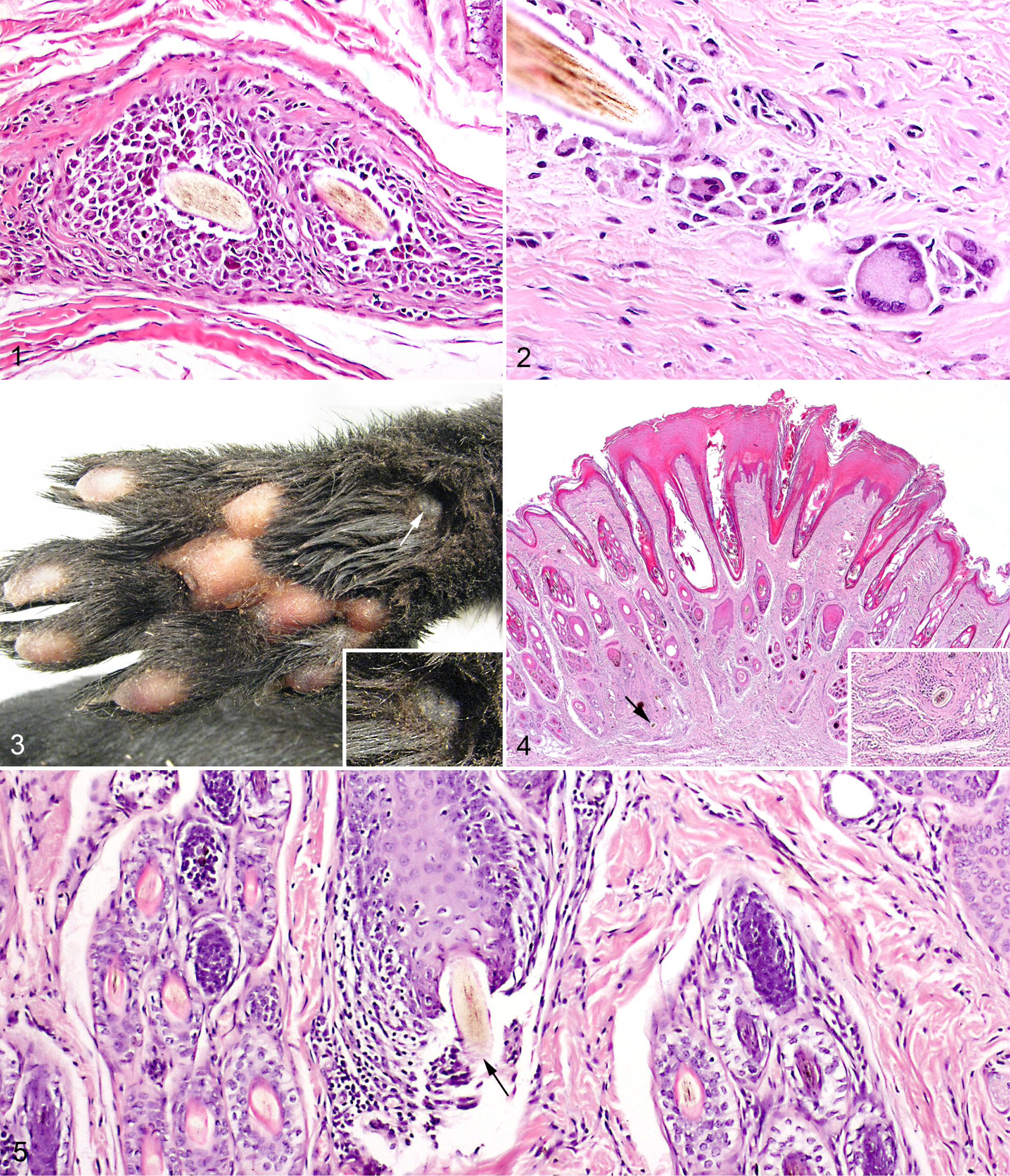
Plantar skin, mink.
Gross lesions (n = 860) characterized as hair loss, hyperkeratosis, or crusts were predominantly located on the plantar surface of the metatarsal area (98.1%). For front feet, 13 mink were unilaterally affected (65%) and 7 mink bilaterally affected (35%). For hindfeet, 199 mink were unilaterally affected (38.6%) and 317 mink bilaterally affected (61.4%).
Gross lesions of hair loss were characterized by focal areas with loss of normal fur density. The skin was otherwise of normal thickness and color and surrounded by skin with normal hair growth (Fig. 3). Histologically, areas with hair loss were characterized by a focally thickened epidermis showing orthokeratotic hyperkeratosis and acanthosis extending into the infundibula of hair follicles (follicular keratosis). Irregular rete peg formation was seen in most cases. The specimens contained trichogranulomas as described above (Figs. 1, 2, 4). In 1 case, furunculosis was seen, characterized by penetration of the base of the hair follicle by a hair shaft, with peri-/mural folliculitis and granulomatous reaction (Fig. 5). One specimen contained an epidermal microulceration. The underlying dermis showed a focal pyogranulomatous cell infiltrate surrounding a hair fragment.
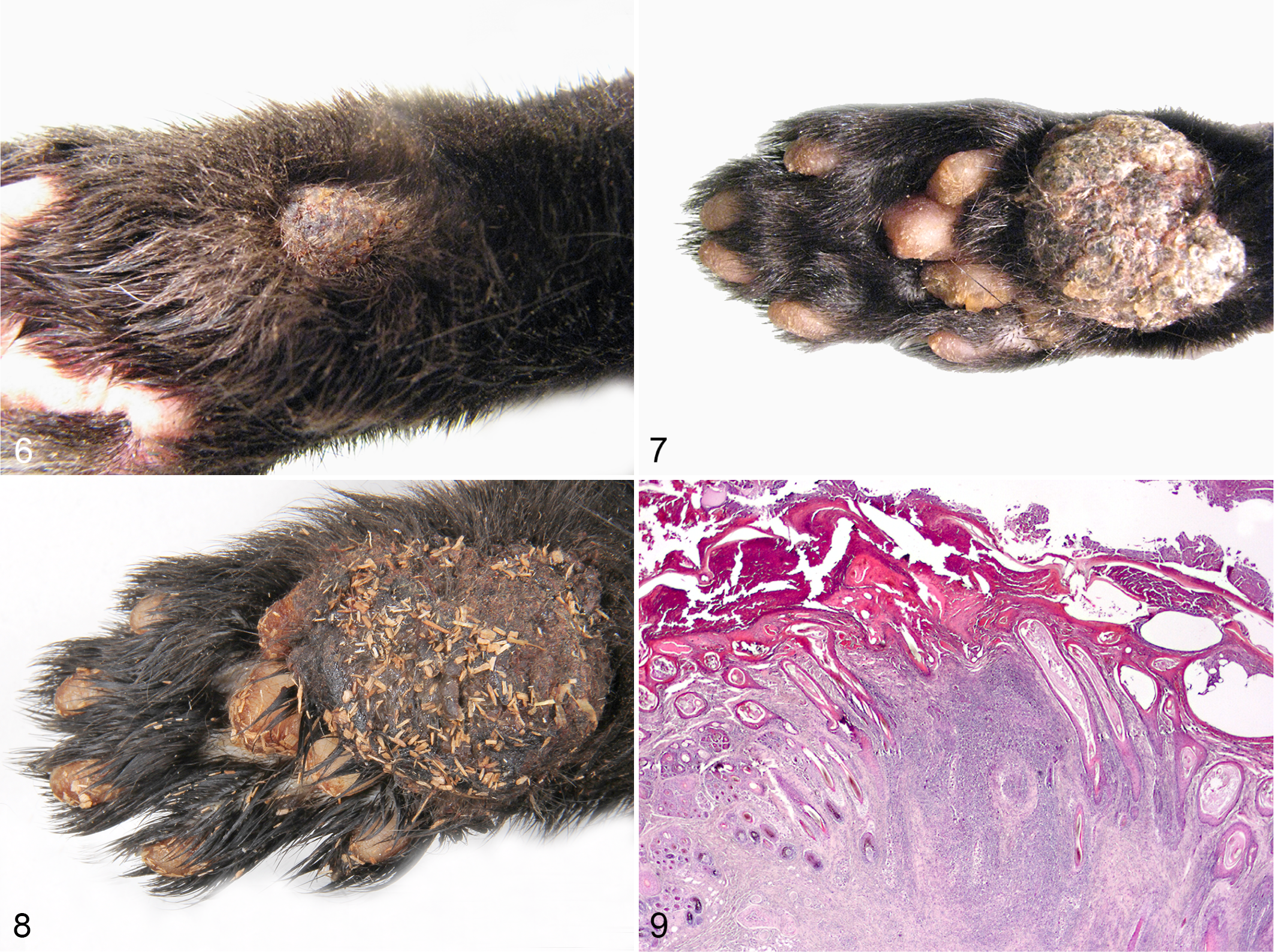
Hyperkeratotic gross lesions were circular or angular, firm, well-circumscribed hairless areas of thickened skin. They had a rough keratinized surface, were flattened, or protruded to varying degrees from the surrounding skin surface (Figs. 6, 7). The lesions were confined to the skin and freely movable. Occasionally, hyperkeratotic lesions were observed on the haired skin adjacent to the torus metacarpeus (large footpad) of the front feet (19 animals). In 3 mink, focal yellowish areas of hyperkeratosis protruded from the surface of ≥1 foot pads. Histologically, lesions included as hyperkeratosis consisted of a marked hyperplastic epidermis characterized by compact orthokeratotic hyperkeratosis and acanthosis (Fig. 4) with irregular rete peg formation. Follicular keratosis and, occasionally, comedones characterized by dilation and plugging of hair follicles (Figs. 4, 9) as well as dilated sweat glands were observed. The hyperkeratotic stratum corneum revealed parakeratosis in a few specimens. Pigmentary incontinence and dermal melanophages were observed sporadically. Two specimens showed focal ulcerations characterized by crust-covered epidermal necrosis. In these cases, a focal subepidermal suppurative tissue reaction with edema was present. The specimens contained multiple trichogranulomas located at the base of hair follicles or occasionally between hair follicles as described above (Figs. 1, 2, 4). Areas of necrotic tissue surrounding hair fragments were typically associated with a sparse inflammatory cell infiltrate. In the most prominent hyperkeratotic lesions, extensive dermal fibrosis predominated, as well as loss of hair follicles and glands. Conglomerates of numerous hairs, macrophages, and multinucleated giant cells were evenly distributed, and pyogranulomas were present in the subcutis. In addition to the well-circumscribed trichogranulomas, 1 case had a diffuse, locally extensive pyogranulomatous cellular infiltrate affecting the entire dermis and subcutis.
Plantar skin, mink.
Feet with grossly visible focal hyperkeratosis of foot pads were microscopically characterized by multilayered compact orthokeratotic hyperkeratosis covering an otherwise normal epidermis, dermis, and subcutis.
Lesions defined grossly as crusts consisted of hairless skin areas with scabs of dried exudate (Fig. 8). In 2 mink, crusts extended from the plantar/palmar, metatarsal/metacarpal area along the lateral side of the feet reaching the dorsal aspect of the feet. These lesions were furthermore characterized by irregular edges and large amounts of dried exudate. Histologically, crusts showed the same range of manifestations as that for lesions defined as hyperkeratosis. Ulcerations were characterized by varying degrees of reepithelialization, fibroplasia, and edema and varied from pinpoint to larger areas of crust-covered epidermal necrosis. Increased amounts of connective tissue in dermis and subcutis were typically associated with a decreased amount of hair follicles and other adnexal structures. Pyogranulomatous dermatitis with a focal to diffuse distribution was also present (Fig. 9). Dense basophilic colonies of coccoid bacteria were sporadically present in the crust material without invasion of vital tissue. No spirochetes or fungi were discovered histologically in any specimen.
Bacterial and fungal cultures revealed the following microorganisms, in order of decreasing frequency: Staphylococcus schleiferi (6 hyperkeratoses; 8 crusts), Staphylococcus intermedius group (5 hyperkeratoses; 2 crusts), Candida famata (2 crusts), Streptococcus canis (1 hyperkeratosis), Psychrobacter phenylpyruvicus (1 hyperkeratosis), nonspecific mix of bacteria (1 hyperkeratosis), and Carnobacterium maltaromaticum (1 hyperkeratosis).
Epidemiology
The proportion of mink with foot lesions on each farm was 152 of 337, 140 of 264, 127 of 262, and 100 of 296. Data for sex, color, age, and weight of mink and size and location of gross lesions are summarized in Table 1. There was a significant association between each variable of sex and age and presence of foot lesions (P < .0001). More males than females were affected (RR = 1.95; 95% CI: 1.62, 2.35), and more juvenile than adult mink were affected (RR = 1.97; 95% CI: 1.45, 2.67). For fur color, there was a significant difference in presence of foot lesions between light and dark color types (P = .023), with more light than dark color types being affected (RR = 1.17; 95% CI: 1.03, 1.33). Furthermore, significantly more light color types had lesions characterized as crusts (P < .0001; RR = 2.53; 95% CI: 1.54, 4.14). There was no significant difference in weight between mink with and without foot lesions (P = .833). Significantly more lesions were found on hindfeet than on front feet (P < .0001). Multivariate logistic analysis showed significant association only between sex and presence of gross lesions (P < .0001; RR = 1.97; 95% CI: 1.45, 2.67).
Distribution of gross lesion size is shown in Table 2. There was a significant difference in size of lesions (area) between hair loss and crusts (P < .0001) and between hyperkeratosis and crusts (P < .0001), with crusts being larger than both hair loss and hyperkeratosis.
Distribution of Lesion Size Among Categories of Lesions Defined as Hair Loss, Hyperkeratosis, or Crusting.
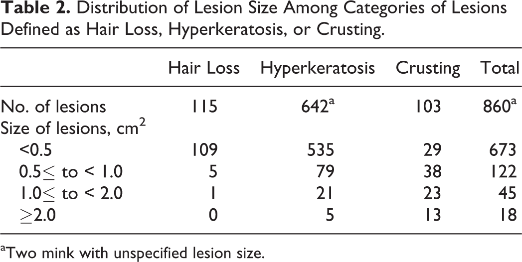
aTwo mink with unspecified lesion size.
Discussion
This is the first study to report foot lesions in Danish farmed mink. Hyperkeratosis as seen in callus represents a normal protective response to persistent mechanical stress applied to skin and was the most frequent gross lesion type discovered on the feet of farmed mink (35.8%). 12,24,28 With their rough keratinized surfaces, the lesions are comparable to different stages of callus as described in other species. 5,7,12,21 –23 Common lesions of rabbits housed in cages with wire mesh floors consist of cutaneous hyperkeratosis and ulcerations on the plantar tarsus, metatarsus, and occasionally the metacarpus. The condition is considered multifactorial; however, physical external pressure resulting from bearing of heavy body weight on wire mesh floors is suspected to play an important role in the pathogenesis. 6,20 The primary localization of lesions in the plantar metatarsal skin of hindfeet (98.1%) may be due to the physiology of mink where the body weight is mainly supported by the hind legs.
Histologically, bacteria were discovered only in crust material with no sign of invasion of microorganisms in deeper vital tissue. Microorganisms are to some extent always present in skin lesions, and the microorganisms cultured from the lesions were probably opportunistic microorganisms residing in the superficial layers of the lesions, such as the Candida found in 2 lesions characterized as crusts. The S. intermedius group is used as a common designation for the closely related S. intermedius, S. pseudintermedius, and S. delphini groups A and B, which are difficult to distinguish. 26 Parasites were ruled out on the basis of no observation of parasitic elements in any of the histologic specimens. Likewise, inclusion bodies and deep necrosis as a sign of viral infection were not discovered.
Lack of certain nutrients is known for causing keratosis of the skin. 17 The pattern of changes induced by, for example, hypovitaminosis A and zinc deficiency, however, does not compare well with the specific location of changes seen in this study. 17 Mink are furthermore fed very controlled and carefully prepared diets including all essential vitamins and minerals. All Danish manufacturers of mink feed are subject to continuous analyses of a number of raw material samples as well as finished product samples, some of which are sampled unannounced.
The dermis of examined specimens contained foci of granulomatous to pyogranulomatous inflammation with centrally located hair fragments. Foreign body reactions have been described in humans and animals in cases where skin or mucosal surfaces were penetrated by free hairs from the environment. 2,8,19,25,29 This is not considered a likely pathogenesis for the trichogranulomas reported here, since no granulomas were encountered superficially in the skin. Compared with callus of human skin, callus in animals affects skin with abundant hair follicles. It is a well-known phenomenon that luminal folliculitis, typically due to follicular infection, can lead to rupture of hair follicles and furunculosis with the formation of granulomatous to pyogranulomatous dermatitis, occasionally with draining tracts to the surface of the skin. 13,15 The specific and localized area of affected skin, which is frequently in contact with the cage floor, as well as the lack of luminal pathogens suggests, however, that mechanical pressure and friction are the initiating event in our cases. Epidermal and follicular hyperkeratosis can facilitate comedo formation when the follicles become severely plugged, eventually leading to rupture and traumatic furunculosis. The release of hair, keratin, and debris into the surrounding dermis provokes a granulomatous foreign body reaction leading to scarring, loss of adnexal structures, and alopecia, which was a consistent feature of the foot lesions. Persistent trauma to callus can lead to callus pyoderma with secondary bacterial furunculosis and ulcerations. 12,13,16 The pathogenesis may thus be viewed as a dynamic series of events, with hair loss followed by hyperkeratosis and fully formed calluses that may eventually ulcerate. This is supported by the significantly larger size of crusts compared with hyperkeratosis and hair loss. Interestingly, trichogranulomas, representing chronic stages of furunculosis, were also found in feet without gross lesions or follicular plugging. It is tempting to speculate that hair shafts in hair follicles of the plantar skin of the feet can penetrate the base of the hair follicle simply as a result of mechanical pressure subjected to the area.
Callosities in humans and other animal species tend to enlarge or worsen with age if mechanical trauma persists. 11,12 It is therefore reasonable to assume that callus seen at pelting would persist or worsen during the winter for animals selected for breeding in the spring. Mink with foot lesions at pelting would therefore be poor candidates for breeding and should be excluded as such from the breeding population. Our results show an overrepresentation of gross foot lesions in juvenile mink (ie, mink born the same year), which is possibly due to farmers’ exclusion of mink with lesions from the breeding population.
There were significantly more males than females with gross foot lesions, which corresponds to the findings of Bröjer. 3 Obesity predisposes to callus formation in dogs and cats. 12 Our results did not show a significant difference in body weight between mink with and without gross lesions; however, the association with body mass index has not been studied.
Wounds in farmed mink are considered an indicator of reduced welfare in mink production. 27 It seems reasonable to believe that mink with ulcerations on their feet feel discomfort to some level. Even callosities are described as potentially painful to humans, and there is no reason to believe that the same is not true for mink. 19,28 The level of pain is presumably associated with factors such as size, severity, and exact location of lesions. In humans, the tendency to form callus varies among individuals. 24,30 This individuality may in part be due to genetic differences affecting foot conformation and movement pattern, which may also predispose to the occurrence of foot lesions in mink. On one other Danish farm (experimental unit), lesions of alopecia and thickening of the skin >5 mm in diameter were rare, and lesions of marked hyperkeratosis were seldom seen (Steen H. Møller, personal communication, November 17, 2014). To shed light on possible epidemiologic factors including genetics, it would be of great interest to study more farms with and without the condition, as well as to conduct parent–offspring studies, as demonstrating heritability would mean a way of controlling the condition through genetic selection for animals without lesions.
In summary, we have described a condition of predominantly plantar foot lesions in farmed mink characterized by different stages of callus formation, dermal trichogranulomas, and scarring. The condition has an apparent mechanical pathogenesis, although certain other risk factors possibly influence the pathophysiologic background. The condition may have an impact on animal welfare in mink production through the increased risk of plantar wound development, secondary infections, and thereby potential loss of breeding animals. Farms experiencing the condition should therefore take steps to minimize the occurrence prospectively, by excluding mink with foot lesions from breeding, apart from maintaining a good management regimen for and good overall health status of their mink.
Footnotes
Acknowledgements
We thank Betina G. Andersen and Elizabeth W. Petersen for their excellent laboratory assistance and the farmers for giving us the opportunity to examine their mink.
Declaration of Conflicting Interests
The author(s) declared no potential conflicts of interest with respect to the research, authorship, and/or publication of this article.
Funding
The author(s) disclosed receipt of the following financial support for the research, authorship, and/or publication of this article: This work was cofunded by Pelsdyrafgiftsfonden, the Danish Fur Breeders’ Association, and the Danish Council for Technology and Innovation.