
Abstract
A single free-ranging common brushtail possum (Trichosurus vulpecula) and 2 captive sibling common ringtail possums (Pseudocheirus peregrinus) from a zoological facility in Sydney, Australia, were diagnosed with multisystemic listeriosis. The brushtail was found dead in an animal enclosure while the ringtails presented with signs of cardiovascular collapse and died shortly thereafter. All 3 animals were culture positive for Listeria monocytogenes and demonstrated focal suppurative lesions within the brainstem in addition to fulminant disease in other areas of the thorax and/or abdomen. Listeriosis in phalangeriformes species has rarely been reported, and brainstem lesions have not previously been described. It is speculated that access to the brainstem by the organism may have occurred hematogenously or via retrograde migration along cranial nerves. Sources of infection and the possibility of transmission between animals are also discussed.
Listeriosis is a well-known food-borne bacterial infection caused most commonly by Listeria monocytogenes and found worldwide in both humans and animals, including over 50 species of mammals as well as birds, reptiles, amphibians, fish, and invertebrates. 4,7,10,12,13 In marsupials, the disease has been previously described in a ringtail possum in addition to a red-necked wallaby (Macropus rufogriseus), a rufous bettong (Aepyprymnus rufescens), a sugar glider (Petaurus breviceps), and in male antechinus (Antechinus stuartii) as a post-mating stress-related disease. 3,5,8,11 Findings have consistently included hepatitis and often other changes suggestive of septicemia but no description of encephalitis. In domestic animals, typical manifestations of listeriosis include septicemia, abortion, and, particularly in ruminants, rhombencephalitis, in addition to various other infections. 9,12,13 In humans, various syndromes are recognized including mild febrile gastroenteritis, septicemia, abortions, and central nervous system (CNS) disease. CNS disease generally manifests as meningitis or meningoencephalitis and is thought to occur secondary to bacteremia. However, rhombencephalitis of a similar description to that in ruminants is being increasingly recognized in humans, with suggestions that it is underreported and is possibly an emerging zoonosis. 12 In these cases, a first phase of disease consisting of headache, lethargy, nausea, vomiting, and fever lasts 4 to 10 days. This is followed by progressive brainstem defects. The pathogenesis of rhombencephalitis in both humans and animals is still not clearly understood.
Brushtail and ringtail possums are common nocturnal species in Australia, found both in urban and rural settings. Both species are primarily herbivorous with a simple stomach (monogastric) and a large cecum. In this report, 3 cases of listeriosis in possums are described in which multisystemic disease with involvement of the brainstem had occurred.
Case No. 1
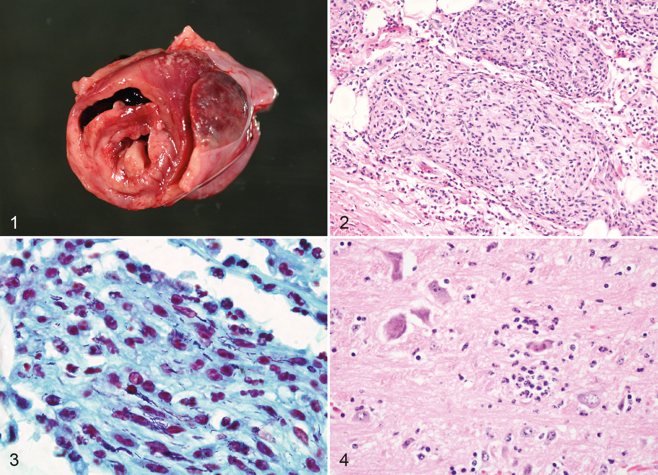
A free-ranging, adult male brushtail possum was found dead in a collection animal enclosure on the grounds of a zoological facility in Sydney, Australia, in early autumn, 2010. The animal had not been noted in the enclosure before being found dead, but many brushtail possums are free-range inhabitants of the zoo grounds. On gross examination, the animal was in poor body condition and dehydrated. The pericardial sac was distended with fibrin, and the visceral pericardium had a granular appearance. Multifocal pale streaks were present throughout the ventricular myocardium (Fig. 1). The pulmonary parenchyma was diffusely firm, consolidated, and tan/brown and contained a 15-mm abscess protruding from the dorsal left lung. The abscess was filled with inspissated green material and surrounded by a white capsule. The pleural cavity contained approximately 5 ml of clear fluid. A selection of tissues were frozen or fixed in 10% neutral buffered formalin for further analysis.
Listeriosis.
On histopathology, 60% of the left and right ventricular myocardium and the interventricular septum were replaced by a vast infiltrate of neutrophils and macrophages. Remaining myocytes in these regions were often hypereosinophilic with shrunken nuclei and basophilic stippling. The mesothelium of the epicardial surface was segmentally effaced by the inflammatory infiltrate with a thick overlying layer of fibrin. The liver exhibited a reticular pattern resulting from loss of hepatocytes and expansion of sinusoids with erythrocytes in the periacinar zones of the tissue. Central lymphocytolysis of the splenic white pulp was noted. The pulmonary interstitium was thickened with macrophages, alveoli were lined with type II pneumocytes, and within alveolar air spaces, moderate numbers of foamy macrophages, multinucleated giant cells, and tangential sections of metastrongyle nematode worms were present (in the author’s experience, these changes are consistent with metastrongyle infection in this species). Focally, there was a large lake of coagulative to liquefactive necrosis immediately beneath the pleura. This lesion was flanked on its deep margin by a band of fibrosis, consistent with the abscess noted grossly. The brain was sectioned sagittally and examined at the level of the cerebrum, hippocampus, and cerebellum. At the level of the fourth ventricle, a small accumulation of neutrophils had collected within the brainstem in what appeared to correspond with the trigeminal nucleus, dorsal to the radix of the facial nerve. No significant changes were detected in tongue, stomach, small intestine, colon, adrenal gland, kidney, and bladder. L. monocytogenes was isolated from samples of heart, lung, and liver. The lung tissue selected for culture was not associated with the abscess. Brain was not cultured in this case.
Case No. 2
In April (autumn) 2013, an adult, neutered male, collection ringtail possum presented with hypothermia, pale foot pads, gingiva, and poor capillary refill time. The animal was treated for cardiovascular collapse with fluids and placed in an incubator overnight. It was found dead the following morning. On gross examination, the carcass was found to be moderately autolyzed as a result of the warm environment of the incubator. The animal was in good body condition but was poorly hydrated. Strands of fibrin were adherent to the serosal surface of the gall bladder. Intestinal contents were thin and watery, not typical of this species. A selection of tissues were frozen or fixed in 10% neutral buffered formalin for further analysis.
On histopathological examination, the gall bladder demonstrated marked autolysis, but despite this, large numbers of neutrophils could be identified within the wall. The small intestine was similarly autolyzed; however, there was a suggestion of blunting and fusion of villi. Focally in the liver, there was infiltration of neutrophils within a portal triad and crossing over into the associated parenchyma, with some necrosis of hepatocytes. Clusters of rod-shaped, gram-positive bacteria were found within neutrophils at this site. Adjacent to the mesenteric lymph node, moderate to large collections of neutrophils were present within adipose tissue. The neutrophils were also focused around and penetrating adjacent nerves (Fig. 2). The nerves were additionally infiltrated by mononuclear cells, often containing rod-shaped, gram-positive bacteria (Fig. 3). In the heart, a single small focus of cardiomyocytes was hypereosinophilic with some loss of cross striations. Low numbers of neutrophils were present in this area. Lungs exhibited edema and congestion. A single parasagittal section of the brain, from forebrain to hindbrain, was initially examined, which revealed small collections of neutrophils within the olivary nucleus, occasionally associated with degenerating neurons (Fig. 4). Subsequent transverse sections of the remaining brain tissue at 4-mm intervals revealed small foci of neutrophils within the nucleus ambiguus and immediately lateral to the dorsal aspect of the medial longitudinal fasciculus. Rod-shaped, gram-positive bacteria were present within neutrophils and possibly mononuclear phagocytic cells at this site. L. monocytogenes was isolated from the liver in pure culture. No significant histopathological changes were detected in the kidney, bladder, spleen, pancreas, stomach, oesophagus, and haired skin. Light growths of mixed organisms were cultured from kidney and gall bladder, and no growth was achieved from lung. Attempts to isolate the organism from the brain were not successful; however, the tissue collected at the time of gross examination (dorsal cerebrum) was not from a site ideal for isolating this organism, as listeriosis was not suspected at the time.
Case No. 3
Forty days after the death of its sibling and cage mate (possum No. 2), an adult female ringtail became inappetant. Two days later, the animal presented in a state of cardiovascular collapse and died shortly thereafter. On gross examination, the possum was in moderate body condition and poorly hydrated. The lungs were mildly congested. The intestines contained watery content. A selection of tissues were frozen or fixed in 10% neutral buffered formalin for further analysis.
On histopathological examination, large numbers of neutrophils were found infiltrating mesenteric fat and connective tissues adjacent to numerous structures including the adrenal gland and pancreas but most severely near the mesenteric lymph node. These neutrophils often surrounded and in some areas penetrated nerves passing through the mesentery. Gram-positive bacilli were noted within inflammatory cells in nerves. The lungs were congested and edematous, and the liver exhibited a mildly increased reticulated pattern as a result of mild vacuolation of hepatocytes in the midzonal and periacinar areas. Transverse sections of the brain were made at the level of the hippocampus/pons/cerebrum and at 2 levels of the cerebellum and midbrain. Within the midbrain, low to moderate numbers of neutrophils formed small clusters in the dorsal nucleus of the vagal nerve and the nucleus ambiguus. Neuronophagia was noted at the latter site. No significant changes were noted in adrenal gland, heart, spleen, pancreas, kidney, bladder, ovary, uterus, stomach, small intestine, ileocecal junction, colon, and eye. L. monocytogenes was isolated in pure culture from the brainstem. Culture of the liver yielded a mixed growth of gram-negative rods. No other tissues were cultured.
Listeriosis has been previously reported in a common ringtail possum, but no reports in common brushtail possums could be found. In the case of the ringtail, the description included pinpoint foci of necrosis in the liver, severe myocarditis, and foci of hemorrhage and inflammation in other tissues such as the kidney. 8 These lesions were attributed to septicemic spread of the pathogen, and there was no mention of involvement of the brain. The cases described here demonstrated some changes similar to those described previously, such as severe myocarditis in possum No. 1 and suppurative hepatitis in possum No. 2, though this was associated with portal tracts, in contrast to the previous description. Most notably different from the previous description was microabscessation in the brainstem in all 3 cases in this series and marked suppurative inflammation involving peritoneal tissues and abdominal nerves in possum Nos. 2 and 3. L. monocytogenes was isolated from multiple blood filtering organs in possum No. 1, confirming a diagnosis of septicaemia; however, this was not found in possum Nos. 2 and 3 where L. monocytogenes was isolated from individual organs but no conclusion of septicaemia could be made based on culture results. However, poor tissue preservation due to autolysis may have impeded culture attempts, and histopathological findings certainly indicated multisystemic disease.
The presence of microabscesses in the brainstem in the absence of meningitis and septicaemia raises questions regarding the route of spread of the L. monocytogenes in these cases. In humans, L. monocytogenes infection leads to bacteremia and meningitis in most cases of infected nonpregnant adults while rhombencephalic encephalitis (brainstem encephalitis) appears to occur in approximately 10% of severe cases of L. monocytogenes infection. 1,2,12 Histopathological review of 9 such cases revealed inflammatory foci in the nuclei and tracts of cranial nerves that innervated the oropharynx, including V, VII, IX, X, and XII. 2 As a result, the authors concluded that L. monocytogenes gained access to the brainstem from the oropharynx via cranial nerves. Rhombencephalitis due to listeriosis has been long recognized by veterinarians as circling disease of ruminants. 6 Classically, the trigeminal nerve has been thought to be the route of entry for the pathogen from the oral cavity to the brainstem via axonal migration; however, recent investigations suggest other cranial nerves are frequently affected and may serve equally well as transport corridors for listerial movement. 12 Furthermore, these investigators conclude that there may be multiple ports of entry in the oropharynx and that the gut and the conjunctiva may also serve as entry points via branches of cranial nerves.
Using domestic animal references, lesions were identified in all 3 cases in brainstem nuclei relating to innervations of the alimentary tract, including the cranial nerves V and VII in possum No. 1 and cranial nerves IX and X in possum Nos. 2 and 3. Furthermore, in both possum Nos. 2 and 3, gram-positive organisms were identified within inflammatory cells in segments of nerves in the abdomen, which was possibly the vagus. These findings raise the question whether development of brainstem lesions in possums may occur via axonal migration along abdominal branches of the vagus or other cranial nerves following oral exposure and development of gastrointestinal disease.
Alternatively, the brainstem lesions may have resulted from hematogenous spread of the pathogen from other areas of the body. Arguments have been put forth to support the possibility of hematogenous spread to the brain, possibly even in cases of classic circling disease in sheep. 15 It is argued that meningitis only occurs in cases of overwhelming bacterial load in the blood but that with established infection in other body locations, intermittent hematogenous showering can lead to multiple, small foci in the brain. Under laboratory conditions, different rodent species demonstrate different manifestations of brain lesions, which the authors suggest indicates variable neurotropism by the listerial pathogen depending on host species. In the cases described here, lesions were also found in a brainstem location not related to cranial nerves (the olivary nucleus), which may further support this hypothesis.
Possum Nos. 2 and 3, sibling possums living together in an outdoor enclosure, died 40 days apart, suggesting a common source of infection or transmission from one to the other. Diet consisted of native browse and freshly cut fruit and vegetables. Although soft cheeses and processed meats are more commonly implicated in human cases of food-borne listeriosis, prepared, fresh cut vegetable and fruit products have also led to the disease. 14 Listeria spp. are nearly ubiquitous in the environment and frequently contaminate the outside of fruits and vegetables. In addition, Listeria spp. are psychrophilic and as such can proliferate in a refrigerated environment. It is these properties that make premade vegetable and fruit salads a risk for exposure to L. monocytogenes. Foods prepared for the possums were cut daily, minimizing this risk, but still must be considered as a possible source. A second potential source of infection was a pile of decomposing leaves caught in a sun shade overhanging the enclosure, as Listeria spp. are often present in decaying vegetation. 13 During rain events, water would pass through these composting leaves and drip into the enclosure. A sample of the leaves was collected shortly following possum No. 3, but culture for L. monocytogenes was unrewarding. Finally, listeriosis can be passed via the fecal-oral route. 13 It is possible that possum No. 3 was infected from the feces of her sibling. Although gastrointestinal manifestations of listeriosis typically occur rapidly following exposure, systemic manifestations have been shown to have 20- to 30-day incubation periods in humans and similar time periods in animals. 15 In this case, the enclosure floor was swept of feces on a daily basis, but nest boxes were cleaned only weekly, providing opportunity for transmission.
In conclusion, this report describes infection due to L. monocytogenes, with brainstem involvement, which has not been described previously, in 3 Australian native possums of two different species.
Footnotes
Acknowledgements
I would like to acknowledge the keepers, veterinarians, and nurses at Taronga Zoo for the impeccable care of the collection possums. I would also like to thank Mr Paul Thompson and Ms Tammy McDonough of the Taronga Wildlife Hospital Laboratory for culturing the tissues. Finally, thank you to Dr Peter Windsor for valuable advice and Dr Robert Johnson for reviewing the manuscript.
Declaration of Conflicting Interests
The author(s) declared no potential conflicts of interest with respect to the research, authorship, and/or publication of this article.
Funding
The author(s) received no financial support for the research, authorship, and/or publication of this article.