
Abstract
Listeria monocytogenes was isolated from the blood, lungs, and liver of a 5-week-old American Quarter Horse filly that presented with a 2-day history of fever, lethargy, ataxia, and seizure activity. The foal was born on a well-managed breeding facility to a multiparous mare with no periparturient complications. At 8 hr of age, the foal had an adequate passive transfer of immunity (immunoglobulin G > 2,000 mg/dl). Since the time of birth, the foal reportedly had mild, intermittent diarrhea that responded to gastrointestinal protectants and probiotics. Despite prompt and aggressive treatment after hospital referral, the foal’s condition deteriorated, and the foal was humanely euthanized. Postmortem gross and histopathologic examination revealed severe hepatitis with necrosis and fibrinonecrotic typhlitis and colitis. In addition to a positive blood culture for L. monocytogenes, immunohistochemistry confirmed the presence of this bacterium in the liver, cecum, and colon. Furthermore, a multiplex polymerase chain reaction identified the etiologic organism as a virulent L. monocytogenes strain.
Listeria monocytogenes is a motile, Gram-positive, facultative, anaerobic rod that has been associated with disease worldwide in human beings, fowl, and a variety of livestock including cattle, llamas, sheep, and goats. 7 Listeriosis is frequently associated with foodborne gastrointestinal illnesses in human beings. 5 Listeria monocytogenes is most commonly associated with encephalitis, septicemia, and abortion in veterinary species 7 ; however, clinical disease in the horse is rare. Previous reports of disease caused by this organism in horses ranging from 6 days to 6 years old include multisystemic infections, septicemia, pneumonia, hepatitis, abortion, and neurologic disease.1,2,12,13,15,16,17,19,25,26,27 Additionally, L. monocytogenes has been implicated in 2 cases of equine ocular disease.4,23 The foal described in the current report developed neonatal septicemia, severe necrotizing hepatitis, colitis, and typhlitis caused by L. monocytogenes. Although diarrhea has been reported as a clinical sign in other equine cases of listeriosis, previously the agent has not been isolated from the colon or cecum.13,26,28 Although a case of typhlocolitis and gastroenteritis due to L. monocytogenes has been previously reported, 18 the current report of L. monocytogenes causing necrotizing typhlocolitis in an equine neonate is unique, and cases such as this have been rarely reported. The clinical presentation, gross necropsy lesions, histopathology, and molecular findings are described.
A 5-week-old American Quarter Horse filly was presented to the Mississippi State University Animal Health Center with a 2-day history of fever, lethargy, ataxia, and seizure activity. The foal was born on a well-managed, but severely overcrowded, Quarter Horse breeding farm to a multiparous apparently healthy mare. The farm produces approximately 40 foals per year, and since 2006, this is the only foal that has succumbed to neonatal diarrhea and septicemia. There were no periparturient complications reported, and the placenta appeared normal. The umbilical stump was immediately tied off, and the cord was dipped in a 3:1 chlorhexidine diacetate solution every 24 hr for the first 3 days. No evidence of omphalophlebitis was identified. The foal was noted to be bright and alert, standing, and vigorously nursing within the first 2 hr. Passive transfer of antibodies was adequate with immunoglobulin G > 2,000 mg/dl 8 hr after birth. Chronic, intermittent diarrhea was reported over the next several weeks.
Upon presentation, the 107-kg foal was in lateral recumbency, severely obtunded, and dyspneic. The foal was febrile at 40.56°C. Mucous membranes were cyanotic and tacky with a capillary refill time in excess of 4 sec. A spot check blood glucose revealed profound hypoglycemia at 21 mg/dl. Thoracic and abdominal ultrasound including the umbilical remnants appeared within normal limits. Laboratory results are reported in Tables 1 and 2.
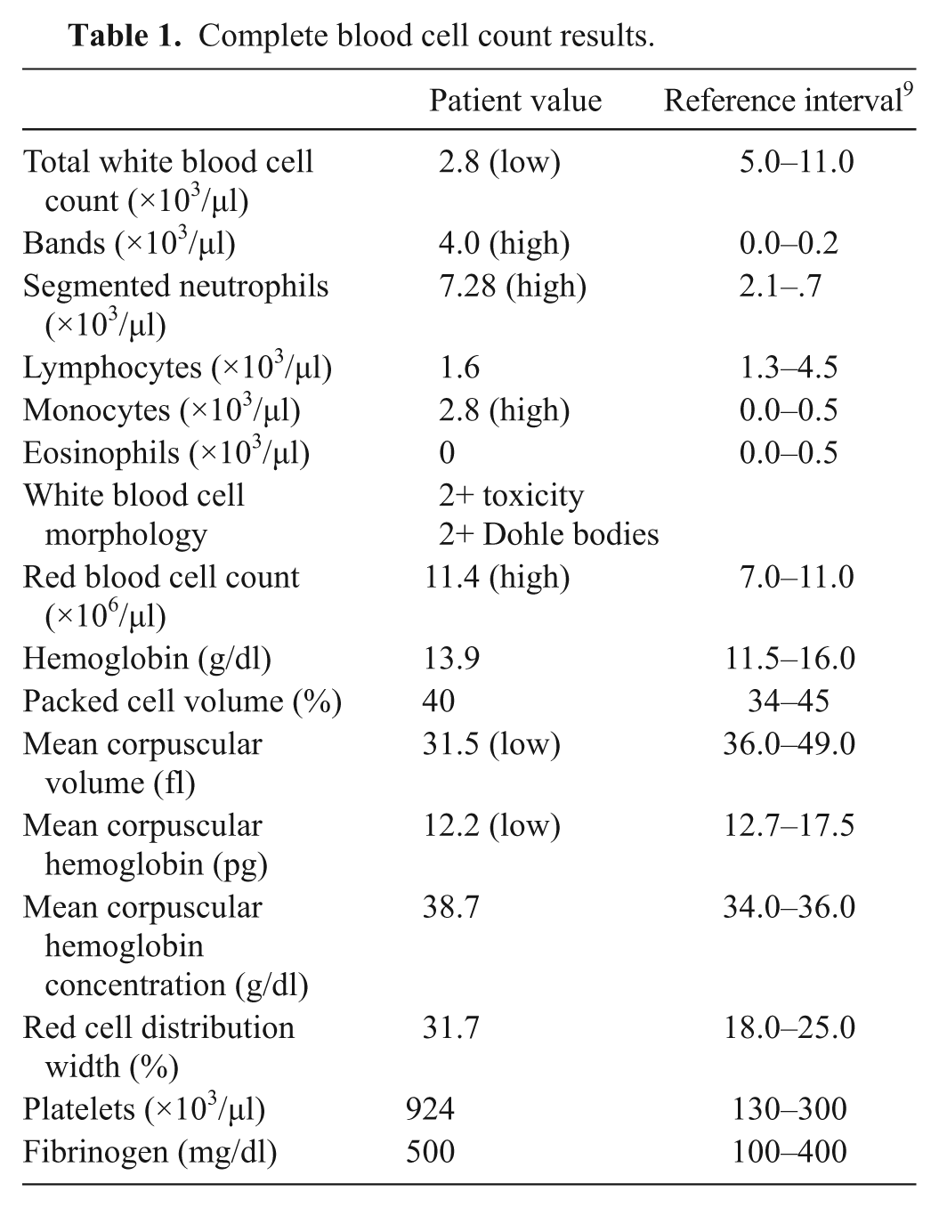
Complete blood cell count results.
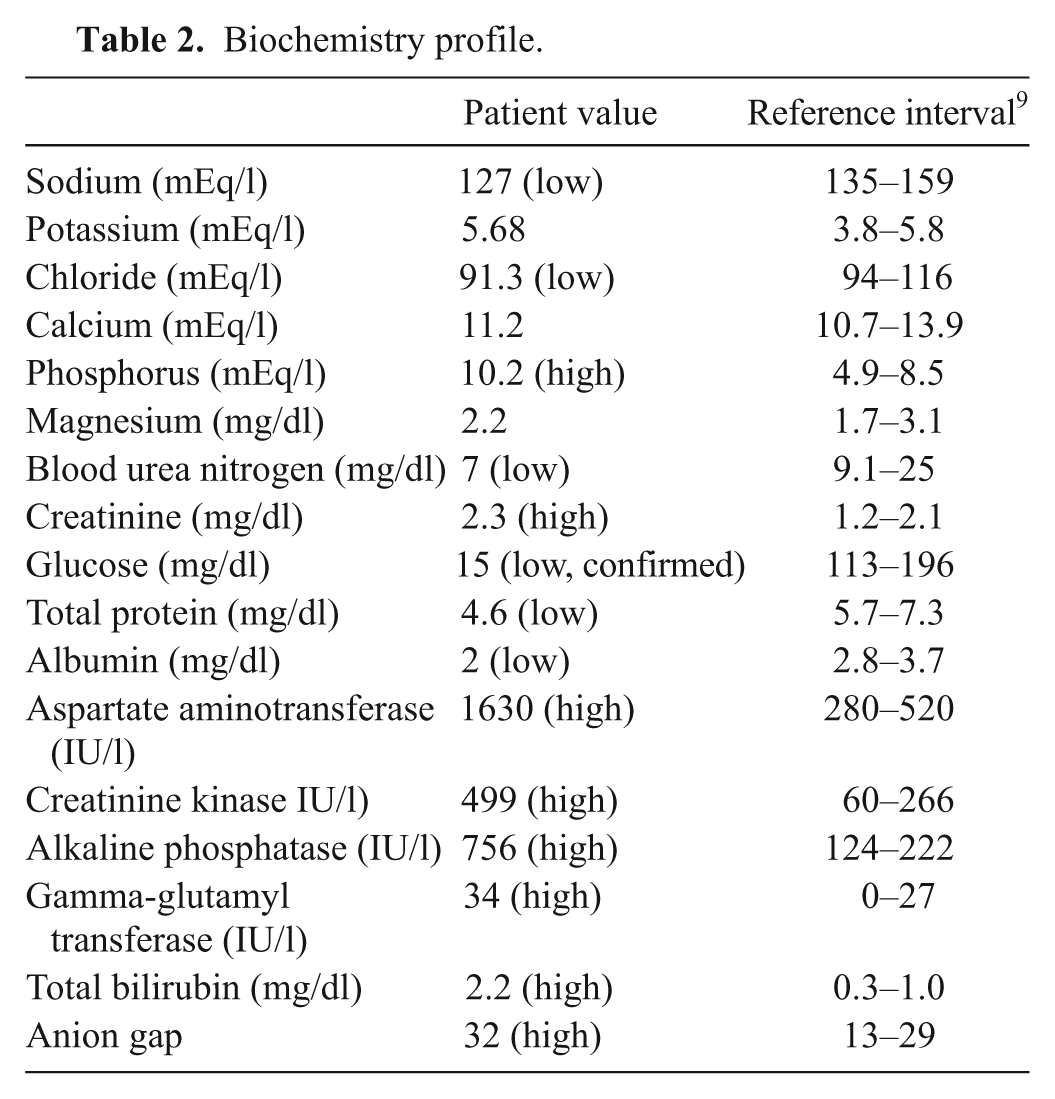
Biochemistry profile.
Whole blood was aseptically collected and submitted for aerobic and anaerobic culture. A small amount of fecal material was collected digitally from the rectum and was submitted for culture and fecal polymerase chain reaction (PCR) for 4 equine enteric pathogens b (genes associated with Clostridium difficile toxins A and B, Lawsonia intracellularis, Neorickettsia risticii, and Salmonella spp.). Based on the history, physical examination, and blood work, a diagnosis of bacterial septicemia was suspected, with subsequent shock, dehydration, and hypoglycemia.
While initial treatments corrected the hypoglycemia, the foal continued to seizure through multiple doses of diazepam. The blood work and clinical picture indicated overwhelming sepsis and poor tissue perfusion despite adequate rehydration. Due to a grave prognosis for improvement, the foal was humanely euthanized 8 hr after admission.
On postmortem examination the following day, a small amount of reddish brown feces was noted around the anus. The abdominal cavity contained a moderate amount of red-tinged watery fluid. Colonic lymph nodes in the mesentery of the large colon were prominent. The cecum and colon were moderately distended. Some hyperemia of mesenteric blood vessels was identified. The contents of the cecum, large colon, and small colon were filled with watery reddish brown fluid. The walls of the cecum and the colon were markedly thickened, gelatinous, and doughy, and clear fluid oozed from the submucosa on cut surface. The mucosa was diffusely covered by yellowish tan to brown fibrillar material that was easily peeled from the mucosal surface (Fig. 1). The underlying mucosal surface was diffusely rough, irregular, and reddened. The liver was light brown and the expected size and consistency.
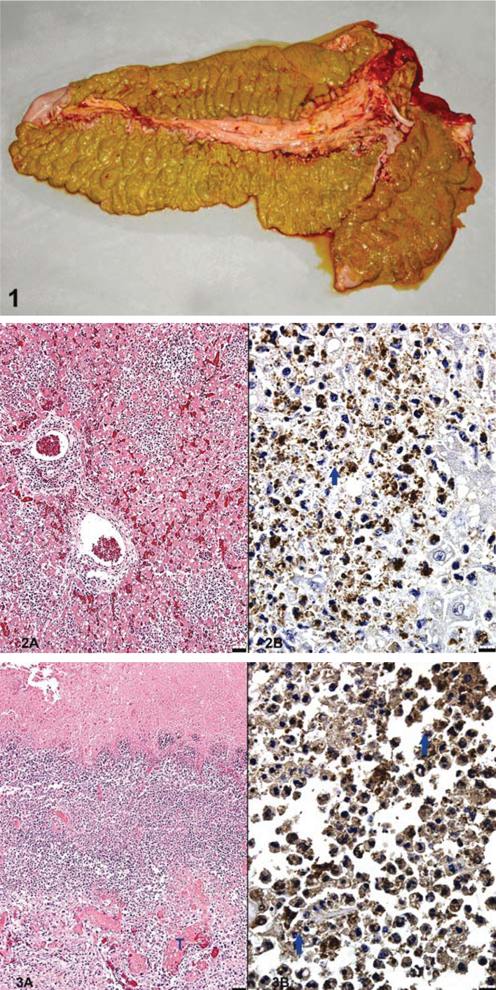
Five-week-old American Quarter Horse foal. Photograph of the mucosa of the large colon and cecum. The cecal and colonic mucosa is diffusely covered by a fibrinonecrotic membrane and thickened by edema and inflammation. The content was reddish brown and watery.
Specimens were collected and routinely fixed in 10% phosphate buffered neutral formalin, dehydrated, and then embedded in paraffin wax. The tissue specimens were sectioned at 5 µm and stained with hematoxylin and eosin for histopathologic examination. Histopathologic examination of the liver revealed extensive multifocal to coalescing random foci of necrosis with abundant necrotic cellular debris and heavy inflammatory infiltration of predominantly neutrophils, intermingled with some lymphocytes, plasma cells, and macrophages. Similar cells in smaller numbers occurred throughout the sinusoids. Kupffer cells frequently contained hemosiderin. The remaining hepatocytes often had wispy cytoplasmic vacuoles. The extensive foci of necrosis and inflammation were disseminated throughout the liver. The mucosal surfaces of the cecum and colon were diffusely necrotic and covered by a layer of fibrin with a wide underlying layer of heavy inflammatory cell infiltrates consisting predominantly of neutrophils, moderate numbers of lymphocytes, and some macrophages. The underlying submucosa was markedly thickened by edema, vascular hyperemia, vascular thrombosis, generalized inflammatory infiltrates, and few fibroblasts. Vasculitis, thrombosis, and luminal neutrophils in venules were prevalent. The thymus was atrophic, with loss of cortical and medullary distinction. Some lymph nodes were enlarged by edema but depleted of primary follicles. The spleen had similar depletion of lymphocytes in splenic follicles, and large numbers of neutrophils were present both in marginal areas of the white pulp and throughout the red pulp. The pulmonary alveolar septa were hypercellular due to prominent septal macrophages and neutrophils. Alveoli contained small amounts of fibrin and proteinaceous fluid. In scattered foci, cerebral cortical neurons were shrunken and hypereosinophilic and had pyknotic nuclei consistent with neuronal necrosis. A tissue Gram stain revealed intracellular Gram-positive, rod-shaped bacilli in neutrophils and macrophages, primarily in the foci of necrosis and inflammation within the liver (Fig. 2).
Aerobic and anaerobic culture isolated L. monocytogenes within 24 hr of inoculation from the blood collected at time of admission. Additionally, L. monocytogenes was cultured from the lung and liver collected at postmortem examination. Aerobic culture from the feces yielded a heavy growth of mixed enteric flora, light growth of Clostridium perfringens with no Salmonella sp. or Listeria sp. isolated. Colon content submitted for enzyme-linked immunosorbent assay c to detect antibodies against the genes for C. difficile toxins A and B was negative. Polymerase chain reaction on feces for C. difficile toxins A and B, L. intracellularis, N. risticii, and Salmonella spp. was negative. Typing of the C. perfringens isolate revealed toxinotype A with beta2 exotoxin and was negative for all other exotoxins and enterotoxin. Using a commercially available anti-Listeria antibody, d immunohistochemistry (IHC) of the liver and colon revealed the presence of L. monocytogenes (Fig. 2B, 3B). The most profound histopathologic lesions were multifocal, widely disseminated, necrotizing hepatitis and diffuse, necrotizing colitis and typhlitis (Fig. 2A, 3A).
Polymerase chain reaction was performed on the Listeria sp. isolate cultured from the lung parenchyma of the foal for further characterization and identification. Using multiplex PCR, 3 reference strains of Listeria sp. along with the isolate from the present case were run with oligonucleotide primers that were designed from the L. monocytogenes internalin genes inlA, inlB, and inlJ. The inlA and inlB genes are necessary for the organism to gain entry into the host cell and were used for species-specific recognition.14,20 The inlJ gene is a well-known virulence factor and was thus used for virulence determination as previously described. 14 The EGD strain is an internationally recognized highly virulent strain of L. monocytogenes.3,14 The F2365 strain is recognized as a virulent strain and has been associated with epidemics of foodborne illnesses in human beings. 29 The avirulent HCC23 strain, which was previously isolated from the brain of a catfish, was used as negative control.14,24 All strains were positively identified as species-specific L. monocytogenes. 14 The inclusion of the inlJ genes further identified the etiologic agent as a virulent isolate (Fig. 4).
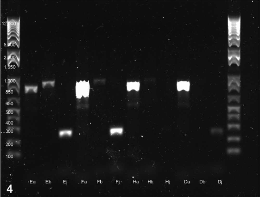
Five-week-old American Quarter Horse foal. Agarose gel electrophoresis of DNA products generated in multiplex polymerase chain reaction using internalin gene inlA, inlB, and inlJ primers. Lane 1: DNA molecular weight marker; lanes 2–4: products amplified from DNA of Listeria monocytogenes strain EGD with genes labeled a, b, and j; lanes 5–7: products amplified from DNA of L. monocytogenes strain F2365 with genes labeled a, b, and j; lanes 8–10: products amplified from DNA of L. monocytogenes strain HCC23 with genes labeled a, b, and j; lanes 11–13: products amplified from DNA of the listeria isolate cultured from the lung of this foal. The expected inlA, inlB, and inlJ gene products are of 800, 884, and 238 bp in size, respectively.
The primary clinical signs noted in the foal were intermittent diarrhea present since birth, depression, recumbency, seizures, and septic shock. The symptoms are nonspecific signs of septicemia in neonatal foals, caused by a variety of etiologic agents, including Gram-negative bacteria such as Escherichia coli and Salmonella sp. and Gram-positive bacteria, including Streptococcus sp., Staphylococcus sp., and Clostridium sp. Listeria monocytogenes, although rarely recognized as a primary agent of neonatal disease, has been reported to cause disease in horses, manifesting as colic, fever, diarrhea, neurologic compromise, and anorexia. 28 In a previous report, L. monocytogenes was isolated from 2% of neonatal blood culture isolates, indicating that this organism may be a clinical reality that should perhaps be considered more readily in a list of differentials for septicemic foals. 16 Listeria monocytogenes was previously isolated from the blood, liver, spleen, kidneys, and lungs of 5 septicemic ponies that were housed in close proximity to cattle. 2 Furthermore, it has been proposed that L. monocytogenes might be a causative agent of neonatal diarrhea, even when not identified on fecal culture, in the absence of other gastrointestinal pathogens. 26 Similarly, while L. monocytogenes was not cultured from the feces in the current case, it was isolated from multiple organs, including the lungs, liver, and blood, consistent with Listeria sp. being the cause of disease in this foal.
The significance of C. perfringens toxinotype A with beta2 exotoxin identified in the current case is uncertain. There are reports in the literature that have identified the beta2 exotoxin to be a significant virulence factor associated with C. perfringens, causing typhlocolitis in adult horses 11 and a primary disease-causing agent of equine neonatal colitis. 10 Traditionally, C. perfringens toxinotype A has not been associated with pathogenicity and is known to be part of the normal gastrointestinal flora of healthy neonates and adult horses and is ubiquitous in the environment. Although somewhat controversial, the presence of the beta2 exotoxin has been reported to contribute to increased virulence of this organism. 11 Similarly, L. monocytogenes is found within the environment in soil, water, and silage, and the pathogen tends to be opportunistic. Clinical disease is speculated to occur when the host’s immune system is decreased as a result of stress, pregnancy, immunodeficiency, concurrent disease, or an overwhelming infective dose.22,28 It is thus possible that co-infection with L. monocytogenes and C. perfringens may have contributed to the overall compromise of this foal. However, the gross and histopathologic lesions identified in the liver and colon are significant and unique. The identification of intracellular L. monocytogenes in phagocytic cells in liver and colon by IHC, confirmation of antemortem Listeria sepsis, and discovery of only very light growth of C. perfringens support a diagnosis of listeriosis with resulting hepatitis and typhlocolitis.
The most common mode of transmission of L. monocytogenes in all species is by ingestion, and most cases arise secondary to the consumption of contaminated foods or silage. 26 Neonatal infection with L. monocytogenes in foals can occur through ingestion of pathogens from the environment or through the mare’s milk. 6 Entry of the organism may occur through the nasal mucosa, through conjunctiva, or via wound contamination. 26 The organism can be transmitted in poor silage, grass, feces, urine, aborted tissues, uterine discharge, surface water, and milk, with the most frequent source being fecal contamination. 12 Additional routes of exposure may include inhalation, transplacental transfer, or via the umbilical remnant. 26 In the foal described herein, the umbilical remnant was subjectively larger than normal, but ultrasound examination at the time of admission was within normal limits. No omphalitis was found at necropsy examination. In the present case, the initial early clinical diarrhea, profound typhlocolitis, and presence of the organism in the cecum, colon, and liver as confirmed by IHC suggest that oral ingestion was the most likely mode of transmission.
While the exact source of contamination in the current case is not known, the referring veterinarian reported a hay feeder in the pasture where the foal and her dam were kept. Upon further inspection it was noted that hay had accumulated below the feed bunk, which was situated at the bottom of a hill in standing water due to frequent rains over the preceding month. The foal had historically eaten hay since 2 weeks of age. The hay was not submitted for analysis. Feeding of silage is a well-known risk factor for listeriosis, particularly when the silage is of poor quality (pH > 5.5).5,8,21 In the present case, it is postulated that the water-soaked hay established an environment similar to that of fermented silage and served as a source of Listeria contamination.
Listeria monocytogenes was not isolated from the feces or ingesta; however, it was cultured from the lungs and from the blood (within 24 hr of inoculation). In addition, IHC positively identified L. monocytogenes in the foci of necrosis and inflammation in the colon and liver. The exclusion of the other gastrointestinal pathogens in conjunction with the positive identification of L. monocytogenes by culture and IHC supports a diagnosis of listeriosis. The InlJ gene is responsible for the entry of the bacterium into the cell, facilitating systemic spread protected from the immune system of the infected host, thus contributing to overall virulence. 14 The presence of this gene was identified on a molecular level by multiplex PCR. Histopathology verified the intracellular location of the Listeria organism. Although L. monocytogenes has been recognized to cause disease in horses, it is potentially a bacterial pathogen that plays a more substantial role in equine foal diarrhea than previously thought.
Footnotes
Acknowledgements
The authors would like to thank Dr. Frank Austin from Mississippi State University, College of Veterinary Medicine, for substantial editorial and intellectual input. The authors thank Dr. Ramos-Vara and the histopathology section of the Purdue Animal Disease Diagnostic Laboratory for Listeria immunohistochemistry.
a.
Nolvasan® solution, Fort Dodge Animal Health, Fort Dodge, IA.
b.
Equine diarrhea panel, Laboratory of Real Time PCR, University of California–Davis, Davis, CA.
c.
ImmunoCard Toxins A&B, Meridian Bioscience Inc., Cincinnati, OH.
d.
Listeria O antiserum poly serotypes 1 and 4, Difco Laboratories, Detroit, MI.
The author(s) declared no potential conflicts of interest with respect to the research, authorship, and/or publication of this article.
The author(s) would like to acknowledge the Department of Clinical Sciences at Mississippi State University, College of Veterinary Medicine, for the financial support and funding for the publication of this article.