
Abstract
Machupo virus, the cause of Bolivian hemorrhagic fever, is a highly lethal viral hemorrhagic fever with no Food and Drug Administration–approved vaccines or therapeutics. This study evaluated the guinea pig as a model using the Machupo virus–Chicava strain administered via aerosol challenge. Guinea pigs (Cavia porcellus) were serially sampled to evaluate the temporal progression of infection, gross and histologic lesions, and sequential changes in serum chemistry and hematology. The incubation period was 5 to 12 days, and complete blood counts revealed leukopenia with lymphopenia and thrombocytopenia. Gross pathologic findings included congestion and hemorrhage of the gastrointestinal mucosa and serosa, noncollapsing lungs with fluid exudation, enlarged lymph nodes, and progressive pallor and friability of the liver. Histologic lesions consisted of foci of degeneration and cell death in the haired skin, liver, pancreas, adrenal glands, lymph nodes, tongue, esophagus, salivary glands, renal pelvis, small intestine, and large intestine. Lymphohistiocytic interstitial pneumonia was also present. Inflammation within the central nervous system, interpreted as nonsuppurative encephalitis, was histologically apparent approximately 16 days postexposure and was generally progressive. Macrophages in the tracheobronchial lymph node, on day 5 postexposure, were the first cells to demonstrate visible viral antigen. Viral antigen was detected throughout the lymphoid system by day 9 postexposure, followed by prominent spread within epithelial tissues and then brain. This study provides insight into the course of Machupo virus infection and supports the utility of guinea pigs as an additional animal model for vaccine and therapeutic development.
Keywords
Treatments and vaccines for viral hemorrhagic fevers are elusive and currently unavailable for most of these often-fatal diseases. At the time of this writing, an outbreak of Ebola hemorrhagic fever is ongoing in West Africa, and an Ebola outbreak of this severity and magnitude is unprecedented. Viral hemorrhagic fevers are enigmatic in when and where they cause outbreaks, and Bolivian hemorrhagic fever (BHF), a viral hemorrhagic fever not unlike Ebola, is no exception. BHF is caused by Machupo virus (MACV), a South American hemorrhagic fever virus that is often hard to distinguish from other hemorrhagic fevers in the region. This reclusive virus first appeared on the Bolivian savannah in 1959 and was subsequently isolated from a fatal human case in 1963 and named BHF after the location where it first occurred. 7,14 –19,22 Between 1959 and 1962, 470 cases were reported in the northeast corner of Bolivia, and a second series of outbreaks transpired between 1962 and 1964, resulting in around 1000 cases and 180 deaths. 7,23 No cases of BHF were reported between 1976 and 1993, but an outbreak in 1994 consisting of 19 cases was the first in a recent string of sporadic outbreaks culminating in 20 cases in 2007 and more than 200 in 2008. 1,22,23 The overall case fatality rate has varied among outbreaks, but up to 30% of those infected with MACV die from it or complications related to infection. 7,19,22,23 Because sporadic outbreaks are so lethal and because aerosol release of hemorrhagic fever agents by a bioterrorist is thought possible, 2,4,5,10,25 a viable animal model is crucial to help explain the course of disease and assist with development and testing of effective medical countermeasures.
The primary reservoir for MACV is a rodent, the large vesper mouse, known locally as the “big laucha” (Calomys callosus). 15,18,23,32 Virus is spread through aerosolization of excreta or from direct contact with infected rodent urine, saliva, or blood. Outbreaks of BHF often coincide with the annual grain harvest that occurs between April and July in northeast Bolivia. 1,23 Occupational exposure to aerosolized virus during grain harvesting is the most likely reason for this spike. 1,19,23 Infection can also occur between caregivers and afflicted patients from close contact, and caregiver transmission has been reported during more than 1 outbreak. 7,20
The incubation period for BHF in humans can vary from 5 to 21 days but generally is between 7 and 14 days. Initial signs of infection include fever, lethargy, anorexia, dehydration, cutaneous hyperesthesia, myalgia, back pain, headache, and dizziness. Patients also often have cutaneous petechia, gingival bleeding, and conjunctival injection. Clinical symptoms progress, and within 3 to 4 days patients may experience nausea, vomiting, abdominal pain, and diarrhea. Clinical laboratory examination often reveals leukopenia, thrombocytopenia, and proteinuria with hematuria. During the second week of illness, neurologic and hemorrhagic symptoms often arise and consist of intention tremors, spasmodic movements, delirium, and convulsions. Hemorrhagic manifestations include hematemesis, epistaxis, gingival bleeding, melena, and hemorrhage from venipuncture sites. In severe, fatal cases, patients often succumb within 7 to 12 days of the onset of symptoms. Survivors begin to improve during the second week of clinical illness, and this improvement is heralded by the appearance of neutralizing antibodies in the serum. Complete recovery takes several weeks to months with convalescence often marred by fatigue, dizziness, hair loss, and Beau lines in the patient’s finger- and toenails. 1,6,7,19,20,27,29
Many aspects of BHF are poorly understood, and an animal model would facilitate pathogenesis studies, therapeutic testing, and vaccines trials. Animal models are needed to study MACV because the sporadic and often secluded nature of outbreaks, in combination with its highly lethal nature, makes it neither feasible nor ethical to do such testing in human subjects. The US Food and Drug Administration recognizes the need for an alternative drug and vaccine approval pathway for highly lethal infectious diseases, and in 2002 it codified CFR 314.600, also known as the “Animal Rule.” 30,31 The Animal Rule allows data from animal studies to be used in lieu of human clinical data for vaccine and therapeutic approval. Part of this rule states that investigators must establish the mode of action for the drug that they are testing in at least 1 animal model that reproduces accurate human disease pathology. The Animal Rule also states that efficacy must be proven in animals of more than 1 species unless the model is sufficiently characterized for predicting the response in humans. 30,31 Cynomolgus macaques (Macaca fascicularis) at this institute were recently studied as an animal model of MACV infection and proved to be an excellent surrogate for understanding the human disease. 3 The objective of the present study was to determine if the guinea pig could be used as a second viable animal model and to better determine the pathologic sequence of events that occurs during an aerosol challenge with MACV–Chicava strain.
Materials and Methods
MACV Aerosol Exposure and Sampling Scheme
An LD50 (lethal dose, 50%) study was conducted prior to the sequential sampling study to determine the optimal exposure dose by the aerosol route in the Dunkin-Hartley guinea pigs from Charles River (Raleigh, NC, USA). Groups of 10 guinea pigs each were exposed to aerosolized target doses of 10, 100, or 1000 PFUs (plaque-forming units) of MACV–Chicava strain. We were unable to calculate the LD50 because all guinea pigs, regardless of dose received, succumbed to infection by 30 days postinfection. We selected 100 PFUs as the challenge dose for this study because it was the lowest lethal dose that could be reliably achieved by the aerosol route. For the serial sampling study, 35 Hartley guinea pigs weighing between 450 and 550 g and of both sexes were randomized into 7 sampling groups (time points) consisting of 5 guinea pigs each. Individual guinea pigs were identified and body temperatures monitored with IPTT-300 temperature transponder chips (BMDS, Seaford, DE, USA). Thirty guinea pigs were aerosol exposed to a target dose of 100 PFUs of MACV–Chicava strain. The physical setup of the aerosol exposure apparatus (ie, automated bioaerosol exposure system) can be viewed in Figure 1 of reference 12. For the aerosol exposures, groups of up to 4 animals at a time were placed in wire mesh baskets inside the aerosol chamber and exposed to aerosolized virus for approximately 15 minutes, which was an estimation of the time required for target exposure based on respiratory rate as a function of an animal’s body weight. 11 Plaque assays were performed on the starting concentrations and samples retrieved from the all-glass impinger for each exposure group. The actual dose received by guinea pigs in each exposure run was determined using calculations previously described. 12 A mean mass aerodynamic diameter of 1–3 microns for arenaviruses was used in the calculations. The actual inhaled doses for each group are listed in Supplemental Table 1. As described earlier, each exposure run consisted of up to 4 guinea pigs of similar weight. For 5 guinea pigs that were housed with the other guinea pigs during acclimation and prestudy manipulations, blood samples were collected on day 0 prior to euthanasia; the data from these animals served as baseline levels for this study. Guinea pigs from all other sampling groups were exposed to aerosolized virus on day 0. After exposure, blood samples and tissues were collected from 5 guinea pigs at each sampling time point. Sampling time points were scheduled at days 0 (negative control, unexposed guinea pigs), 5, 9, 13, 16, 19, and 21 postexposure. Complete blood counts and blood chemistry analyses were performed on samples collected from each animal at each sampling time point. Animals were observed, weighed, and scanned for body temperature using a BMDS DAS-7007R transponder scanner daily to record disease progression. On each scheduled sampling day, 5 randomly selected guinea pigs were anesthetized by intramuscular injection; blood was collected; and the animals were euthanized by intraperitoneal administration of a barbiturate overdose.
Body weight, temperature, and morbidity score for guinea pigs aerosol exposed to Machupo virus–Chicava strain (MACV). Each color represents a group of 5 guinea pigs from each of the 6 sample days (5, 9, 13, 16, 19, and 21 days postexposure).
Guinea pigs were group housed and provided water and certified guinea pig feed ad libitum. Daily enrichment was also provided. Research was conducted under a protocol approved by the Institutional Animal Care and Use Committee in compliance with the Animal Welfare Act, Public Health Service Policy, and other federal statutes and regulations relating to animals and experiments involving animals. The facility where this research was conducted is accredited by the Association for Assessment and Accreditation of Laboratory Animal Care International and adheres to principles stated in the National Research Council’s 2011 Guide for the Care and Use of Laboratory Animals.
Virus Isolation, Preparation, Plaque Assay, Exposure, and Sampling Scheme
The MACV–Chicava strain used in these studies was isolated from a fatal human case of BHF occurring in the Beni province in 1994. The serum sample was sent to the Centers for Disease Control and Prevention (CDC), confirmed to contain MACV–Chicava strain, and designated as CDC#806786. The serum sample was passaged once (P1) on Vero E6 cells at the CDC by personnel of the Special Pathogens Branch. Cell culture supernatant from the infected flask was retrieved and used to infect a second Vero E6 monolayer (P2). A sample of supernatant from P2 was sent to the US Army Medical Research Institute of Infectious Diseases (USAMRIID). Detailed description of (1) the isolation and preparation of the virus and (2) the animal inoculation and sample collection is provided in the Supplemental Materials.
Clinical Chemistry and Hematology
For blood sample collection, guinea pigs were anesthetized, and samples of whole blood were collected from the cranial vena cava and distributed into tubes containing EDTA for complete blood counts or serum separator tubes for blood chemistry analysis. Complete blood counts were performed using a HemaVet 950 FS Hematology Analyzer (Drew Scientific, Oxford, CT, USA) in accordance with manufacturer’s guidelines. Serum was separated from whole blood by centrifugation at 1800 × g. An aliquot of 100 μl was analyzed for glucose, creatinine, blood urea nitrogen, calcium, albumin, total protein, alanine aminotransferase, alkaline phosphatase, total bilirubin, amylase, globulin, potassium, sodium, and phosphorous via the veterinary comprehensive diagnostics rotor on a VetScan VS2 Blood Chemistry Analyzer (Abaxis, Union City, CA, USA). Results were compared to day 0 samples used as a baseline. Five samples were collected from each time point immediately prior to euthanasia, and guinea pigs were randomly assigned to each time point. Hematology and chemistry data collected during this study are provided as Supplemental Tables 4 and 5.
Histopathology and Immunohistochemistry
Necropsies were performed on each guinea pig immediately following euthanasia in the USAMRIID biosafety level 4 laboratory. Tissues from all major organ systems were collected, immersion fixed in 10% neutral buffered formalin, and held in biocontainment for a minimum of 21 days. Histopathology samples were routinely processed, embedded in paraffin, sectioned, and stained with hematoxylin and eosin.
Immunohistochemistry was used to identify cells containing MACV antigen via an immunoperoxidase assay using rabbit polyclonal antisera specific for MACV. This antibody was produced at the USAMRIID by injecting rabbits with ultraviolet-inactivated MACV virus (Malale strain). Serum was collected and determined to be cross-reactive to MACV strains Caravallo, Chicava, and Malale. Sections of normal guinea pig liver served as the negative control. The positive control was liver sections from a guinea pig that had died from experimental MACV–Chicava strain infection at this institute. Normal mouse IgG was used as the negative serum control for the control slides. Details of the immunohistochemistry methods are provided as Supplemental Materials.
Statistical Analysis
Dependent variables were screened for outliers, normality, and homogeneity of variance. Dependent variables met assumptions of normality and homogeneity of variance. No outliers were detected. Analysis of variance with Dunnett post hoc test was used to compare chemistry and hematology values and maximum temperature between the day 0 group and the other 6 time point groups. Paired t tests were used to compare day 0 temperature with maximum postchallenge temperature within each group. Stepdown Bonferroni adjustment was then used to adjust for multiple comparisons. Analyses were conducted using SAS 9.4 (SAS OnlineDoc 9; SAS Institute Inc, Cary, NC, USA).
Results
Clinical Presentation of MACV Infection
In this sequential sampling study and previous LD50 studies, clinical signs among groups of guinea pigs did not vary significantly by dosing group or by exposure route. Clinical signs first appeared around 1 week postexposure and included piloerection, fever, loss of appetite, erythema of the haired skin in the axillary and inguinal areas as well as the ear tips, dyspnea, and intermittent diarrhea. At times, the diarrhea appeared to be bloody, but this was difficult to determine on an individual guinea pig basis because of group housing arrangements. In group Nos. 3–7 (sampled at days 9, 13, 16, 19, 21), maximum temperatures were statistically significant when compared to the day 0 control group (Suppl. Table 2). Neurologic signs appeared around 16 to 20 days postexposure and included a head tilt and ataxia in some guinea pigs. Once neurologic signs began they were generally progressive in severity. Rapid breathing was observed in moribund guinea pigs, and in some cases, respiratory difficulty was noted. Guinea pigs aerosol exposed to MACV began to lose weight starting approximately 11 to 13 days postexposure with a trend of decreasing weight after day 13 postexposure (Fig. 1).
Clinical Chemistry and Hematology
Hematology results revealed a statistically significant reduction in lymphocyte number and percentage on days 9 and 13 and a statistically significant reduction in total numbers of leukocytes on days 13 and 16. Platelets were also significantly decreased in number on days 13, 16, and 21, with a nadir of 52.40 × 10³/μl on day 16 (Suppl. Tables 3, 4). Chemistry results were largely unremarkable (Suppl. Table 5). Chemistry analyzers for rodent specimens were not available in the biosafety level 4 environment for these studies, although veterinary-specific instruments were used.
Gross Pathology
Gross lesions consisted of multifocal petechial hemorrhages on the mucosal and serosal surfaces of the gastrointestinal tract and the uterus as early as day 9 postexposure, with similar hemorrhages observed throughout the remaining time points. Hepatic pallor (Fig. 2) was noted as early as day 9 postexposure and continued to progress in the degree of pallor and friability at each time point. Lymph nodes were generally enlarged by day 13 postexposure, and the lungs were often wet, heavy, and failed to completely collapse (Fig. 3).
Machupo virus infection, guinea pig.
Histology and Immunohistochemistry
The organs primarily affected in guinea pigs exposed to aerosolized MACV–Chicava strain virus were lymphoid tissues, lung, liver, bone marrow, epithelial cells of the respiratory and gastrointestinal tracts, the adrenal gland, the urinary epithelium, and brain (Suppl. Table 6; a complete list of epithelial tissues affected is provided at the bottom of the table). Inflammatory changes were consistently present in the heart albeit minimal in severity. At later time points, inflammation became more widespread, affecting a variety of tissues.
Lymph nodes had mild to moderate paracortical lymphoid hyperplasia on day 5 postexposure. In most guinea pigs, this was accompanied by cortical lymphoid atrophy characterized by a decrease in the numbers of lymphoid follicles and lymphocytes within the cortex. By day 9 postexposure, this was accompanied by hyperplasia of macrophages and fibroblastic reticular cells, which was generally mild to moderate but sometimes severe. The thymus had mild cortical atrophy of lymphocytes by day 13 postexposure, which increased in severity with time, becoming moderate to severe by day 19 postexposure (Figs. 4, 5). This finding may have been partially related to stress. In the thymus, there was also a proliferation of cells in the medullary region, which appeared to be a combination of macrophages and large lymphocytes. Hemorrhage/erythrophagocytosis was occasionally present in the lymph nodes and thymus, but overall hemorrhage was not a major finding. In the interstitium of the lungs of 2 guinea pigs on day 5 postexposure, there was minimal to mild mixed mononuclear cellular infiltration, which increased in severity to moderate or marked thereafter and was considered to be related to viral exposure (Figs. 6, 7). By day 16 postexposure, inflammation was generally marked by perivascular orientation. In a few guinea pigs, there was eosinophilic, beaded material suggestive of fibrin within affected areas of the lung; however, this was infrequent and minimal, and fibrin deposition was therefore not considered a major feature in the lung.
Lung, guinea pig No. 1, day 0 (nonexposed). Hematoxylin and eosin (HE).
By day 13 postexposure, virus-associated findings were also observed in the liver, heart, and bone marrow. In the liver, there was minimal mixed inflammation consisting of macrophages and lymphocytes associated with hepatocellular necrosis. The severity of the inflammation increased to moderate in some animals by day 19 postexposure (Figs. 8, 9). Hepatocellular inclusion bodies were not seen by light microscopy; however, immunohistochemical staining revealed distinct circular collections of antigen similar to that seen in primate experiments with MACV conducted at this institute (Fig. 9). 3
From day 13 postexposure, there was also lymphohistiocytic inflammation in the heart, which remained generally minimal to mild throughout the course of the study. The inflammation occasionally involved the endocardium, myocardium, pericardium, or a combination of the 3 with multifocal mild myocardial degeneration and necrosis. In bone marrow, there was minimal to mild cellular depletion in an unusual multifocal or pockmarked pattern with the remainder of the bone marrow exhibiting normal cellularity; in certain instances, there was leukocyte and megakaryocyte hyperplasia within the bone marrow adjacent to areas of cellular depletion. By day 16 postexposure, there was minimal to mild gliosis/perivascular inflammation in the brain with mononuclear perivascular cuffing, occasional neuronal cell death near affected vessels, and meningitis, and on day 19 and 21 postexposure neurologic lesions progressed in severity (Figs. 11–13). From day 16 postexposure, there were also inflammatory changes in a wide variety of other tissues, including the eye. In some examined eyes, the inflammation in the sclera and choroid was quite prominent and consisted of mixed mononuclear inflammation admixed with cellular debris. In numerous guinea pigs, multiple epithelial cells within various organs were affected (Suppl. Table 6, see notes). The adrenal gland, renal pelvis, urinary bladder, pancreas, tongue (Figs. 14, 15), esophagus, small intestine (Figs. 16, 17), large intestine (Fig. 18), urethra, epididymis, and gall bladder were all affected in the majority of guinea pigs from day 13 onward. The inflammation in the esophagus was associated with erosion/ulceration in a few guinea pigs.
Cerebrum, guinea pig No. 1, day 0 (nonexposed). Hematoxylin and eosin (HE).
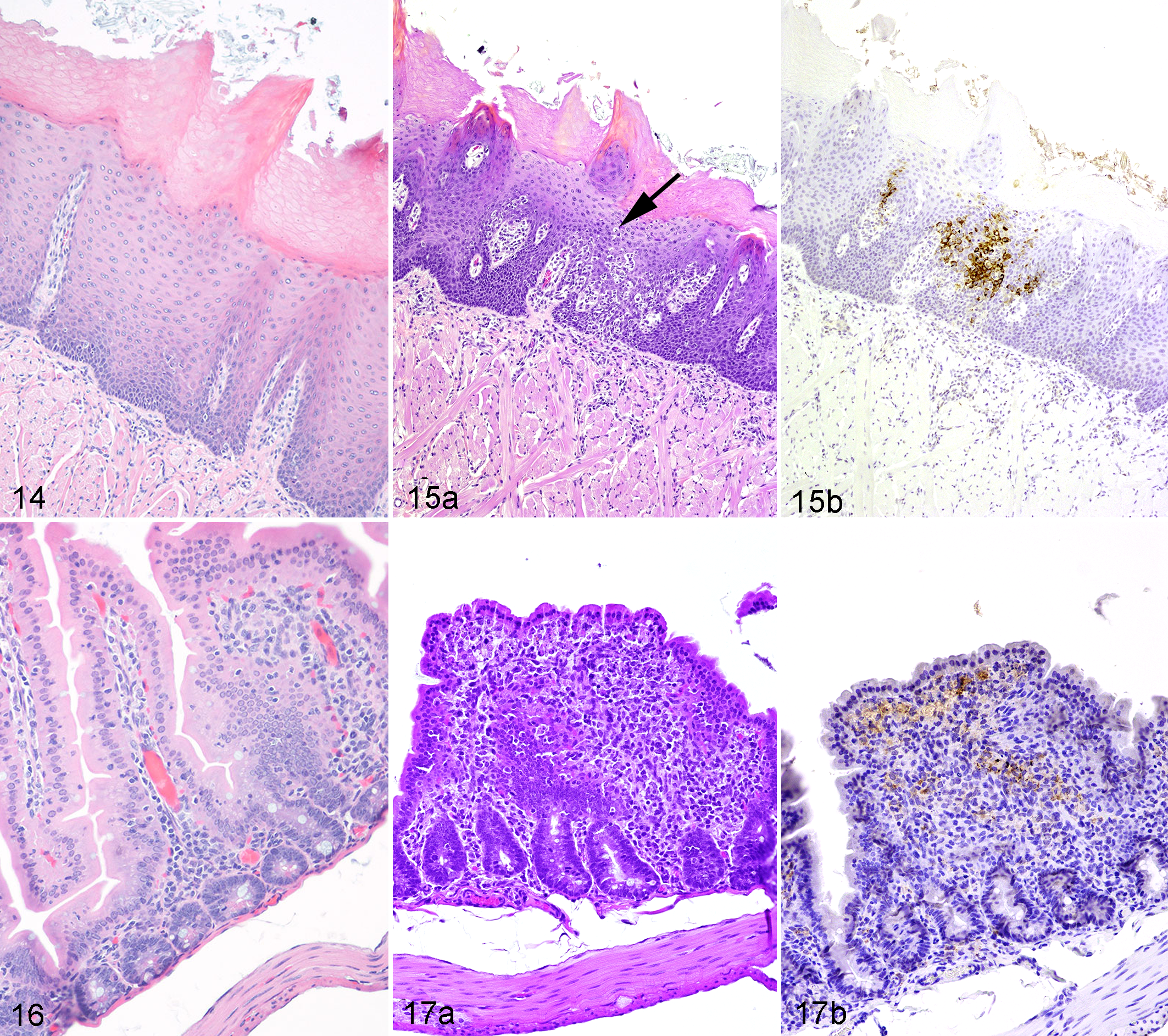
Tongue, guinea pig No. 1, day 0 (nonexposed). Hematoxylin and eosin (HE).
(a) Proximal colon, guinea pig No. 1, day 0 (nonexposed). Immunohistochemistry for Machupo viral antigen. (b) Machupo virus infection, proximal colon, guinea pig No. 21, day 16 postexposure. Numerous epithelial cells are strongly immunoreactive for Machupo viral antigen.
Immunoreactivity for MACV (Suppl. Table 7) generally colocalized with histologic lesions, with the macrophage being the primary cell containing viral antigen. The earliest definitive labeling occurred at day 5 postexposure in alveolar macrophages within the lungs and macrophages in the tracheobronchial lymph nodes. Thereafter, increased labeling of macrophages occurred in lymph nodes and multiple organs. Fibroblastic reticular cells in lymph nodes also exhibited immunoreactivity but less frequently and with less intensity. The intensity of labeling of macrophages and epithelial cells increased through day 16 postexposure but in the final 2 groups (days 19, 21) appeared much less intense and was sparse. Immunoreactivity of parenchymal cells for MACV antigen generally occurred at a later time point than that of macrophages. There was minimal labeling of esophageal squamous epithelium at day 5 postexposure. By day 9, there was noticeable staining in liver, intestinal tract, spleen, all lymphoid tissues, and adrenal cortical cells. By day 13 postexposure, there was increased staining intensity and prevalence in hepatocytes, squamous epithelium of the tongue, esophagus, pancreatic acinar and/or islet cells, adrenal cortical epithelial cells, epithelial cells in the gastrointestinal tract, lung, accessory sex glands, and the renal pelvis. Day 16 was of special note because it was the first time point with clearly evident antigen staining in the brain, as noted in neurons multifocally throughout the cerebrum. Several other organs showed labeling of parenchymal cells in at least 1 guinea pig from day 16 through day 21 postexposure.
Discussion
This study was performed in the hope of further elucidating the pathogenetic mechanisms and temporal sequence of infection following MACV aerosol exposure, in addition to assessing the suitability of the guinea pig as an animal model. Further studies with this animal model could be used under the Food and Drug Administration’s Animal Rule to develop countermeasures against BHF resulting from outbreaks, laboratory accidents, or intentional exposure as a biowarfare or biothreat agent. Approval of medical countermeasures against diseases that must use the Food and Drug Administration’s Animal Rule may be required to demonstrate proof of efficacy in more than 1 animal model, and this model, in combination with the nonhuman primate model, 3 is a platform for such testing. Previous MACV studies performed at the USAMRIID and elsewhere used the Caravallo strain. In our studies, the Caravallo strain was not lethal in Hartley guinea pigs without adaption by serial passage of the virus through guinea pig spleen; conversely, Chicava strain exhibited lethality without adaption to guinea pigs or serial passage through suckling mouse brain. Our aim for development of animal models of arenavirus infection is to use the same virus strain for rodents and nonhuman primates to enable more direct comparisons between these models. Serially passaging virus in cell culture or in animals to enhance its virulence in animal models can cause changes in the virus sequence that are not present in the human isolates. Ideally, the virus strain used in animal model development studies should resemble, as closely as possible, the viruses that circulate in the human population. Based on the data gathered from this study, the guinea pig model resembles both human disease and the disease in cynomolgus macaques 3 in several ways. The incubation period in guinea pigs ranges from 5 to 12 days, with minimally elevated temperatures first appearing on day 5 postexposure, and by day 12 postexposure, all guinea pigs in the study had elevated temperatures, clinical signs of illness, or both. The incubation period is similar to that seen in humans and in macaques, with 7- to 14-day and 6- to 10-day incubation periods, respectively. 1,7,9,28,29
Many similar clinical signs seen in both human and nonhuman primates also appear in guinea pigs and include the initial nonspecific signs of fever and anorexia. In guinea pigs, a macular rash was not present, but erythema was noted in not only the axillary and inguinal regions but also the ear tips. This erythema did progress in a few cases to culminate in blackened ear tips. Intermittent bloody diarrhea was also often present after a few days of illness, and by day 16 to 20, guinea pigs often began to show neurologic signs of disease, including ataxia and head tilt. Appearance of neurologic signs correlates temporally with lesion development; the progression from initial illness to neurologic disease in guinea pigs mirrors the disease course seen in nonhuman primates and humans. 3,22
Guinea pigs that were exposed parenterally or via aerosol had clinical signs of respiratory difficulty, and this was in contrast to nonhuman primates, with only those aerosol exposed having noticeable respiratory distress. 3 These observations were subjective and performed in the challenging biosafety level 4 environment, so future studies should continue to assess these clinical findings for corroborative evidence.
Hematologically, similar trends were also observed. In humans and macaques, thrombocytopenia and lymphopenia often occur, and in this study guinea pigs displayed similar trends. 1,7,9,28,29
As with macaques, monocytes, macrophages, and dendritic cells appear to be a primary site of MACV infection. The examined data suggest that these cells are initially infected and allow viral transit throughout the body. These cells demonstrate strong cytoplasmic immunoreactivity in lymph nodes, spleen, vasculature of almost all organs (monocytes), occasional Kupffer cells, and lung (alveolar macrophages) as early as day 5 postexposure. Fibroblastic reticular cells and macrophages in lymphoid organs also appear to be commonly affected with a hyperplastic response noted as early as day 5 postexposure and with progression over the course of infection. Similar findings have been reported in both human and macaque cases in the literature. 3,8,28 In situ hybridization was not performed but would be useful as a confirmatory test to rule out confounding factors such as background/nonspecific staining and to identify infected cell types with an even higher degree of confidence. It appears that the MACV causes destruction of some select lymphoid cells in the cortex with a concomitant hyperplastic response in paracortical regions in guinea pigs. Fibroblastic reticular cells also appear to be affected, which may alter the immunologic environment as occurs with other viral hemorrhagic fevers. 26 Of interest, immune dysregulation from aberrant cytokine release is a common finding observed with other viral hemorrhagic fevers, 13 and further studies to determine the exact mechanisms behind immune dysregulation in cases of MACV infection may be warranted.
Histologic changes in bone marrow indicate cellular depletion and loss, which correlate directly with the hematology results; histologic depletion of bone marrow and statistically significant decreases in leukocyte and platelet numbers both occurred by day 13 postexposure. Certain sections of bone marrow taken at later time points had hyperplasia of leukocyte progenitors, and there was also very prominent megakaryocytic hyperplasia. MACV in guinea pigs also infected multiple epithelial tissues beginning around day 9 postexposure and becoming prominent by day 13 postexposure. Epithelial cells of the gastrointestinal and respiratory tracts were notably affected. Epithelial cells in numerous other organs were also affected but with a decreased incidence and severity (Suppl. Table 6). This propensity for infection of epithelial cells was also seen in macaques challenged with MACV at this institute, 3 and this epithelial localization correlates directly to the known entry receptor of MACV, transferrin receptor 1, found in large numbers on rapidly dividing epithelial cells. 21,24 This viral distribution also correlates with disease progression and the associated clinical findings. The pattern of infection, subsequent inflammation, and resultant clinical signs of MACV infection in guinea pigs is very similar to the disease course previously reported in macaques. 3 Given the reported progression of clinical signs and symptoms in humans, 22 we believe that the course of disease is very similar in humans, guinea pigs in this study, and macaques in previous studies. In patients with MACV infection, initial signs include fever, lethargy, and anorexia, and these initial signs of infection are also observed in the guinea pig model. Clinical symptoms progress, and within 3 to 4 days, patients may experience nausea, vomiting, abdominal pain, and diarrhea; in guinea pigs, clinical signs are also generally progressive, and these correlate with the increasing incidence and severity of histopathologic findings in lymphoid organs and epithelial cells lining mucosal surfaces. In patients, neurologic symptoms, if present, occur during the second week of infection, 22 and again guinea pigs mimicked this response with histopathologic lesions manifesting around day 16 of infection and becoming progressively more severe with each time point examined.
Conclusion
The guinea pig model is a viable animal model for MACV infection. The Animal Rule states that efficacy must be demonstrated in animals of more than 1 species unless a model is sufficiently well characterized to predict response in humans. Based on the results of this study, the guinea pig model can be used as an additional model in combination with the cynomolgus macaque model to help satisfy this regulatory requirement. The incubation period of 5 to 12 days; the febrile response; clinical signs of anorexia, diarrhea, and later-onset neurologic signs; and the hematologic changes all parallel those seen in humans and macaques infected with MACV 3 ; gross and histopathologic lesions are also similar in nature, distribution, and progression. In nonhuman primates and guinea pigs studied at this institute, aerosol and parenteral exposure routes seem to mimic one another in their clinical disease manifestations, with the exception of respiratory distress and more severe lung lesions generally present in nonhuman primates aerosol exposed to MACV. 3 Disease progression in guinea pigs—from initial malaise and fever with progression to end-stage neurologic disease—is also consistent with the course seen in humans and macaques, and this analogous disease course could be vitally helpful in future diagnostic, vaccine, and therapeutic development.
Footnotes
Acknowledgements
We gratefully acknowledge the numerous contributions of the pathology personnel of the US Army Medical Research Institute of Infectious Diseases (USAMRIID), including William Aguilar, Neil Davis, Phil Fogle, Angela Grove, Gale Krietz, Christine Mech, Nancy Twenhafel, and Brandon Wilke. We also thank members of the USAMRIID’s Virology Division—Heather Esham, Christopher Reed, and Ashley Keeney—for help with phlebotomy and/or animal observations, as well as and members of the Center for Aerobiological Sciences at the USAMRIID for assistance with the aerosol exposures. Cindy Rossi of the Diagnostic Systems Division, USAMRIID, kindly provided the polyclonal rabbit serum for immunohistochemistry. We also thank Sarah Norris and the Statistics Department for their help on the statistical analysis. Finally, we acknowledge the Special Pathogens Branch of the Centers for Disease Control and Prevention for providing the USAMRIID with the original sample containing Machupo virus–Chicava strain.
Authors’ Note
The views, opinions, and/or findings contained herein are those of the authors and should not be construed as an official Department of Army position, policy, or decision unless so designated by other documentation.
Declaration of Conflicting Interests
The author(s) declared no potential conflicts of interest with respect to the research, authorship, and/or publication of this article.
Funding
The author(s) disclosed receipt of the following financial support for the research, authorship, and/or publication of this article: The research was performed under US Army Medical Research Institute of Infectious Diseases project No. 195743 and funded by a Defense Threat Reduction Agency, Joint Science and Technology Office / Translational Medical Technologies grant (TMTI_0045_09_RD_T).