
Abstract
Feline mammary carcinomas are highly aggressive neoplasms. Several mechanisms are thought to be involved in their progression, including the loss of epithelial adhesion molecules. The present study was carried out on 21 adenomas and 139 mammary carcinomas. Of the carcinomas, 66 were not reported to have metastasized, while the remaining 73 had evidence of regional lymph node metastasis at the moment of diagnosis. The relationship was examined between the expression of the E-cadherin–β-catenin complex and basal (CK5/6, CK14) and luminal (CK8/18) cytokeratin expression. In the medical literature, carcinomas expressing basal cytokeratins are reported as having a poor prognosis in human breast cancer. Results revealed that preservation of the expression of E-cadherin and β-catenin is a significant feature of carcinomas without metastasis, whereas carcinomas with metastasis reveal the loss of one or both adhesion molecules. Additionally, basal cytokeratin expression was statistically associated with the presence of regional metastasis. Furthermore, the expression of E-cadherin–β-catenin was significantly correlated with the high expression of CK18 and low expression of CK5/6.
Mammary neoplasia is the third-most-common tumor type affecting female cats, following hemopoietic neoplasms and skin tumors. Malignancy is frequently reported and presents with local and distant metastases and high mortality rates. 42 Based on age, incidence, risk factors, histopathology, prognostic aspects, metastatic pattern, and response to therapy, feline mammary carcinomas have been proposed as a good model for human breast cancer. 2,42 Similar to reports in women, many veterinary studies describe regional lymph node involvement as one of the most important prognostic factors for feline mammary tumors. 11,16
Recent studies of human breast cancer by means of gene expression have defined different subgroups of breast tumors with distinct molecular signatures (luminal A, luminal B, Her-2+, or basal-like). 28 The same classification has also been made by immunohistochemistry. 25 Some of the subgroups, especially those described as basal-like, are correlated with an unfavorable prognosis. 27 This basal-like subtype includes carcinomas expressing markers that are characteristic of the myoepithelium of the normal mammary gland, such as cytokeratins CK14, CK5/6, and CK17 or epidermal growth factor receptor. 14 Additionally, these carcinomas have been noted to display decreased levels of luminal cytokeratins (CK8/18). 13 Several theories have been offered to explain the ability of epithelial tumoral cells to infiltrate regional circulation and establish new neoplastic colonies distant from the primary mass. Carcinomas may invade in the form of multicellular aggregates in a process known as collective cell migration, in which the carcinoma cells may retain their epithelial characteristics. 8 Also, epithelial mesenchymal transition (EMT) may play a role in tumor invasion and metastasis. 39 EMT is an essential developmental process in which cells of epithelial origin lose their epithelial characteristics and polarity and acquire a mesenchymal phenotype with increased migratory behavior. This change is characterized by a loss of intercellular adhesion molecules (eg, E-cadherin) and the upregulation of mesenchymal markers, mainly vimentin. 40 Other changes, such as changes in location of β-catenin, have been associated with EMT. 4 Coming full circle, a recent study has established that the basal-like phenotype (expressing CK5/6 or CK14 and with decreased expression of CK8/18) is especially prone to undergoing EMT in human breast cancer. 34
In the field of veterinary pathology, the loss of E-cadherin expression and the loss of its cytoplasmatic molecular anchorage, β-catenin, are well-established prognostic factors in canine mammary carcinomas. 1,5,10 However, few studies have been published on the expression of EMT markers (E-cadherin, β-catenin, or vimentin) in feline mammary carcinomas. 6,18,38 Those reports in the literature point to a high percentage of mammary carcinomas expressing vimentin as well as having a reduction of E-cadherin in the cell membranes of the neoplastic cells. Both findings could point to the existence of EMT phenomena in feline mammary carcinomas. However, no studies have been performed in feline carcinomas to establish the existence of molecular subtypes similar to those described in human breast cancer. The aim of the present work was to study a wide panel of feline mammary tumors, with and without metastasis at the moment of diagnosis, to ascertain the expression of basal and luminal cytokeratins as molecular subtype biomarkers. The relationship of the expression of these cytokeratins was analyzed in correlation with degree of expression of EMT markers, such as E-cadherin, β-catenin, and vimentin.
Material and Methods
Tumors
In sum, 160 feline mammary tumors (21 adenomas and 139 carcinomas) were selected for the present study from among the 6113 feline mammary tumors (1054 adenomas and 5059 carcinomas) submitted to the Histovet diagnosis laboratory over a period of 6 years (2004–2009). Cases of multiple tumors from the same animal or recidivant neoplasms were discarded from the study. Diagnosis of adenoma or carcinoma was performed independently by 2 board certified veterinary pathologists (J.S. and M.V.). Carcinomas with diagnoses of metastasis were selected only where there was sufficient tissue in the lymph node to conclusively demonstrate the presence or absence of metastasis. Lymph node metastases were assessed by evaluation of hematoxylin and eosin–stained slides as well as immunohistochemistry using a pancytokeratin monoclonal antibody, as described below. 20 Among the selected carcinomas, 73 displayed regional metastasis at the moment of diagnosis, while in another 66 carcinomas, there was no regional metastasis. Carcinomas were classified according to their histologic pattern 24 as tubulopapillar (46 of 139), cribiform (62 of 139), or solid (31 of 139). Among the metastasizing carcinomas, the histologic pattern distribution consisted of the following: tubulopapillar, 31.5%; cribiform, 46.6%; and solid, 21.9%. Among the nonmetastasizing carcinomas, the breakdown of histologic patterns included tubulopapillar, 34.9%; cribiform, 42.4%; and solid, 22.7%. The gradation of the carcinomas was carried out following the modification of the Nottingham method for human breast cancer 7 described by Castagnaro et al. 3
Immunohistochemistry
Selected tissues were sectioned at 4 μm from formalin-fixed and paraffin-embedded samples from primary adenomas and carcinomas as well as from those tumors with lymph node metastases. All tissues were stained by the avidin–biotin–peroxidase complex method (ABC) as previously described 32 with several primary antibodies, whose clone, source, and pretreatment for antigen retrieval are summarized in Table 1. Counterstaining involved Mayer’s hematoxylin. Normal feline mammary gland and skin served as positive control for each immunohistochemical series. Negative controls were carried out by replacing the primary antibody with phosphate buffered saline.
Characteristics of the Primary Antibodies
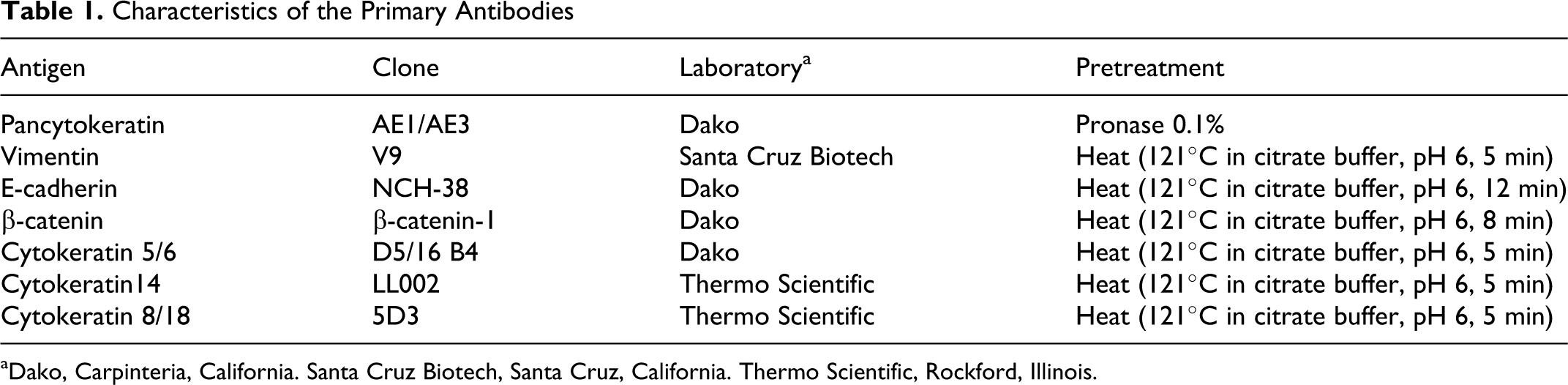
aDako, Carpinteria, California. Santa Cruz Biotech, Santa Cruz, California. Thermo Scientific, Rockford, Illinois.
Quantification of Immunolabeling
E-cadherin and β-catenin immunoreactivity was classified as membranous (localized in the cell–cell boundaries, in cases in which the immunoreactivity was stronger than in the cytoplasm), cytoplasmatic (uniformly distributed through the cytoplasm, with no recognizable distinction between membrane and cytoplasm), or nuclear. Cases were grouped according to Brunetti et al 1 as preserved, when positivity was membranous and occurred in more than 75% of the neoplastic epithelial cells, and reduced in all the remaining samples, including negative tumors. A third variable was obtained by integrating the data from both proteins and so named E-cad/β-cat. The preserved expression of this variable included cases in which both proteins had a preserved status, whereas E-cad/β-cat was classified as reduced when the expression of at least one protein was reduced.
Basal (CK5/6 and CK14) cytokeratin expression was assessed semiquantitatively according to the percentage of immunoreactive cells: negative, <1% of positive cells; mild, 1%–10%; moderate, 10%–50%; and strong >50%. To obtain dichotomic variables to compare with other tumor features and following the definition of “basal-like” proposed by Rakha et al 29 in human breast cancer, carcinomas were classified as low expression (<10%; ie, negative or mild labeling) or high expression (>10%; ie, moderate or strong labeling).
Luminal (CK8/18) cytokeratin was assessed semiquantitatively according to the percentage of immunoreactive cells: negative, <10% of positive cells; mild, 10%–50%; moderate, 50%–75%; and strong, >50%. As with the basal cytokeratin expression assay, dichotomic variables were achieved to compare these with other tumor features, and in accordance with Sarrio et al, 34 carcinomas were classified as low expression (<50%; ie, negative or mild labeling) or high expression (>50%; ie, moderate or strong labeling).
Statistical Analysis
The association between the different markers was examined using the Pearson χ 2 test. For each association, the odds ratio was calculated with a confidence interval of 95%. In the particular case of tumoral grade (1–3), its association with the other markers was examined with a Mann-Whitney U test. Analyses were performed with SPSS 15.0. A P value < .05 was considered to be statistically significant.
Results
Immunohistochemical Analysis of the Adenomas
Immunolabeling with the markers used in the present study showed similar results for the 21 adenomas analyzed. The labeling pattern showed the preserved membranous expression of E-cadherin and β-catenin. Both basal CKs (CK5/6 and CK14) stained basally located cells as well as a limited number of luminal cells, while the luminal CK8/18 stained only luminal cells, predominantly in their apical cytoplasm. Vimentin expression was found in the epithelial cells in 1 of the adenomas studied.
Tumoral Grade
The 139 carcinomas studied were graded using the modified Nottingham method. This revealed 38 grade 1, 65 grade 2, and 36 grade 3 carcinomas. Among those that displayed metastases at diagnosis, 12 of 73 (16.4%) were grade 1, 40 of 73 (54.8%) were grade 2, and 21 of 73 (28.8%) were grade 3 carcinomas. Among those without metastasis, 26 of 66 (39.5%) were grade 1, 25 of 66 (37.8%) were grade 2, and 15 of 66 (22.7%) were grade 3 carcinomas. A statistically significant association (P = .019) was established between the existence of metastasis and grade of the carcinomas.
Immunohistochemical Analysis of E-cadherin and β-catenin Expression in Carcinomas
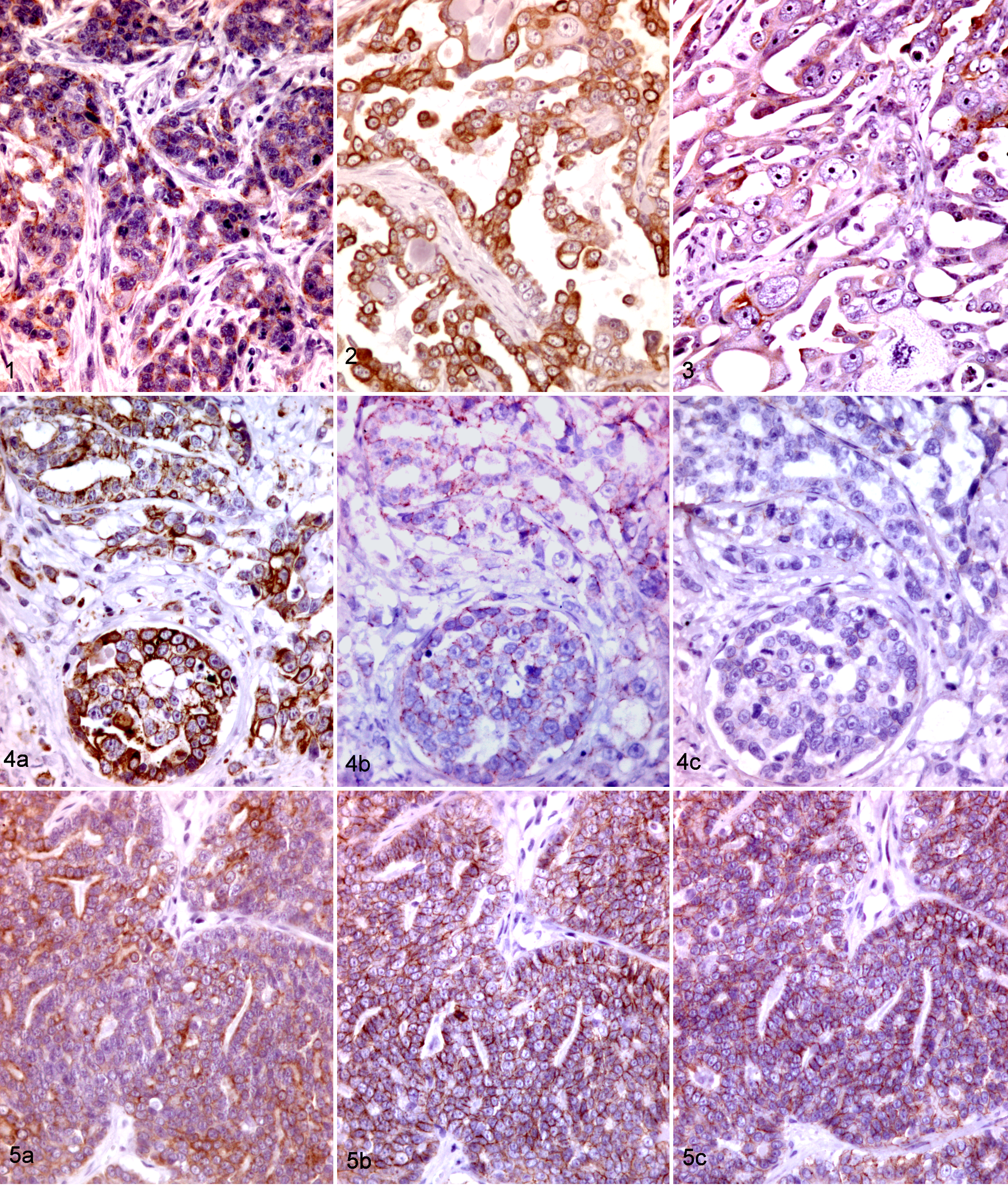
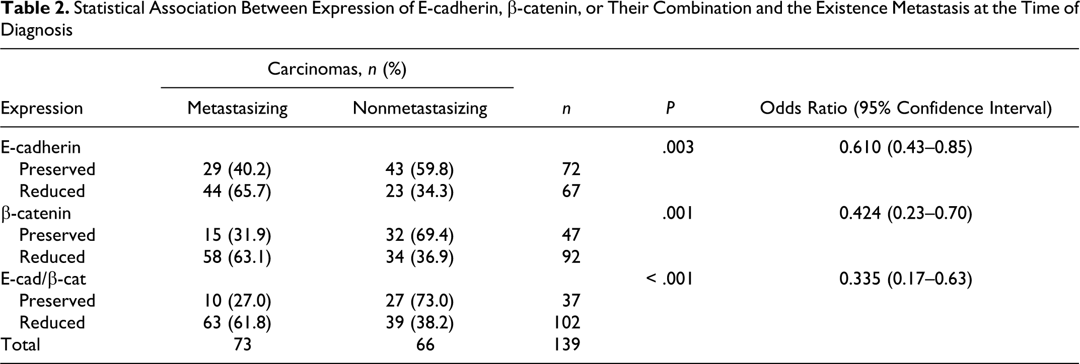
Analysis of the results obtained by labeling both molecules showed that E-cadherin expression was preserved in 51.8% of the carcinomas while β-catenin expression was preserved in 33.8% of the carcinomas. Only 26.6% of the carcinomas preserved the combined expression of E-cad/β-cat. When the data relating to adhesion molecules in the primary tumors were evaluated in tandem with the presence of regional metastasis, a significant association between the existence of metastasis in the carcinomas and the preserved/reduced expression of both molecules could be established using a χ 2 test (Table 2, Fig. 1). Analysis of the odds ratio demonstrated a negative correlation between the expression of E-cadherin, β-catenin, or their combined expression and the existence of metastasis, with this negative association being stronger for the combined expression of E-cad/β-cat (Table 2).
Statistical Association Between Expression of E-cadherin, β-catenin, or Their Combination and the Existence Metastasis at the Time of Diagnosis
Additionally, when data about molecular adhesion expression were related with the grade of carcinomas using the Mann-Whitney U test, a statistically significant correlation was found between the preservation of β-catenin and E-cad/β-cat and tumoral grade (P = .023 and P = .026, respectively). Grade 1 carcinomas displayed significantly higher preserved expression of both β-catenin and E-cad/β-cat than grade 2 and grade 3 carcinomas. No statistically significant association was found between E-cadherin and tumoral grade (P = .243).
Immunohistochemical Analysis of CK8/18, CK14, and CK5/6 Expression in Carcinomas
Analysis of the results obtained for CK8/18 pointed to negative labeling in 11.5% of the carcinomas, mild labeling in 48.2%, moderate labeling in 22.2%, and strong labeling in 18.1%. Using the terms of low and high expression, the percentage of low-expression carcinomas was 59.7% and high-expression carcinomas, 40.3%. When the labeling of basal cytokeratins was analyzed, the results for CK14 were as follows: negative labeling in 7.2% of carcinomas, mild labeling in 15.8%, moderate labeling in 54.7%, and strong labeling in 22.3% (ie, 23% low-expression carcinomas and 77% high-expression carcinomas). Results for CK5/6 were 38.1% for negative labeling, 27.3% for mild labeling, 23.1% for moderate labeling, and 11.5% for strong labeling so that 65.4% of the carcinomas displayed low expression of CK5/6 while 34.6% of the carcinomas displayed high expression.
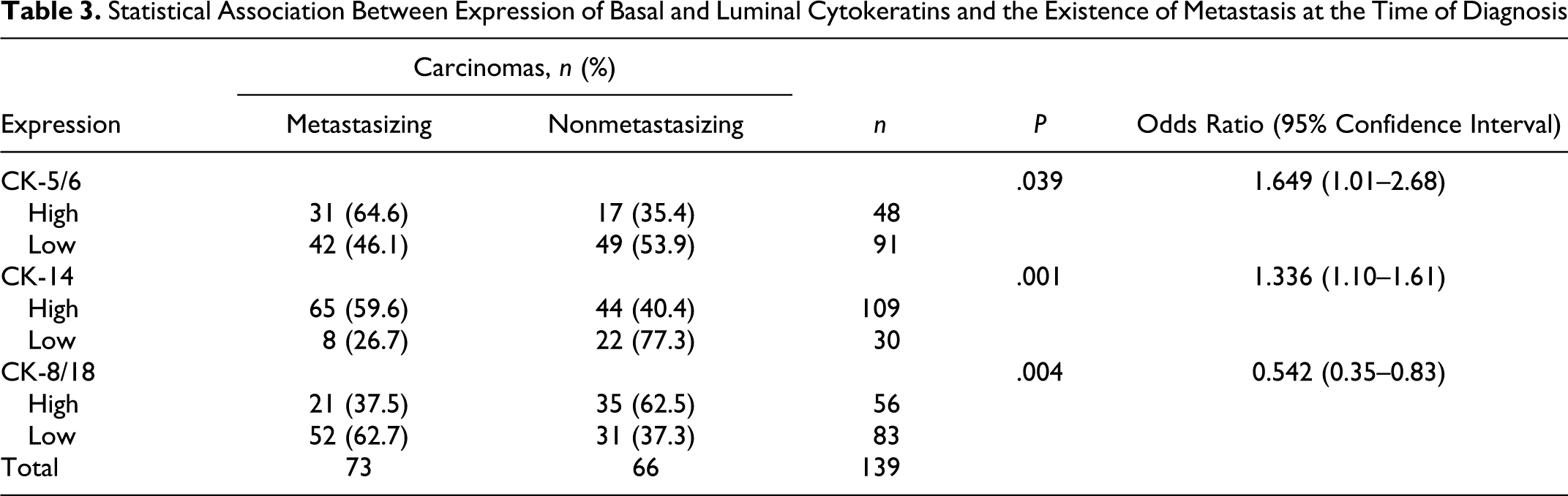
Data for basal cytokeratin displayed a significant association between the existence of metastasis in the carcinomas and the expression of both CK5/6 and CK14 using the χ 2 test (Table 3, Fig. 2). Analysis of the odds ratio demonstrated a positive correlation between the high expression of CK5/6 or CK14 and the existence of metastasis (Table 3). A significant association between the existence of metastasis in the carcinomas and the expression of the CK8/18 luminal cytokeratin was established using a χ 2 test (Table 3, Fig. 3). Analysis of the odds ratio demonstrated a negative correlation between the high expression of CK8/18 and the existence of metastasis (Table 3).
Statistical Association Between Expression of Basal and Luminal Cytokeratins and the Existence of Metastasis at the Time of Diagnosis
Additionally, when data for CKs expression were related with the grade of carcinomas using the Mann-Whitney U test, an association was found between the CK5/6 and CK14 positive labeling and tumoral grade (P = .005 and P = .037, respectively). Grade 1 carcinomas displayed significantly lower labeling of both basal CKs than grade 2 and grade 3 carcinomas. No statistically significant association was found between CK8/18 labeling and tumoral grade (P = .237).
Relationship Between E-cadherin and β-catenin Expression and Basal and Luminal Cytokeratins
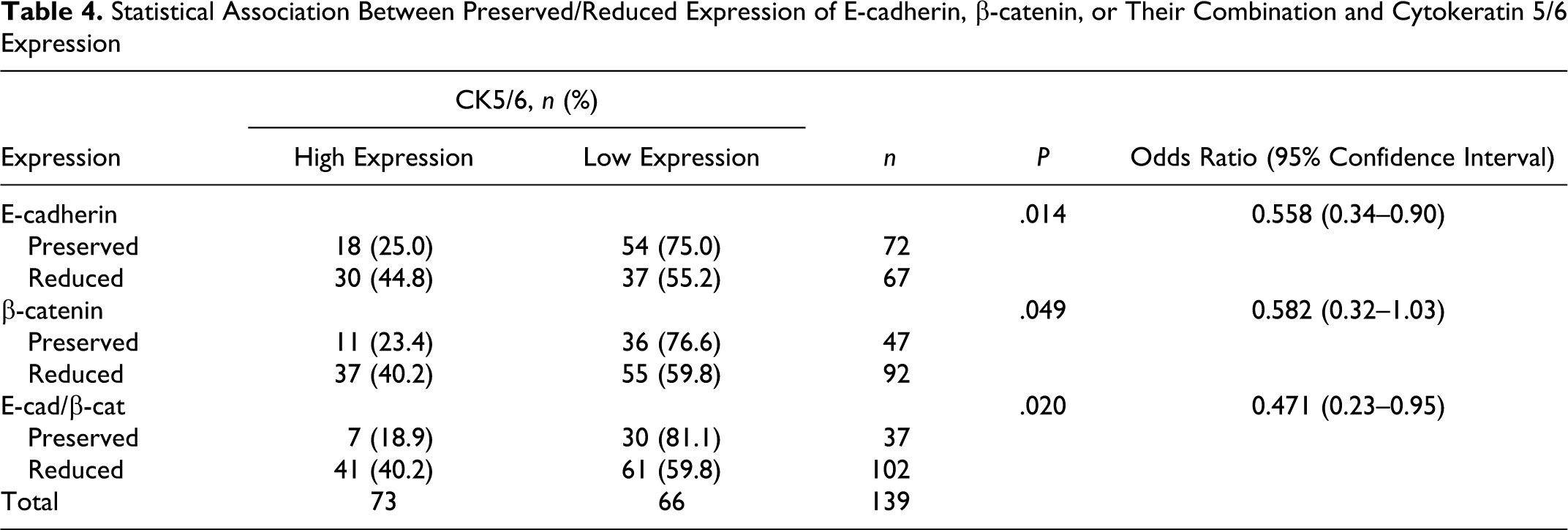
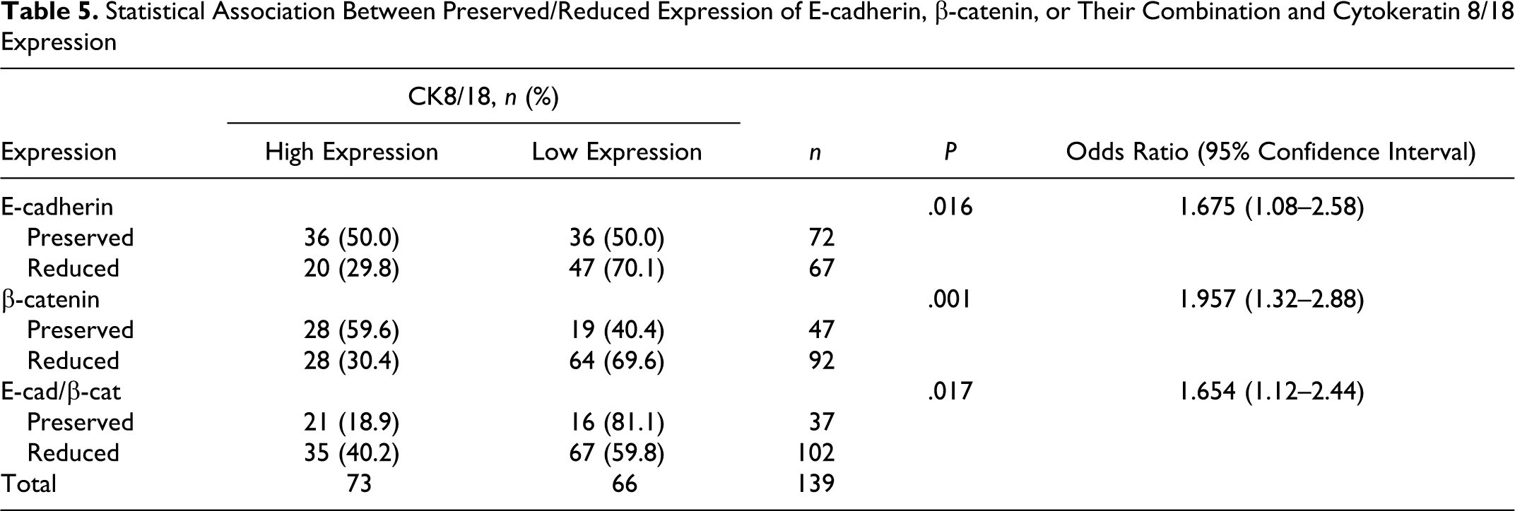
The relationship between the E-cadherin–β-catenin complex and luminal and basal cytokeratin expression was analyzed using the χ 2 test. A significant association was observed between the existence of positive labeling for CK5/6 in the carcinomas and the preserved/reduced expression of both E-cadherin and β-catenin and their combination using a χ 2 test (Table 4, Fig. 4). Analysis of the odds ratio demonstrated a negative correlation between the expression of E-cadherin and the combined expression and the existence of positive labeling for CK5/6 (Table 4). Analysis of labeling for CK8/18 in the carcinomas also demonstrated significant association with the preserved/reduced expression of both E-cadherin and β-catenin and their combination (Table 5, Fig. 5). Analysis of the odds ratio demonstrated a positive correlation between the expression of E-cadherin and β-catenin and the existence of positive labeling for CK8/18 (Table 5).
Statistical Association Between Preserved/Reduced Expression of E-cadherin, β-catenin, or Their Combination and Cytokeratin 5/6 Expression
Statistical Association Between Preserved/Reduced Expression of E-cadherin, β-catenin, or Their Combination and Cytokeratin 8/18 Expression
Immunohistochemical Analysis of Vimentin Expression in Carcinomas
Vimentin expression was found in the epithelial cells of 70.5% of the studied carcinomas (98 of 139). Coexpression of cytokeratin and vimentin could be observed in most of tumors (Fig. 6). No statistically significant association could be established between the expression of vimentin and metastasis or between the expression of vimentin and the other biomarkers analyzed in the present study.
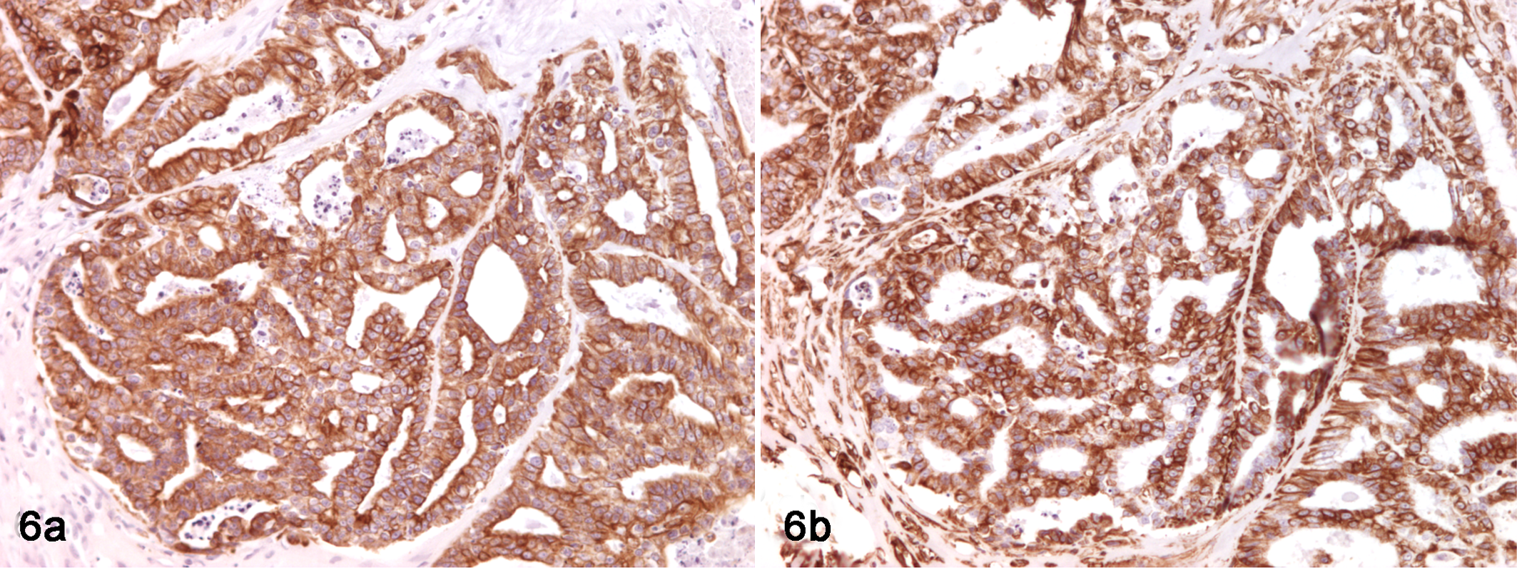
Mammary gland; cat. Presence of coexpression of cytokeratin and vimentin in nonmetastasizing feline carcinomas. (A) Labeling with anti-pancytokeratin antibody. (B) Labeling with anti-vimentin antibody. ABC technique with Mayer's hematoxylin.
Discussion
The present study points to the correlation of 2 groups of biomarkers, E-cadherin–β-catenin complex and basal and luminal cytokeratins, with the presence of regional metastasis, a well-established feature of malignancy in feline mammary carcinomas. Obviously, every carcinoma has the potential to metastasize, and the absence of metastasis described in this study should be understood as a phase in the process of tumor progression, which does not preclude possible future changes in antigen expression or in metastasis status. Notwithstanding, expression of the studied antigens and their association with the presence of metastases provides valuable knowledge for understanding the evolution of mammary neoplasms. Furthermore, expression of the antigens studied in the present work was associated not only with the presence of metastasis at the moment of diagnosis but also with tumoral grade, recently described as an independent prognostic factor associated with overall survival and disease-free survival in feline mammary carcinomas. 36 In fact, an association between the expression of basal cytokeratins and tumoral grade was found in which grade 2 and 3 carcinomas showed higher expression of these cytokeratins. An association was also found between combined E-cadherin and β-catenin preservation and tumoral grade, with grade 1 carcinomas having shown the preservation of both molecules.
The loss of E-cadherin, which permits the detachment of the epithelial tumoral cells and their movement, has been associated with tumor invasiveness and metastasis as part of the EMT process. 39 Several studies of canine mammary tumors have reported that loss of E-cadherin, alone or in combination with loss of β-catenin, is associated with the presence of metastasis or with a shorter overall survival period. 1,5,10 Studies on E-cadherin expression in feline mammary tumors are scarce. An earlier study examining normal mammary tissue, hyperplastic lesions, and a number of neoplasms (4 adenomas and 14 carcinomas) reported that the loss of E-cadherin and abnormalities in the pattern of membranous location were associated with some of the carcinomas but not with adenomas. 6 Another study, using 8 cell lines from feline mammary tumors, showed the reduced expression of E-cadherin in cell lines obtained from a regional lymph node metastasis. 38 Our findings for E-cadherin confirm these previous reports: The highest percentage of reduction was observed in carcinomas with metastasis, while no reduction was observed in adenomas. The present study shows that a high percentage of carcinomas, mainly in those with regional metastasis, had a reduced expression of β-catenin, which suggests the presence of nonfunctional E-cadherin in some of the tumors that preserve the expression of this adhesion molecule. Loss of β-catenin has been described in human invasive lobular carcinomas. 30 Indeed, the association observed between the combined preservation of E-cad/β-cat and metastasis was stronger than in each of the individual biomarkers, suggesting that this variable would be a better prognostic factor that the study of E-cadherin preservation alone.
Basal cytokeratins are high molecular weight cytokeratins associated with the basal layers of the stratified epithelium. In the mammary gland, these cytokeratins are also expressed in the basally located myoepithelial cell layer, in a small proportion of luminal gland cells and in the so-called mammary stem or a progenitor cells. 13 Thus, “basalness” has a dual meaning and could be related with a myoepithelial or progenitor/stem cell origin. The discovery in human breast cancer of in situ carcinomas expressing basal cytokeratins seems to rule out the myoepithelial origin of basal-like tumors. 15 Mammary progenitor or stem cells are also characterized by a low expression of CK8/18. 37 The possible association between EMT and the existence of a basal-like phenotype has recently been demonstrated in a study of human breast cancer, suggesting that EMT may not be a sign of overall tumor dedifferentiation but, rather, the manifestation of a specific phenotype of stem cell origin showing more aggressive behavior. 34 In fact, cells with a basal phenotype probably have a higher proclivity to develop EMT changes and subsequently undergo invasion. This feature has not been studied in feline mammary tumors, and our results point to another interesting similarity between human and feline mammary tumors.
The results related with the immunolabeling of vimentin demonstrated that a high percentage of carcinomas expressed this intermediate filament in contrast with the negative labeling found in most of the adenomas. The percentage of vimentin-positive carcinomas is similar to that found in other studies carried out in feline mammary carcinomas. 18,33 However, no association was found between the expression of vimentin and well-established features of malignancy, such as regional metastasis or tumoral grade, unlike other EMT markers, such as the reduced expression of E-cadherin or β-catenin. The results described in the present study suggest that vimentin expression could be considered as a hallmark of carcinoma but not as a marker of invasiveness. In reports studying human breast cancer, some authors claim that vimentin is one of the first steps in carcinoma progression and that these vimentin positive cells are prone to develop subsequent EMT changes, such as loss of cadherins. 17
In the field of veterinary pathology, the carcinogenesis of canine mammary tumors has been widely studied, 12 and several attempts have been made to establish a model of a molecular-based subgrouping of malignant tumors similar to that developed for human breast cancer. 9,35 To our knowledge, no studies on molecular subgrouping have been performed in feline mammary tumors, although the loss of hormone receptors in tumor progression is well known, 19,21,23 and several reports have evaluated the role of HER-2 overexpression in mammary carcinomas of cats. 22,26,31,41 A more profound study of feline carcinoma molecular subtypes would be valuable, since these carcinomas present a number of similarities with the disease in humans, such as the highly malignant behavior or the almost complete absence of myoepithelial proliferations (a common feature of mammary tumors in dogs, the so-called complex and mixed tumors).
Although this work is not a prognostic study, the relatively high number of cases studied, the findings regarding the association between different markers, and the presence of metastasis or tumoral grade and the similarities found with some aspects of breast cancer in women open an interesting field for future prognosis studies.
Footnotes
Acknowledgement
C. Peñafiel-Verdu is the recipient of a predoctoral grant from the Fundacion Seneca (Regional Govern of Murcia).
Declaration of Conflicting Interests
The authors declared no potential conflicts of interest with respect to the research, authorship, and/or publication of this article.
Funding
The authors received no financial support for the research, authorship, and/or publication of this article.