
Abstract
Intravascular nematodes were considered the cause of death of 14 captive callitrichids. All animals were captive born at zoos in France and died with little or no premonitory signs of disease. No consistent gross lesions were observed at necropsy, although in certain cases intracardiac adult parasites were noted. The most significant histologic findings were verminous pneumonia and pulmonary endarteritis. In all cases except one, intravascular adult nematodes were observed with eggs and larvae in the lungs. Adult nematodes were obtained from 8 animals and in all cases were identified as Parastrongylus dujardini. To the authors’ knowledge, this is the first report of intravascular angiostrongylosis with primary cardiopulmonary location in callitrichids in France.
The Metastrongyloidea superfamily consist of strongylid nematodes grouped according to the site specificity of adults in mammals (primarily in the respiratory system), a reduced copulatory bursa, and heteroxenous life cycles. 1 Most species for which life cycles have been determined use gastropods as intermediate hosts. However, nongastropod intermediate hosts also exist, such as fish and earthworms. Thus, a wide variety of mammalian hosts and life cycle strategies are known among the Metastrongyloidea. 6 Nematodes of the genus Angiostrongylus are those that classically reside in the pulmonary or mesenteric arteries of their hosts. They are mainly parasites of rodents and carnivores. 2,4 Two species are well-known pathogens in humans and nonhuman primates: Angiostrongylus cantonensis causes eosinophilic meningitis or meningoencephalitis, 5,10 and Angiostrongylus costaricensis is responsible for abdominal angiostrongylosis. 3,9,12 –17,19,20 The purpose of this report is to describe gross and histologic lesions and morphologic characteristics of nematodes in 14 callitrichids with cardiopulmonary angiostrongylosis due to Parastrongylus dujardini.
Materials and Methods
All institutions within the Francophone Association of Zoological Park Veterinarians were invited to participate in the study. When available, medical records were reviewed for sex, age, clinical signs, and gross findings. All animals were captive born and housed at 4 geographically distant zoos in France (Réserve zoologique de Calviac and Zoo de la Palmyre in the southwest, Safari de Peaugres in the southeast, and Zoo de Pont Scorff in the northwest). All cases occurred between October 2008 and December 2011. Gross necropsies were performed at the zoos or in neighboring veterinary clinics. Tissue samples from different organs were collected during necropsy and fixed in 10% buffered formalin. Formalin-fixed tissues were routinely processed, embedded in paraffin, sectioned at 4 μm thick, and stained with hematoxylin and eosin. Slugs and rodents found in the enclosure of animals in the different zoos were examined. For detection of nematode larvae, fresh whole slugs were dissected under a stereomicroscope. Over the course of 13 months, 430 rodents were trapped on the premises of one of the zoos (Zoo de la Palmyre), necropsied, examined for the presence of adult worms in the heart, and tested for fecal larval excretion via Baermann’s test. Formalin-fixed samples of rodent lung and heart were submitted for histology when nematodes were visualized. Formalin-fixed and alcohol-fixed samples of adult nematodes collected from 8 primates (Nos. 1, 3, 5, 9, 10, 12–14), and 14 rodents were sent to the University of Reims for identification. The anterior and posterior aspects of the worm (especially copulatory bursa from males) was cleared and preserved in Amman lactophenol between slide and cover slide and examined by light microscopy. Morphologic features were compared with those proposed in keys from the review by Ubelaker. 21
In groups where cases were identified, remaining callitrichids were screened for fecal larval excretion via Baermann’s test at each zoo.
Results
Among the 14 callitrichids included in the study were 4 cotton-top tamarins (Saguinus oedipus), 5 Goeldi’s tamarins (Callimico goeldii), 1 white-lipped tamarin (Saguinus labiatus), 3 white-headed marmosets (Callithrix geoffroyi), and 1 pygmy marmoset (Callithrix pygmaea). The average age was 2.7 years (range 3 months–8 years). The sex distribution was 70% female and 30% male (see Supplementary Table 1, available online at http://vet.sagepub.com/supplemental). In most cases, animals died spontaneously with no premonitory signs of disease. Lethargy was reported in 3 animals 12 to 24 hours before death (Nos. 9, 12, 13), and episodes of blepharedema were noted in 2 individuals (Nos. 9 and 10).
Gross Lesions
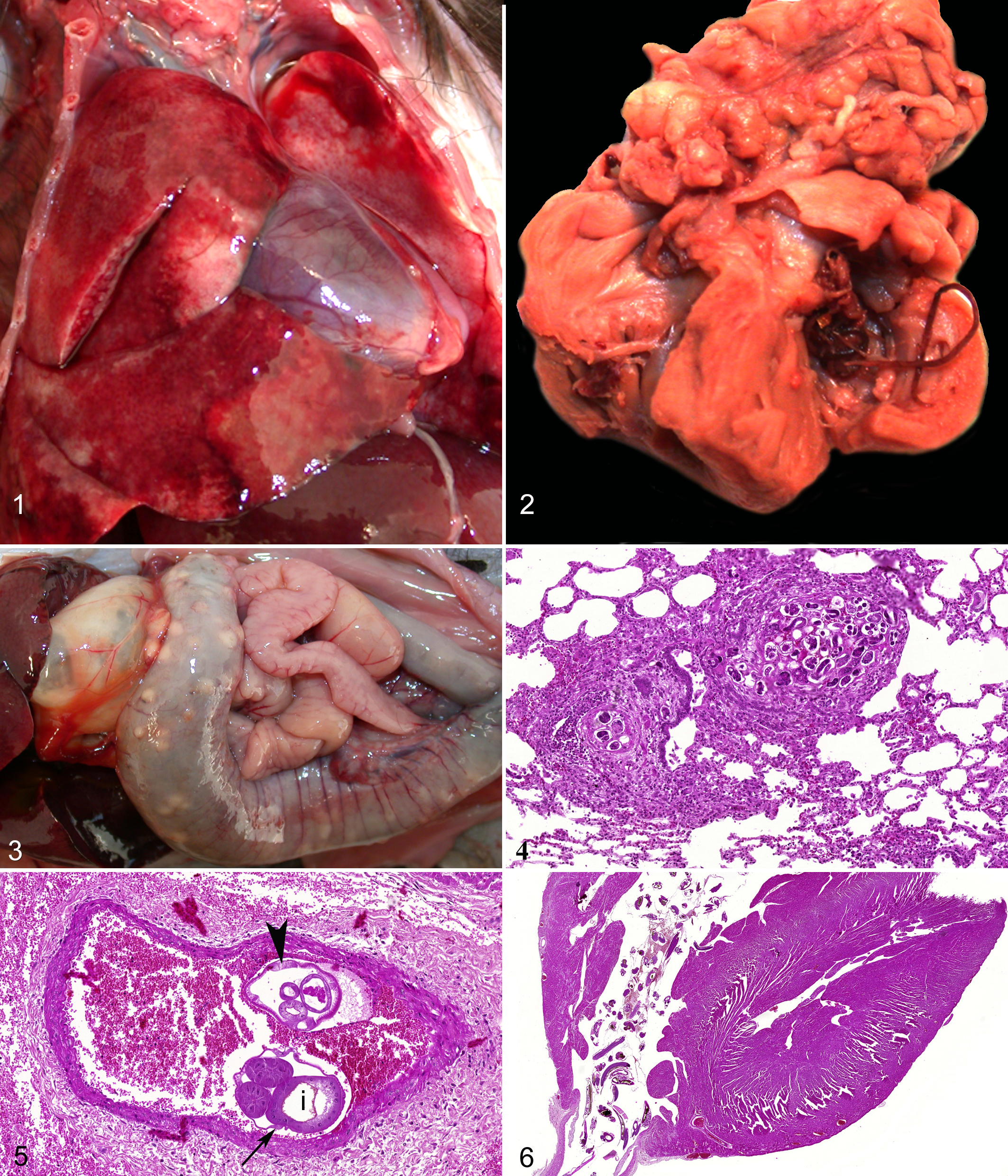
Gross lesions are summarized in Supplementary Table 2. The most prominent finding was moderate to severe congestion of the lungs (Fig. 1). In 5 cases, adult nematodes were noted in the right ventricle of the heart (Fig. 2). These were associated with right cardiomegaly. In a single case, numerous 5- to 10-mm-diameter firm white nodules were noted in the wall of the large intestine (Fig. 3).
Histology
Histologic lesions are summarized in Supplementary Table 3. All animals except 1 had verminous pneumonia and pulmonary endarteritis. The pulmonary parenchyma contained multifocal areas of granulomatous inflammation composed of macrophages, multinucleated giant cells of foreign body type, fibroblasts, lymphocytes, and plasma cells arranged concentrically around eggs and larvae (Fig. 4). Very few eosinophils were noted. Larvae were elongate with a thin eosinophilic cuticle and a primitive 10- to 20-μm-diameter enteron. Ova were 50 to 80 μm in diameter, ovoid, and thin shelled and contained basophilic and eosinophilic granules or a coiled larva. Lumens of some pulmonary arteries contained 150- to 250-μm-diameter adult nematodes (Fig. 5) with a thin outer smooth cuticle, a thin hypodermis with lateral cords, and a coelomyarian-polymyarian musculature surrounding the pseudocoelom containing digestive and reproductive tracts. The intestine was large and composed of few multinucleated cells (syncytous, oligocytous intestine). Intravascular nematodes were often surrounded by thrombi, which were partially adhered to the vessel wall. These thrombi were composed of fibrin or fibrous connective tissue containing multiple small blood-filled channels (organized and recanalized thrombi). Numerous adult nematodes in the right ventricular lumen were another hallmark lesion (Fig. 6). A single evidence of mural vasculitis in the colon with intralesional nematode eggs and larvae was noted. Adult nematodes were not observed in this lesion.
Parasitology
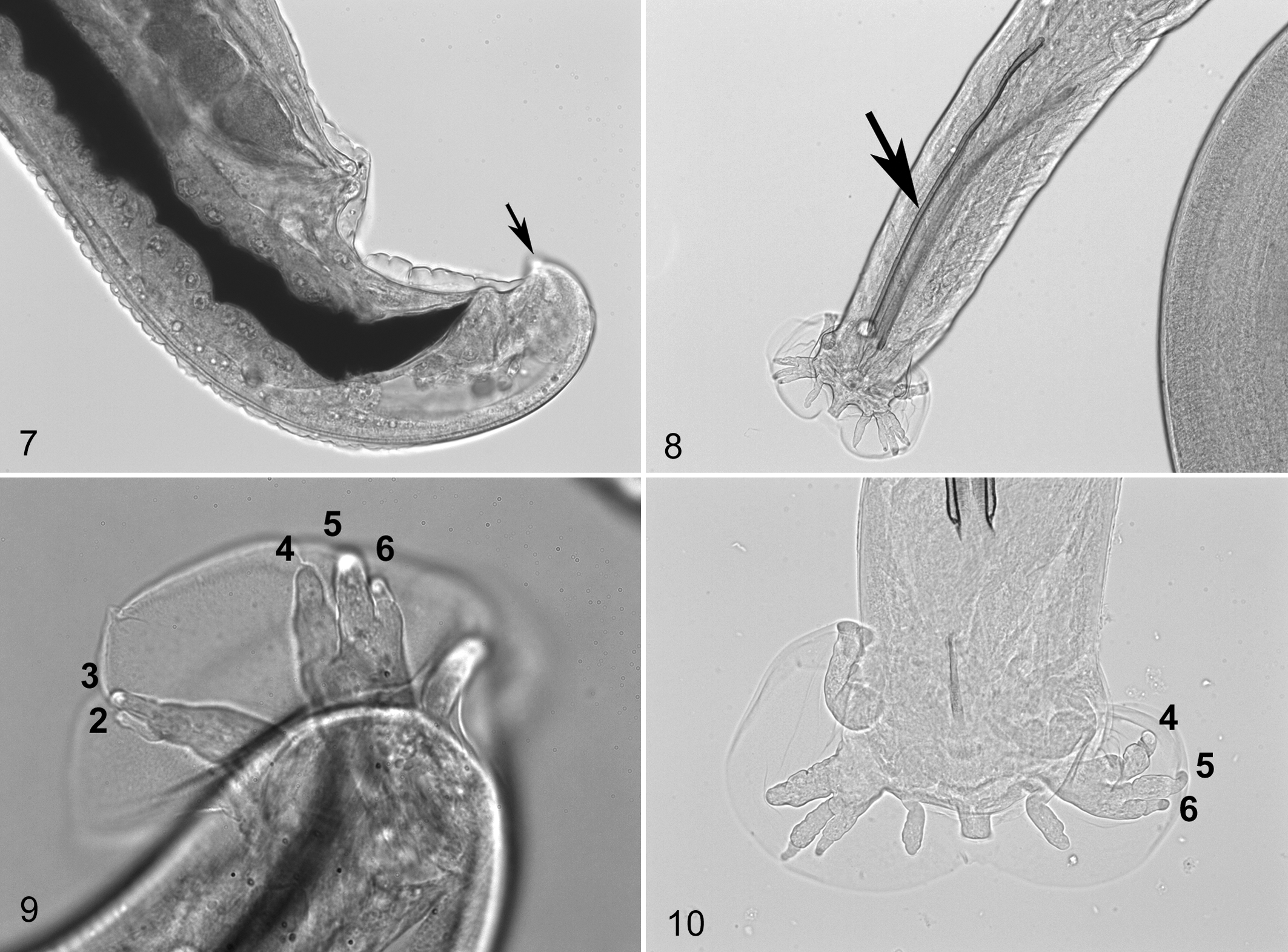
Parasites from cases Nos. 1, 3, 5, 9, 10, and 12–14 were examined. All female and male parasites were identical according to observed morphologic features. Males and females were determined to belong to the subfamily of Angiostrongylinae. 1 The papilla on the extremity of tail of the female is a common feature of the genus Parastrongylus (Fig. 7). The specific identification was based on features of the copulatory bursa from male specimens. All lateral rays of the copulatory bursa arose from a long common trunk, compatible with Parastrongylus and distinct from closely related Angiostrongylus. 21 The length of the spicules (Fig. 8) (351–430 μm) and the longer length of the mediolateral ray in comparison with the other lateral rays (externo and postero lateral rays) (Fig. 9) were consistent with Parastrongylus dujardini. 7,13 –15
Dissection of slugs has not revealed any larvae to date. Baermann’s tests on callitrichids were all negative, including animals that died of verminous pneumonia.
Adult nematodes were observed in 12 wood mice (Apodemus sylvaticus) and 2 voles (Microtus agrestis). Among the wood mice, 2 were not trapped but were found dead with verminous pneumonia similar to the callitrichids. Histologic changes were identical to those reported in the primates of our study. Nematodes from rodents were also identified as P. dujardini (Fig. 10). Furthermore Baermann’s testing allowed for identification of L1 larvae in the feces of the 14 rodents.
Discussion
In all cases described in this report, the gross, microscopic, and parasitologic findings indicated that infection by P. dujardini was the cause of death. All animals except 1 had similar histologic changes of pneumonia due to larvae and eggs and pulmonary proliferative arterial endarteritis and thrombosis in response to adult worms. Intestinal lesions were observed only in 1 case (No. 9). Adult nematodes were never observed in the mesenteric arteries.
Morphologic characteristics of nematodes found in primates and in rodents in our study are those reported for P. dujardini. 8 This is the first report of this parasite in primates.
P. dujardini was first described in rodents of the species Clethrionomys glareolus and A. sylvaticus in southwest France. At one facility, wood mice (A. sylvaticus) and voles (M. agrestis) from the enclosures housing callitrichids were confirmed infected and shedding L1 larvae and are therefore considered definitive hosts.
The lack of L1 larvae identified on Baermann’s tests in primates supports their status as accidental hosts, as described in humans. 19 This suggests that the primates became infected by contact with the local fauna.
Several intermediate hosts have been identified for P. dujardini, 8 but this aspect of the life cycle was not elucidated in our study and requires further investigation. Nevertheless, infection of primates by ingestion of infected intermediate hosts or consumption of feed contaminated with excreta from gastropods remains plausible. Remote possibility for a direct cycle exists. Transmission through nursing could also be possible given the occurrence of the disease in very young individuals (Nos. 3 and 14).
Cases of angiostronglylosis in humans and nonhuman primates are distinct from those described here. Most reported cases are of mesenteric 3,17,20 or cerebral 5,10 angiostrongylosis due to A. costaricensis and A. cantonensis, respectively.
A single case in our study had colonic mural vasculitis similar to lesions described in mesenteric angiostrongylosis due to A. costaricensis. 3,17,20 The pathogenesis of this lesion could not be elucidated in the absence of visualization of adult nematodes in the mesenteric arteries, but these may have been overlooked at necropsy.
One previously reported case of pulmonary angiostrongylosis in a cotton-top tamarin (S. oedipus) 11 seemed closest to the case series described here, but intracardiac nematodes were identified as Angiostrongylus vasorum. Pulmonary angiostrongylosis due to A. vasorum is the disease most closely resembling our cases. 2 It has been described in several exotic species in Europe. 18 However, nematodes were ruled as morphologically distinct from A. vasorum.
Death of animals in our study was suspected to be a combination of right heart failure and verminous pneumonia of variable severity in most cases. Heart failure was attributed to pulmonary endoarteritis and/or presence of adult nematodes in the right ventricle leading to secondary cardiomegaly. The absence of verminous pneumonia in case No. 14 could be explained by the lack of representative lesion in submitted pulmonary formalin-fixed samples or could represent an early stage of the infection with premature death of unknown origin.
Preventive treatment with ivermectin and albendazole have been attempted, but it is too early to make any meaningful conclusions on the efficacy of the prophylactic treatment. Also subject to further investigation are the possible factors that lead to the clustering of these cases over a short period, as no specific factor seemed present at the time.
As this new form of angiostrongylosis represents a significant health risk for New World primates in a zoological setting and could be a public health concern, further investigations are needed to understand the biological life cycle (source of contamination, intermediate hosts) and migratory pathway in callitrichids.
Footnotes
Acknowledgements
We wish to acknowledge Drs Nicolier, Rietschel, Caviccio, Wenker, Hoby, and Sasseville for their assistance and insight on these cases and Nicolas Gadot and Armelle Paquet for preparation of histologic slides.
Declaration of Conflicting Interests
The author(s) declared no potential conflicts of interest with respect to the research, authorship, and/or publication of this article.
Funding
The author(s) received no financial support for the research, authorship, and/or publication of this article.
References
Supplementary Material
Please find the following supplemental material available below.
For Open Access articles published under a Creative Commons License, all supplemental material carries the same license as the article it is associated with.
For non-Open Access articles published, all supplemental material carries a non-exclusive license, and permission requests for re-use of supplemental material or any part of supplemental material shall be sent directly to the copyright owner as specified in the copyright notice associated with the article.