
Abstract
Wounds were created by incision in skeletal muscle of 2 mixed-breed canine cadavers at multiple time points from 0.5 to 74.5 hours postmortem and were exposed to artificial seawater (35 parts per thousand), 0.9% saline (8 parts per thousand), or freshwater for 24 hours before fixation for histology. Discoid and segmental disintegration of myofibers deep to the severed edges was observed in injuries inflicted within 6.5 hours of death and exposed to 0.9% saline and seawater and was not observed in injuries made at later time points or in other treatments. Exposure to artificial seawater had pronounced effects on histomorphology that markedly diminished with increasing postmortem wounding interval. In a third cadaver, these changes were shown to be detectable with confidence following up to 10 days of submergence in seawater at 22.2°C despite decomposition. These findings are important for evaluation of skeletal muscle injuries that are exposed to seawater, such as those occurring in marine animals, and may assist in recognizing wounds inflicted either antemortem or within the supravital period.
Determining whether traumatic injuries occurred before or after death can be one of the most difficult challenges faced by pathologists. Some physiologic processes and morphologic changes occur after circulatory arrest and irreversible tissue injury within what is referred to as the surpravital period. 3 Mechanical and electrical excitability of skeletal muscle is an example of supravital reaction, and familiarity with these phenomena and the attendant histologic changes is critical to evaluation of traumatic injuries.
Mechanical and electrical stimulation of skeletal muscle has been used to estimate time since death. 2,8 Response to mechanical stimulation declines rapidly during the postmortem interval but may be elicited for up to 13 hours postmortem (HPM). 8 Study of histologic changes resulting from this type of stimulation has shown that structural changes in myofibers previously interpreted as antemortem also occur within the supravital period. 1,7 Although these microscopic changes cannot be used to determine whether trauma occurred antemortem, their presence can help define the timing of injuries. This information can be useful in evaluation of wounds in some circumstances and when considered with concurrent findings.
The impetus for the current study was application in evaluation of traumatic injuries in marine mammals and sea turtles based on observations in forensic cases; however, basic principles and relevance may be more broadly applicable. Injuries in marine animals are, by nature, often immersed in seawater for prolonged periods and frequently exhibit various degrees of decomposition. Duration of survival following injury may be insufficient for development of detectable inflammatory response. Additionally, immersion in water often washes away hemorrhage and leaches blood from wound margins. 4 Decomposition and current lack of validated methodology for various animal taxa limit forensic application of ancillary methods, such as immunohistochemistry; thus, assessment of morphologic changes is paramount.
There have been relatively few experimental studies on supravital structural changes in traumatized muscle. 1,7 To our knowledge, the effects of seawater on wound histomorphology have not been described. In marine animals, we have frequently observed very prominent, often widespread disruption of the sarcoplasm of myofibers within the margins of wounds known or suspected to have occurred antemortem and that were naturally immersed in seawater under field conditions. Similar changes have not been seen in clearly postmortem injuries caused by scavengers. We hypothesize that seawater exposure enhances structural changes in damaged skeletal muscle in wounds that are inflicted antemortem or within the supravital period. The objective of this study was to test this hypothesis using canine cadavers by creating incisions in muscles at different time points following cardiac arrest and exposing muscle to aqueous solutions of different salinities. As a related objective, the histologic detectability of muscle changes during decomposition while submerged in seawater was also examined.
Materials and Methods
Animal protocols were approved by the University of Florida Institutional Animal Care and Use Committee (study No. 201207479). Three similarly muscled mixed-breed canine cadavers were obtained from a local animal shelter after being euthanized for animal control purposes. Intravenous pentobarbital sodium solution (Fatal-Plus Solution, Vortech Pharmaceuticals Ltd, Michigan, USA) was administered (approximately 1 ml per 4.5 kg), and time of cardiac arrest was documented. The cadavers were kept at room temperature (22.2°C) throughout the entire study. Rigor mortis was evaluated by gentle manipulation of the limbs and jaw.
In 2 cadavers (Nos. 1 and 2), postmortem incisions were created at multiple time points with a surgical scalpel to transversely transect major muscle bellies of the proximal and distal limbs perpendicular to the predominant orientation of muscle fibers. The initial incisions were made at 0.5 HPM, before the onset of rigor mortis. All incision time points relative to rigor mortis are given in Table 1. Only 1 incision was created per muscle or individual muscle head to eliminate interactions between neighboring incisions, and all incisions were made in the middle of muscle bellies. Two muscles, one larger muscle of the proximal limb and a smaller distal muscle, were sampled for each time point through full rigor to consider effects of muscle size on timing and extent of myofiber changes. Sampling continued through complete dissipation of rigor and onset of putrefaction, as characterized by abdominal bloating and generalized gas formation within soft tissues. Quadruplicate samples were collected from each postmortem incision. Each sample was approximately 1.0 cm wide, 2.0 cm deep, and 2.0 to 2.5 cm in length from the incised wound margin. Efforts were made to have associated connective tissue elements, such as the epimysium, consistent among replicates to the degree possible. The superficial aspect of most samples included the epimysium. Replicate samples from each wound received 1 of the following 4 treatments: immediate fixation in 10% neutral phosphate buffered formalin (NBF), submersion in freshwater for 24 hours, submersion in 0.9% saline (8 parts per thousand [ppt]) for 24 hours, and submersion in artificial seawater (35 ppt) for 24 hours. Artificial seawater was prepared with Red Sea Salt (Red Sea USA, Houston, TX). All solutions were kept at 22.2°C. Samples were submerged in 120-ml plastic specimen cups at a depth of approximately 4.5 cm. Following the freshwater, saline, or seawater submersion treatments, muscle samples were transferred into NBF.
Timing of Postmortem Wounds for Cadaver Nos. 1 and 2 Relative to Progression and Dissipation of Rigor Mortis.a
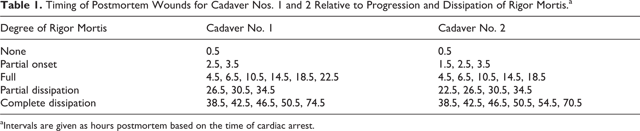
aIntervals are given as hours postmortem based on the time of cardiac arrest.
Cadaver No. 3 was used to study detectability of myofiber changes during decomposition of wounds submerged in seawater. One set of 14 wounds was created at 0.5 HPM (before onset of rigor mortis) and a second set at 6.5 HPM. The latter time point was selected according to the results from cadaver Nos. 1 and 2 as a time when rigor mortis was well developed and supravital function was expected to be greatly diminished or absent. Duplicate muscle samples from each incision were immediately placed into artificial seawater (35 ppt) kept at 2 different temperatures, 22.2°C and 4.4°C, to produce 2 rates of decomposition. A set of samples from each temperature group and time point was randomly selected and transferred into NBF every 24 hours for 14 days.
Muscle samples from all cadavers were sectioned along the longitudinal axis of the fascicles. Only longitudinal sections were examined, because the primary interest was lengthwise continuity of myofiber sarcoplasm relative to the severed ends. Tissues were processed into paraffin, and sections were stained with hematoxylin and eosin through routine methods. Muscle sections from cadaver Nos. 1 and 2 were reviewed (nonblinded) to examine histologic changes in series across the different treatments and time points. The degree of discoid and segmental disruption of sarcoplasm deep to the incised wound margin was given a semiquantitative score from 0 to 5 based on the relative degree and maximum depth of histologic changes (Table 2). Maximum depth was rounded to nearest 100 μm. Slides of experimental wounds from cadaver No. 3 were evaluated by blind histologic review for presence or absence of sarcoplasmic disintegration.
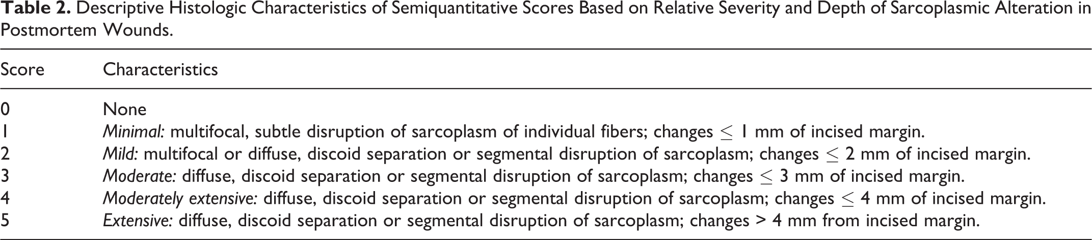
Descriptive Histologic Characteristics of Semiquantitative Scores Based on Relative Severity and Depth of Sarcoplasmic Alteration in Postmortem Wounds.
Results
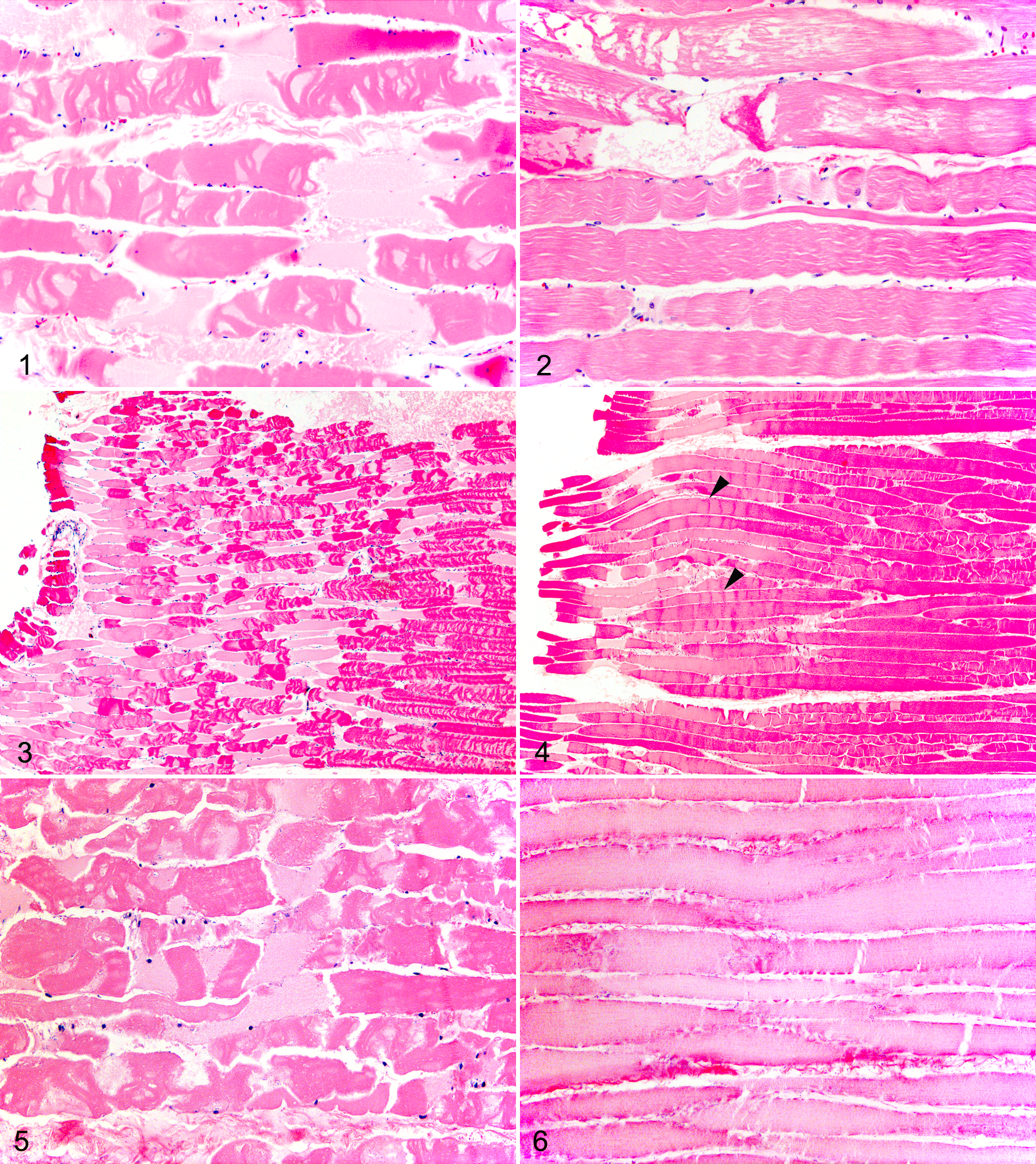
Onset of rigor mortis occurred within 2.5 HPM in cadaver Nos. 1 and 2 and was well developed within 6.5 HPM. Rigor had significantly dissipated by 26.5 HPM. Strong muscle contraction occurred upon incision at 0.5 HPM and was unapparent by 2.5 HPM. Formation of a gap between severed margins was evident in wounds created before development of full rigor. Histologic changes in postmortem incisions in both cadavers and in duplicate samples from muscles of proximal and distal limbs were very similar. Visualization of the wound margins and orientation of continuous myofibers were inadequate for consistent review of some muscle sections for some time points, especially smaller distal muscles; thus, a combined score was given for each time point and immersion treatment based on all examinable sections (Table 3). Variation in myofiber diameter, visibility of cross striations, formation of contraction bands, and presence of wavy fibers were highly variable across treatments and time points. In all treatments, there was bulging and cupping of the severed ends of at least some myofibers, most apparent at time points through 6.5 HPM. In muscle exposed to 0.9% saline and seawater, there was disruption of myofiber continuity deep to the incisions, which was characterized by loss of cross striations, multiple repeated partial and complete separations of short disclike segments of sarcoplasm, and longer segmental separations of sarcoplasm (Figs. 1, 2). These changes were not observed in muscle exposed to freshwater or immediately fixed in formalin. The extent of myofiber disruption was substantially greater in muscle exposed to artificial seawater as compared to saline (Table 3) (Figs. 1, 2). In the saline treatment group, myofiber changes were more superficial, subtle, and patchy, whereas disruption was widespread and extended deeper in muscle exposed to seawater. In addition, the extent of distribution and depth of myofiber disruption in both saline and seawater treatments generally declined with increasing postmortem wounding interval (Table 3). This decrease was remarkable in muscle exposed to seawater where myofiber changes within wounds created at 0.5 HPM were dramatically more extensive than those observed at later wounding intervals (Figs. 3, 4). These myofiber changes were not observed after 4.5 HPM in cadaver No. 1 and 6.5 HPM in cadaver No. 2 (Table 3). The degree of alteration observed at 6.5 HPM in cadaver No. 2 was minimal and limited to a small area within the peripheral margin of the sampled muscle. In both cadavers, these time points coincided with well-developed rigor mortis.
Degreea and Maximum Depthb of Sarcoplasmic Disruption of Myofibers in Wounds Created at Different Postmortem Intervals and Exposed to Aqueous Solutions of Different Salinities for 24 Hours Before Fixation.
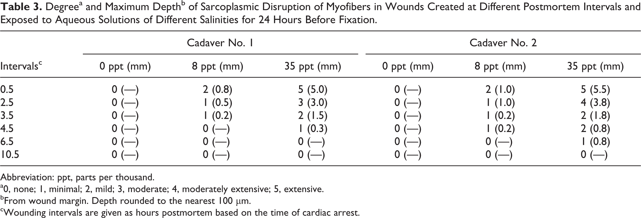
Abbreviation: ppt, parts per thousand.
a0, none; 1, minimal; 2, mild; 3, moderate; 4, moderately extensive; 5, extensive.
bFrom wound margin. Depth rounded to the nearest 100 µm.
cWounding intervals are given as hours postmortem based on the time of cardiac arrest.
Findings were similar in cadaver No. 3. Discoid and segmental disruption of myofiber continuity deep to the incised margin was detected by blind review in all incisions created at 0.5 HPM and was not found in any created at 6.5 HPM. These myofiber changes were observed in 0.5-HPM incisions in all submergence temperatures and time points up to 14 days postmortem (Figs. 5, 6), despite histologic changes resulting from autolysis and putrefaction, including loss of cellular detail, microcavitation, and bacterial overgrowth. After 10 days, however, decomposition of samples kept at 22.2°C substantially diminished the degree of confidence in evaluation. Myofibers began to exhibit artifactual fragmentation that was difficult to distinguish from sarcoplasmic changes observed in the early postmortem interval. In addition, muscle from incisions made at 0.5 HPM and kept at 22.2°C had sarcoplasmic disruption (scores 4 to 5), similar to cadavers Nos. 1 and 2, whereas myofiber changes within 0.5-HPM wounds kept at 4.4°C were diffuse but milder and most prominent within 2 mm of the margin (scores 2 to 3). As expected, the state of preservation was much better in samples maintained at 4.4°C.
Discussion
The structural changes observed in incisions created during the early postmortem interval and exposed to 0.9% saline and seawater are similar to the processes of discoid and segmental disintegration described as supravital changes in human cadaver studies. 1 Respective histologic descriptions of these terms include “disc-like separation of sarcoplasm and interruption of myofiber continuity at short distances” and “multiple interruptions of fiber continuity with formation of separate groups of sarcoplasm.” 1 Discoid and segmental disintegration have been demonstrated by histology in percussed muscle for up to 8.5 HPM but not in human cadavers tested from 10 to 60 HPM. 1,7 As occurred in the current canine study, the degree of myofiber disintegration in human cadavers decreases with increasing postmortem interval and is theorized to be directly related to contractile potential. 1
There is little consideration of supravital phenomena in the veterinary literature beyond the general principle that contraction of muscle occurs postmortem and may be seen by histology. 6 Although the supravital period is considered more extensively in the human forensic literature, relatively few studies include correlative histology. 1,7 Supravital myofiber changes and their similarity to necrosis present questions with respect to correct terminology in cases where timing of injury relative to death is unknown. A diagnosis of muscle necrosis could erroneously imply that injuries occurred antemortem. The term disintegration is not a commonly used morphologic diagnosis in veterinary pathology, but we suggest that it is the most correct term given precedence for use in characterizing supravital myofiber changes in the human literature.
The effect of seawater on the histologic changes of muscle traumatized within the early postmortem interval was pronounced. Disintegration observed in wounds inflicted at 0.5 HPM was comparable to our observations in antemortem injuries in marine mammals and sea turtles (Fig. 7); however, changes are often much more extensive in known antemortem wounds. A similar but much reduced effect in muscle exposed to saline suggests that the mechanism is osmotic. The reduction in these changes over time and the absence in wounds inflicted after 6.5 HPM indicate a relationship between environmental conditions and supravital processes, including contractile potential, rather than mere histologic artifact resulting from exposure to a saltwater. Although myofiber disintegration alone cannot be considered proof of antemortem injury, presence or absence can have substantial value when assessment of a wound is limited by decomposition, damage by scavengers, and other factors, as is often the case in marine wildlife. The ability to estimate the timing of injuries to within antemortem or supravital periods can be very informative in some scenarios, such as application in support of other evidence or in recognition of postmortem injuries inflicted after the supravital period.
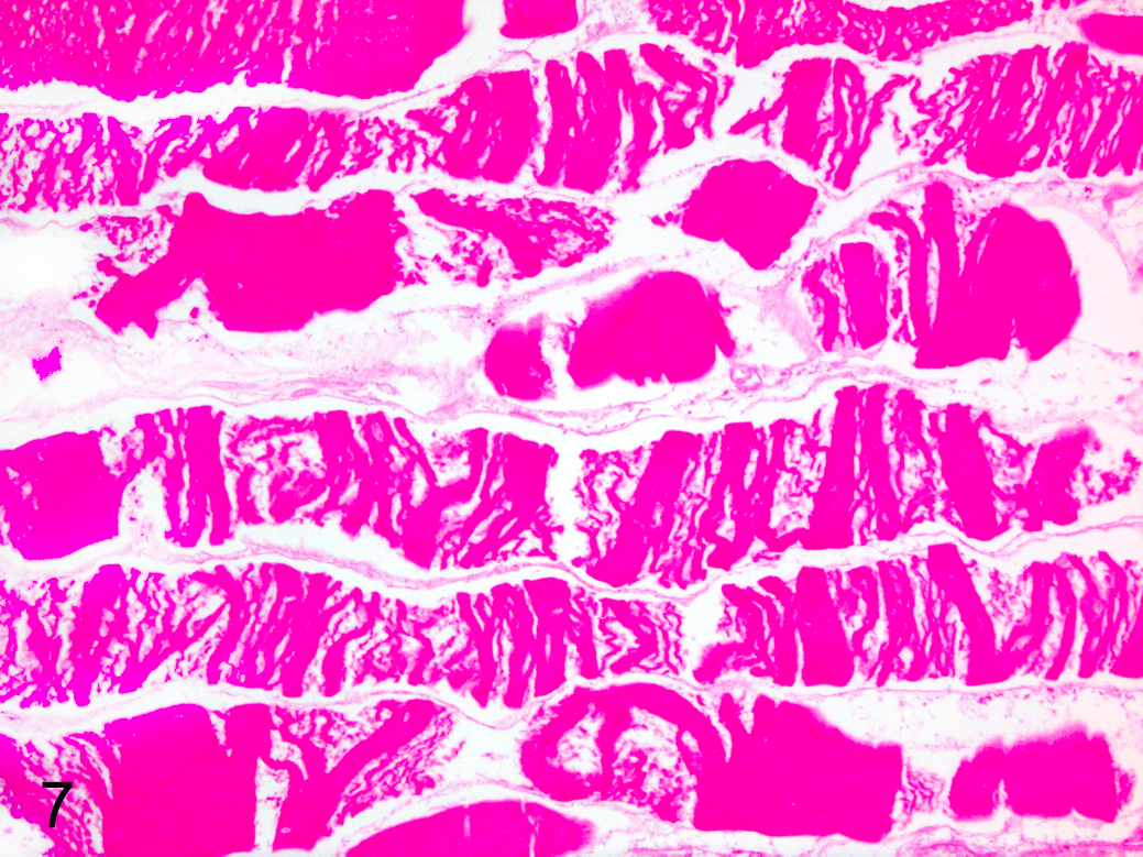
Skeletal muscle; green sea turtle. Muscle composing the margin of an antemortem chop wound caused by a watercraft propeller. The wound was immersed in seawater for an unknown period before recovery of the moderately decomposed carcass. There is discoid and segmental disintegration of myofibers as well as loss of cellular detail from autolysis. HE.
In cadaver No. 3, incisions inflicted at 0.5 HPM were consistently distinguished from those inflicted at 6.5 HPM based on the presence of discoid and segmental disintegration. Potential-reduced detectability in wound margin samples maintained at 4.4°C warrants additional study and could be relevant to the examination of wounds exposed to colder conditions, either in the natural environment or during preservation. The cold-exposure treatment was primarily intended to examine detection of structural muscle changes at 2 different decomposition rates and was not a variable studied in the initial 2 cadavers. Although rapid cooling of the wound margin samples, as done in the current study, may somewhat replicate exposure of injuries to colder environmental conditions, taxon-specific characteristics, such as cooling rates and internal temperature differentials, may be important variables. Other important factors of interest in future studies include the potential differences in different wound types (eg, sharp trauma, chop wounds, blunt trauma), as a well as factors that affect the onset of rigor mortis, such as muscle type, myoglobin content, nutritional condition, and state of exertion.
Acknowledged potential limitations of this study include low sample size and euthanasia by barbiturate overdose. The number of cadavers was considered adequate to fulfill the study objectives given the conserved nature of the processes studied, as demonstrated by considerable similarity of results among replicate samples from all cadavers. The barbiturate could have had some degree of a pharmaceutical effect on muscle response to injury, as pentobarbital is a relaxant at the administered dose. 5 Nonetheless, contraction and fasciculation produced upon incision were visibly strong in all canine cadavers within the early postmortem interval and grossly similar to that observed in animals that die by causes other than chemical euthanasia.
This study contributes to the understanding of supravital phenomena in injured skeletal muscle and the effects of environmental conditions on histomorphology of external wounds. Submergence in seawater markedly enhances histologic alteration of skeletal muscle that is injured antemortem or within the supravital period, and these changes are detectable despite advanced decomposition. Further consideration of these findings in actual case studies is necessary to understand the forensic applications and potential interpretative value in the evaluation of traumatic injuries.
Footnotes
Acknowledgements
We thank Dr Jon Thogmartin for his contributions to conceptual design, Dr Jeffrey Abbott for manuscript review, and the histology laboratory at North Carolina State University for processing of histology samples.
Declaration of Conflicting Interests
The author(s) declared no potential conflicts of interest with respect to the research, authorship, and/or publication of this article.
Funding
The author(s) received no financial support for the research, authorship, and/or publication of this article.