
Abstract
Intestinal adenocarcinomas were identified in 76 adult deer from a closed herd of 193 breeding animals grazing pasture heavily infested with bracken fern (Pteridium aquilinum). Tumors were observed postmortem in 32 animals with rapid weight loss, and similar neoplasms were detected in a further 44 clinically normal deer at “cull.” Tumors were located in distal ileum, cecum, and proximal colon and presented as single (26%) or multiple (74%), variably sized, pale-gray, firm, poorly circumscribed neoplasms with associated intestinal strictures. Histopathologically tumors were well-differentiated, locally infiltrative, low-grade adenocarcinomas of tubular (51%), mucinous (33.5%), or mixed (15.5%) types. Extraintestinal metastases were not observed. The high incidence of intestinal adenocarcinoma within this herd suggests a specific and novel syndrome, and genetic and/or environmental factors may be involved in the pathogenesis.
Intestinal adenocarcinomas are rare in domestic animals. 16 Surveys based on necropsies have reported prevalences of 0.7% to 1.1% in cats and dogs. 38,51 In cattle and horses, 0.5% to 1.0% of neoplasms are intestinal adenocarcinomas. 3,50 Sheep have the highest prevalence of intestinal adenocarcinomas, the prevalence of which increases with age but varies with geographical region. 14,30,32,52 Intestinal adenocarcinomas in domestic animals mainly arise in the jejunum 3,14,32,39,51,52 and are often poorly differentiated 32 with widespread lymph node invasion and frequently metastasis. 3,14,32,52 The ingestion of bracken fern in cattle with contemporaneous bovine papillomavirus infection has been proposed as a cause, 16 while herbicides have been implicated in sheep. 33,52,53 A genetic influence is suspected in Siamese cats, German Shepherd dogs, and some sheep breeds, 17,26,38 while in humans, up to 30% of cases of colorectal cancer have an identifiable familial component. 22 To date, intestinal adenocarcinoma has not been reported in deer. The present study describes an unusually high prevalence of intestinal adenocarcinomas in a closed herd of Sika deer over a 10-year-period.
Materials and Methods
Farm Details
The affected farm is located in the southeast of Ireland and comprises approximately 70 acres of dry hilly grassland heavily infested with bracken fern. The Sika deer herd was established in 1988 and operated as a closed herd, with the last new stag introduced in 1997. The herd consists of approximately 180 breeding hinds, 13 breeding stags, and their offspring. Deer are free-roaming on pasture all year round. Citrus pulp is used as the main supplementary winter feed, complemented with hay and/or dried beet nuts fed in small amounts once daily. Panacur (fenbendazole) drenches were given to the deer at regular intervals. As a result of 2 separate outbreaks of tuberculosis caused by Mycobacterium bovis (in 1993 and 2002), approximately 50% of the herd was culled at each of these time points. The herd size was reestablished in each instance by breeding existing stock. 36
Animals
Two distinct groups of animals were presented for necropsy. Group A comprised 38 deer submitted over a 10-year period (2003 to 2012) because of weight loss or unexplained death. The entire carcass was present for postmortem examination in each case. Group B comprised 50 apparently clinically normal adult older stock, which was culled in 2011. Only gastrointestinal tracts were submitted from these animals. For both groups, the gastrointestinal tract was opened along its entire length, and grossly visible tumors, the ileocecal lymph node, and a section of ileum were fixed in 10% neutral buffered formalin.
Histopathology
Following fixation, tissues were embedded in paraffin wax, sectioned at 4 μm, and stained with Gill-2 hematoxylin and eosin. A selection of additional sections was stained using periodic acid–Schiff for mucin and Ziehl-Neelsen for acid-fast bacteria. 1 The intestinal adenocarcinomas were classified according to the World Health Organization international histological classification 16 and were graded as well, moderately, or poorly differentiated when >95%, 50%–95%, and <50% of the neoplastic cells formed glands. 15
Parasitology and Bacteriology Evaluations
Fecal samples from 27 deer in group A were subjected to McMaster, zinc sulphate flotation, modified Baermann, and Kinyouns stain assessments. 49 Mesenteric lymph nodes from 23 cases (group A) were cultured for mycobacterial and other bacterial species. 41,42 Direct smears of feces were also carried out, and feces was cultured for bacterial species from 20 cases (group A). 42
Results
Of 38 deer from group A, intestinal adenocarcinoma was diagnosed in 32 (30 hinds, 2 stags). The remaining 6 deer showed evidence of wasting, and 2 of these had parasitic enteritis. Of the 50 culled deer (group B), 44 had intestinal adenocarcinoma. Group A animals ranged in age from 3 to 11 years (8.45 ± 2.11 years), and clinical signs included weight loss over 4 to 6 weeks despite good appetite (n = 32), as well as intermittent episodes of diarrhea (n = 15).
Gross Pathology
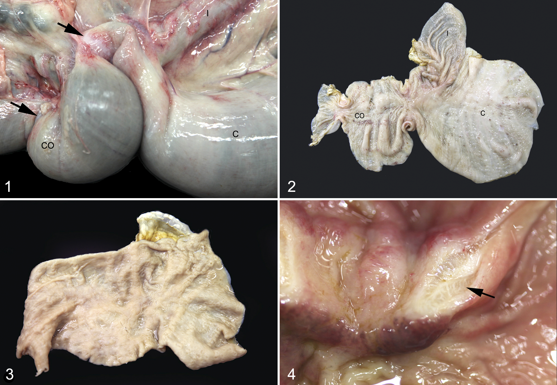
Macroscopic changes were similar in group A and B deer, consisting of single or multiple intestinal lesions located within a region of the intestinal tract from approximately 30 cm proximal to 50 cm distal to the ileocecal junction (Figs. 1, 2). In group A animals, the ileocecal junction was most commonly affected (n = 22), followed by the proximal colon (n = 20), cecum (n = 12), and terminal ileum (n = 7). Neoplasms at the ileocecal junction (n = 15) and in the ileum (n = 3) were much less common in group B deer, where lesions were typically detected in the proximal colon (n = 38) and cecum (n = 36) (Table 1).
Macroscopic Findings of Intestinal Adenocarcinoma in Deer in Respect to Clinically Affected (Group A) and Unaffected (Group B) Animals, n (%).
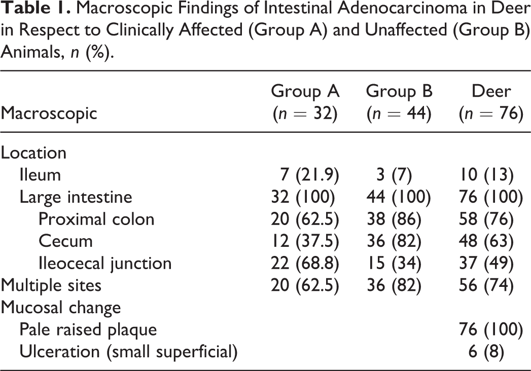
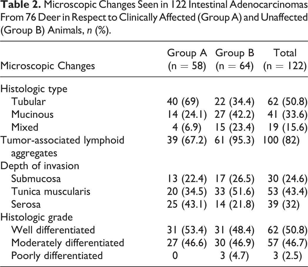
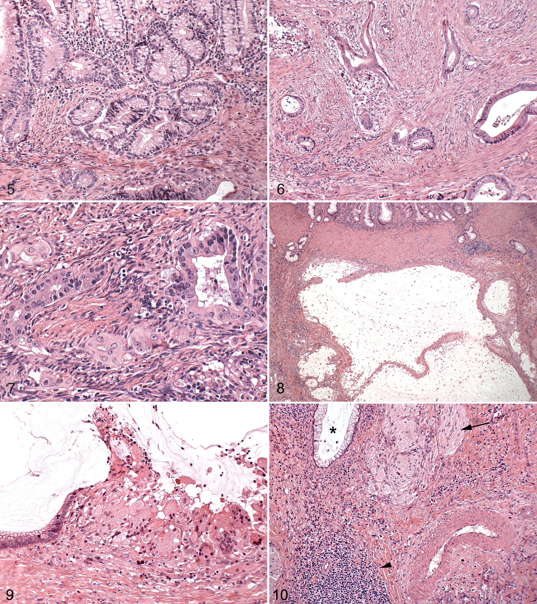
The least conspicuous changes consisted of poorly circumscribed, variably sized (between approximately 2 cm and 8 cm), plaquelike, smooth or corrugated, pale-gray mucosal and submucosal thickenings with rare small overlying ulcers (n = 6) (Fig. 3). In more severe cases, white firm confluent masses extended transmurally and circumferentially. In all deer from group A, these masses had resulted in intestinal wall thickening of up to 1.5 cm over a length of up to 3 cm, leading to severe luminal stenosis (luminal diameter < 1 cm). In severe cases, localized mesenteric-serosal adhesions were present. The cut surface of the masses had a fibrous appearance, and multiple small cystic cavities were a feature in some cases (Fig. 4). Proximal and distal to the sites of constriction, the intestine was markedly distended, and the wall of the terminal ileum was diffusely thickened with marked lymphangiectasia in 7 cases.
Additional macroscopic findings in clinically affected animals were the lack of body fat reserves and, frequently, a mild to marked increase in serous fluid within the abdominal cavity and pericardial sac (n = 17). Chronic rumenitis was noted in the caudal ventral sac in 7 animals characterized by multifocal fibrosis and puckering of the ruminal wall with accompanying firm mesenteric adhesions. Small numbers of mature liver fluke were observed in the bile ducts in 1 animal. Individual deer had renal cysts and vaginal cysts, and 1 had a moderate mandibular osteomyelitis. Multifocal caseous granulomas were observed in the retropharyngeal lymph node of 1 deer and in the lung and bronchial lymph node of a second animal.
Histopathology
The following microscopic description is based on the examination of 122 intestinal adenocarcinomas (58 from the 32 deer in group A; 64 from the 44 deer in group B) (Table 2).
Microscopic Changes Seen in 122 Intestinal Adenocarcinomas From 76 Deer in Respect to Clinically Affected (Group A) and Unaffected (Group B) Animals, n (%).
Tubular, mucinous, and mixed subtypes of adenocarcinoma were identified. The tubular form was most commonly seen in group A (40 of 58): tubules extended from the crypts through the lamina propria muscularis and were lined by single or multiple layers of cuboidal to high columnar epithelioid cells with elongated to oval, large, mainly basally located nuclei. These cells had indistinct cell borders and cytoplasm that was either moderately eosinophilic or mucin filled (Fig. 5). The tubules were variably sized and often irregularly shaped, infiltrating as isolated structures with associated mild desmoplasia (Fig. 6). Infrequent degenerate neutrophils and/or other cell debris were noted within tubular lumens. Occasionally, neoplastic cells were poorly regimented, either attenuated or cuboidal in shape with large centrally located and vesiculated nuclei (Fig. 7). These cells infiltrated the submucosa or tunica muscularis in small groups eliciting marked desmoplasia. The mitotic index varied among individual tumors, from rare up to 4 mitotic figures per 10 high-power fields.
In group B, the mucinous tumor subtype predominated, characterized by large cystic spaces filled with pale basophilic mucin (periodic acid–Schiff positive) and few foamy macrophages (Fig. 8). The cystic spaces were lined by neoplastic epithelial cells with abundant mucin-rich cytoplasm and basally located oval nuclei. In some instances, these cells were flattened or necrotic or were replaced by multinucleated giant cells and fibroblasts (Fig. 9). Extracellular mucin “lakes” encircled by multinucleated giant cells were also noted. Mixed-type adenocarcinomas composed of approximately equal numbers of neoplastic tubules and mucin-containing structures were observed less frequently in both groups.
In all but 3 cases, the adenocarcinomas were either well differentiated (62 of 122) or moderately differentiated (57 of 122). The accompanying desmoplasia was generally mild (69 of 122) or moderate (44 of 122). Neoplasms typically extended farther through the intestinal wall in group A than in group B, infiltrating as far as the serosa in 43% (25 of 58) from group A but in only 22% (14 of 64) in group B (Table 2).
A diffuse mild to moderate lymphocytic infiltrate was frequently observed surrounding the neoplastic tubules and occasionally within the neoplastic tubular lining. Single or multiple nodular lymphoid aggregates with occasional germinal centers were frequently seen at the advancing border of the tumor. Other inflammatory changes noted in association with the adenocarcinoma were multifocal granulomas that often centered on dilated lymphatics.
Fibroplasia of the submucosa and serosa, hypertrophy of the tunica muscularis and lamina propria muscularis, angiogenesis, moderate hypertrophy of large vessel walls, and hypertrophy and hyperplasia of intramural ganglia were often significant changes noted in the intestinal wall in the immediate vicinity of, and immediately proximal to, tumors (Fig. 10).
Mild to moderate villus stunting and fusion were evident in 27 sections of ileum irrespective of the presence of tumor. In these sections, the lamina propria mucosa was infiltrated by eosinophils and macrophages or lymphocytes, and in some cases (n = 7), this was accompanied by lymphangiectasis and lymphangitis.
There was no evidence of metastasis in the ileocecal lymph nodes (n = 76). Multifocal small granulomas were frequently seen (n = 29), characterized by aggregations of multinucleated giant cells that often contained or surrounded irregular gritty basophilic material (calcium deposits). Ziehl-Neelsen staining of the affected lymph nodes did not reveal acid-fast bacteria.
Parasitology
Fecal worm egg counts were low in all cases (<100 eggs per gram: Strongylus spp, Nematodirus spp). Fasciola hepatica eggs were detected in a single case, where few adult fluke had been found in the bile ducts.
Bacteriology
M bovis was isolated from 2 deer— from a granulomatous lesion in the retropharyngeal lymph node of one and from granulomatous lung lesions of the other. Mycobacterium avium subspecies avium was isolated from the ileocecal lymph node of a further deer in the absence of any lesions. No mycobacteria were cultured from any of the other ileocecal lymph nodes. Direct smears of feces did not reveal any significant organisms, and no significant bacterial pathogens were cultured from the feces.
Discussion
This report details a novel intestinal neoplasm in adult farmed Sika deer from an inbred herd grazed on bracken infested pastures. The tumors were locally infiltrative, slow-growing, low-grade adenocarcinomas of mainly tubular or mucinous subtypes predominantly located in the cecum and proximal colon. Intestinal strictures associated with the tumor masses correlated with clinical signs. The diagnosis of adenocarcinoma was based on the identification of proliferating tubules exhibiting varying degrees of differentiation infiltrating through the lamina propria muscularis into the underlying intestinal layers. 7,20
Submucosal herniation of glandular structures in adenomas in the large intestine, termed pseudoinvasion, can lead to erroneous diagnosis of malignancy. 17,37 This is often associated with polypoid tumors 48 ; the lamina propria characteristically still surrounds the proliferative tubules 37 ; and these structures are continuous with the overlying mucosa. 48 None of these features were evident in the present study. Other benign diseases where crypts are displaced into the submucosa, muscularis, and/or serosa—such as diverticular disease, 9 adenomatosis/adenomyosis associated with proliferative enteritis, 11 or enteritis cystica profunda 2,34 —could equally be ruled out, as continuity with the overlying mucosa was not evident and proliferative, necrotizing, or suppurative enteritis extending from the mucosa into the underlying intestinal layers was not found.
Although 74% of the deer had multiple tumors at distinctly different sites along the intestinal tract, implantation, serosal spread, or lymphatic or vascular invasion was not evident. In addition, there was no apparent correlation between the number of individual tumors and whether or not an animal displayed clinical signs. Different tumor subtypes were observed within individual animals, suggesting multiple independent foci of carcinogenesis rather than metastatic spread.
The anatomic and morphologic features of intestinal adenocarcinoma in Sika deer differed considerably from those observed in other species. Involvement of the cecum and proximal colon as seen in this study has rarely been observed in sheep, cattle, cats, and dogs. 3,6,14,32,38,39,51,52 In humans, adenocarcinoma is most commonly seen in the colon and rectum, but only 20% of these tumors arise in the proximal colon. 21,24 Furthermore, the mucosal changes in the deer typically manifested as slightly thickened or raised plaques. In contrast, in humans, the majority of intestinal adenocarcinomas arise out of benign adenomatous polyps, 21 and in sheep, about 50% of reported cases had 1 or more polyps. 32,52 Similarly, the mucinous subtype of adenocarcinoma observed in one-third of the deer in the present study has not been reported in sheep or cattle 16,32 and is infrequently seen in cats (20%) 39,51 and humans (10%). 18 In the deer, there was far less infiltrative growth by this tumor subtype compared with the tubular subtype.
Peritumoral lymphocytic infiltration, as described in the current study, has been widely reported in human colorectal cancers. 18,20 It is considered a host response to the neoplasm and has been associated with a better prognosis. 18,20 Interestingly, lymphoid aggregates were more commonly seen in clinically normal deer. In animals, there has been only 1 report of a peritumoral inflammatory infiltrate, in a single case of intestinal adenocarcinoma in a goat. 40 Intestinal adenocarcinomas generally show highly malignant behavior with widespread lymph node invasion and frequent distant metastasis, 3,6,14,32,52 features not observed in the current study.
As reported in other animal species with intestinal adenocarcinomas, the more extensive neoplasms were associated with a marked narrowing of the intestinal lumen. 17 In the present study, the occurrence of intestinal strictures strongly correlated with clinical signs of weight loss, presumably because of restrictions on gastrointestinal throughput. Muscle hypertrophy and hyperplasia of mural ganglia were observed in the deer proximal to the tumor mass, features thought to be induced by the increased workload required to overcome the obstruction. 4,5
Spontaneous intestinal neoplasia in animals is considered a rare event, 23 strongly suggesting that particular predisposing risk factors must be at play to account for such a high incidence of neoplasia in the study herd. This may reflect a genetic predisposition and/or environmental exposure to carcinogens.
Given that this herd was subject to veterinary investigation 36 after a herd outbreak of tuberculosis in 1993 and that this intervention necessitated the culling of deer and postmortem examination of viscera, it is unlikely that many cases of neoplasia were occurring at that time that escaped detection. It thus seems reasonable to conclude that the high incidence of neoplasia has been a relatively recent phenomenon.
The Sika deer in the present study were exposed to, and were observed to consume, bracken fern, which contains the potent carcinogen ptaquiloside. 54 Although this fern can induce intestinal adenocarcinomas in the ileum and cecum in rats, 46 in cattle it typically causes tumors of the upper alimentary tract and small intestine, generally in conjunction with bovine papilloma virus infection. 28,54 In cattle, tumors induced by bracken fern show a variety of morphologies of mesenchymal or epithelial origin, and these can arise in different locations. 43,46 Although bracken fern has always been present on the study farm, it is only since 2003 that cases of intestinal adenocarcinoma have been reported. Thus, while this potential carcinogen may have played a role in tumorigenesis in this case, it seems likely that at least 1 other cofactor was involved.
While there may be other feed-associated carcinogens present on the farm, the main supplementary winter feed used is citrus pulp, which, if anything, has been found to reduce the incidence of neoplasia. 47 Other environmental toxins, such as herbicides implicated in intestinal neoplasia in sheep, 45,52,53 have not been used on the study farm.
Hereditary predisposition to intestinal carcinoma is suspected in Siamese cats, German Shepherd dogs, and individual bloodlines of sheep. 17,26,39 It is recognized in humans as familial adenomatous polyposis and heritable nonpolyposis colorectal cancer. 13,20 Interestingly, there are marked morphologic similarities between intestinal adenocarcinomas in Sika deer and heritable nonpolyposis colorectal cancer in humans. 10,13 No new animals have been introduced to the herd in the center of this study for many years. The herd was established from wild Sika deer introduced into Ireland in 1860. 15,29 Furthermore, the culling consequent on 2 outbreaks of tuberculosis had considerably reduced the number of deer in the herd. 36 Limited genetic diversity might have allowed for 1 or more genes predisposing to the development of neoplasia to have become widely distributed within this herd. 24
Chronic intestinal inflammation is considered to increase the risk of colon cancer. 31 Furthermore, infectious agents are suggested to cause about 18% of all neoplasms in humans annually, and common pathogens associated with this carcinogenesis are Helicobacter pylori, papilloma viruses, herpes viruses, and retroviruses. 35 The high incidence of neoplasia in a single herd suggests the involvement of an infectious agent; however, if this were the case, younger animals would likely be affected. 25
Johne disease (paratuberculosis) caused by infection with Mycobacterium avium subspecies paratuberculosis is the most common chronic enteritis occurring in deer, and the ileocecal lymph node is considered the optimum site to detect mild disease. 8 This site was examined histologically in all cases in the present study, and no evidence for Johne disease was found. The occasional focal granulomas and calcified areas were negative for acid-fast bacteria and negative on culture for M avium subsp paratuberculosis. These lesions were thus interpreted as postreactive changes or parasite induced, as suggested in a study on normal deer lymph nodes. 19 While there was no histopathologic evidence of papilloma 44 or herpes virus 12 infection, its potential causative role cannot be ruled out without further molecular analysis. 27
In conclusion, the present study is the first to report intestinal adenocarcinoma in Sika deer. The disease occurred at an unusually high incidence in a single herd, and the degree of uniformity in tumor morphology differs markedly from findings in other animal species, suggesting a novel syndrome. A common carcinogenesis is proposed, with involvement of environmental carcinogens including bracken fern and genetic predisposition, and future studies will aim at investigating the molecular carcinogenesis and possible involvement of infectious and toxic agents.
Footnotes
Acknowledgements
We would like to thank Dr Hugh Bassett and Dr Joe Cassidy for critically reviewing the manuscript. Technical assistance was given by Brian Cloak (photomicrographs), Alex Fawcett, Joe Brady, and Sheila Worrall from the Pathobiology Section of the School of Veterinary Medicine, University College Dublin, Ireland, and by the staff of the Regional Veterinary Laboratory, Kilkenny, Ireland. We are grateful for the interest, support, and excellent herdsmanship of Helena and Rory Harrington, Waterford, Ireland, without which this study would not have been possible. We are indebted to the late Dr Eddie Weavers, who diagnosed the first case with us and sparked our interest in conducting this study.
Declaration of Conflicting Interests
The author(s) declared no potential conflicts of interest with respect to the research, authorship, and/or publication of this article.
Funding
The author(s) received no financial support for the research, authorship, and/or publication of this article.