
Abstract
Advancement in electrophoresis and mass spectrometry techniques along with the recent progresses in genomics, culminating in bovine and pig genome sequencing, widened the potential application of proteomics in the field of veterinary medicine. The aim of the present review is to provide an in-depth perspective about the application of proteomics to animal disease pathogenesis, as well as its utilization in veterinary diagnostics. After an overview on the various proteomic techniques that are currently applied to veterinary sciences, the article focuses on proteomic approaches to animal disease pathogenesis. Included as well are recent achievements in immunoproteomics (ie, the identifications through proteomic techniques of antigen involved in immune response) and histoproteomics (ie, the application of proteomics in tissue processed for immunohistochemistry). Finally, the article focuses on clinical proteomics (ie, the application of proteomics to the identification of new biomarkers of animal diseases).
Keywords
Historical Perspective
In the last few years, there has been an awakening interest in using proteomics and the complementary, essential advances in bioinformatics, to address problems of veterinary pathogenesis. However, the application of proteomics in veterinary medicine has been limited in comparison to studies that have explored the potential of these advanced protein-analytic technologies in human clinical medicine. Historically, analysis of serum protein has been an essential part of the armory of veterinary diagnostic investigations for disease pathogenesis but has been limited to measurement of total protein, albumin, globulin, and albumin: globulin ratio and serum protein electrophoresis (SPE) on agarose. 39 The latter method separates serum into around 8 fractions from albumin to the γ-globulin fraction, but the protein bands seen on SPE hide a multitude of proteins that, if identified 46 and measured with valid procedures, could provide a treasure trove of pathologic and diagnostic biomarkers. 6 Proteomics holds the key to unlocking this vision of advancing veterinary pathology and diagnostics.
Although the application of proteomics in veterinary medicine has lagged behind human medical uses, there has been increased activity recently, especially for investigation of farm animal health and disease. 42 A number of relevant and informative reviews have appeared 12,38,39,41,53 that lay the groundwork for further participation of veterinary laboratories in this exciting and rapidly advancing field. In this review, we look at the current uses of proteomics in veterinary medicine with particular emphasis on infectious disease, pathogenesis, and diagnostics.
Proteomic Techniques
The term proteomics refers to the large-scale study of proteins, including their structures and functions, whereas proteome can be defined as the set of proteins expressed by the genetic material of an organism under defined environmental conditions. 99 Proteomics has emerged as a field of research in less than 2 decades 108 and has developed rapidly, driven by improvements in technology and by the need for analytic approaches that can deliver global protein characterization. The ability to sequence entire genomes and to collate the resulting data into genome sequences has enabled proteomics, but the global characterization of the proteins that compose even relatively simple biological systems is still not achievable. 21 A proteome is generally more complex than the encoding genome, and the proteins are present across a broad dynamic range. 31 These issues are compounded by regulation of protein expression, in response to developmental and environmental stimuli, which results in a dynamic proteome. Nevertheless, the importance of proteins as the primary effector molecules of biology, which are also the major drug targets and antigens, has promoted strong interest and investment in proteomics, and the field continues to develop rapidly.
Proteomics involves the resolution of a complex mixture of proteins into components that can then be characterized. Characterization always involves matching protein to encoding gene (identification) but can also involve relative quantitation or further characterization to reveal posttranslational protein modification. Protein characterization is performed by mass spectrometers, which are generally fed proteins after some initial fractionation. The type of fractionation employed is determined by the complexity of the proteome and by the specific research question but must be compatible with the downstream mass spectrometry (MS). Currently, 2 major MS platforms are employed for proteomics, differentiated by the mechanism through which ions are generated: these ion sources are termed matrix-assisted laser desorption/ionization (MALDI) and electrospray ionization (ESI). MALDI instruments receive analytes in the solid state, while the sample is delivered to ESI instruments in a volatile solvent. These different platforms have advantages and disadvantages but are complementary, and most proteomics labs will operate both systems. Whatever the MS system that is used, the optimum goal is to deliver peptides as homogeneous populations that can be characterized without competition from other species, such as different peptides, other polymers, or salts. Given that even simple prokaryote proteomes comprise thousands of proteins and multicellular species may comprise greater than 100 000 proteins, this is a tall order and requires extensive sample fractionation.
The protein fractionation systems that are employed in proteomics are either electrophoretic or chromatographic: the former is typically applied to intact proteins and the latter to peptides generated by protein cleavage. Orthogonal separation approaches are often utilized to enhance resolution, and the archetypal orthogonal separation in proteomics is 2-dimensional electrophoresis (2DE). 48 Conventional 2DE involves separation by isoelectric focusing in the first dimension, followed by sodium dodecyl sulphate electrophoresis in the second. Both dimensions are performed in a polyacrylamide gel matrix, and the proteins migrate on 2-dimensional gels as spots according to isoelectric point and apparent molecular weight. The resulting spot map, which can be visualized by protein staining, can resolve several thousand protein species, and spots can be excised directly from the gel for characterization by MS.
2DE is a powerful approach for descriptive and comparative proteome analysis, as it remains the highest-resolution protein separation approach and is inherently quantitative. The separation of intact proteins by charge and mass can highlight posttranslational modifications that would not be evident in 1-dimensional electrophoresis or in peptide-based separations. 96 2DE tends to underrepresent some classes of protein, including those that are of relatively low abundance, very large, or highly charged. Some of these issues can be circumvented by prefractionation to enrich proteins of interest or by focusing 2DE on specific charge and/or mass ranges. Very hydrophobic proteins may be refractory to solubilization in the nonionic conditions that are required for isoelectric focusing, and alternative detergents or 2-dimensional separations, such as the BAC/SDS-PAGE system, 18,51 have proven useful in this context. Nevertheless, 2DE is often considered a time-consuming and technically challenging approach that has limited advantages over more readily automated chromatographic methods.
The heterogeneity of intact proteins, which is exploited in 2DE, limits the resolution of chromatography for proteomic workflows. Instead, proteins are typically fragmented to peptides before chromatographic separation. Reversed-phase chromatographic separation of peptides is ideally suited to proteomics because peptides can be trapped and desalted before elution and because the mobile phase comprises volatile solvents that can be evaporated in the ESI source. Chromatography can thus be directly coupled to ESI-MS, facilitating automation and minimizing sample loss. Multidimensional chromatographic separation is increasingly employed 117 because it can be automated and because even highly charged or hydrophobic proteins will likely generate some peptides that are amenable to MS analysis. Ion exchange is mostly used as a first dimension, giving separation based on orthogonal biophysical characteristics to reversed-phase chromatography and taking advantage of the latter to desalt ion exchange fractions before MS. Chromatographic approaches can be more sensitive than electrophoresis because there is no requirement to recover proteins or peptides from a gel matrix. However, the conversion of each protein in an already complex proteome to a large number of peptides is counterproductive when the goal is to maximize proteomic coverage, because there are limits to the resolution of multidimensional peptide chromatography, so prefractionation or proteomic material is often important. Combinations of electrophoresis and chromatography are among the most efficient fractionation systems, 116 but targeted approaches such as subcellular fractionation or affinity purification of protein complexes can result in greatly enhanced coverage of a subproteome.
Typically, proteins will be obtained from a biological source and fractionated by electrophoresis. Protein fractions will then be trypsinized to generate peptides that can be further fractionated by high-performance liquid chromatography (LC) before analysis by an ESI-MS that is fed directly with the chromatography eluate (LC-MS). The MS will generally be capable of recording the mass of analytes and able to isolate and fragment peptide ions (MS/MS, or tandem MS) to generate information about structure. 21 The resulting data are fed to a search engine, such as Mascot (Matrix Science Ltd), which generates in silico MS data for the specified genome sequence database and looks for statistically significant matches with the experimentally generated MS data. The data output is typically a list of potential matches, ranked by confidence, to proteins that may be components of the sample. It is important to recognize that this will not be a complete list of protein components, largely because some will be present at levels below the threshold of detection but also because some proteins are refractory to analysis or are not represented in the genome database. It is also a qualitative data set, but quantitative data are often desirable.
Absolute protein quantitation is difficult to undertake in an -omic context, but relative quantitation can be achieved by a diversity of comparative approaches. Relative quantitation of intact proteins is generally performed by gel-based methods, such as 2DE using semiquantitative protein stains, or protein-labeling strategies, such as difference gel electrophoresis (DiGE).
3
The advent of DiGE technology has greatly enhanced the utility of 2DE for quantitative proteomic analysis, enabling the direct comparison on a single gel of samples that are differentially labeled by fluorophores that are mass and charged matched but spectrally discrete. For example, DiGE has been exploited to reveal changes in the expression of discrete isoforms of tryparedoxin peroxidase in virulent and attenuated strains of Leishmania.
34
Quantitation at the peptide level can be achieved by stable isotope-labeling approaches or by label-free comparison. Isolated proteins or tryptic peptides can be chemically labeled before separation [iTRAQ]
97
,
Label-free approaches, which involve serial LC-MS analysis of multiple unlabeled samples, are becoming commonplace as the robustness of chromatographic separation improves and facilitates the alignment of data sets that is essential for comparison. Label-free approaches are more costly in instrument time, as unlabeled samples cannot be multiplexed—an important consideration, as LC-MS instrumentation is costly to maintain. Regardless of the quantitation approach, comparative proteomics experiments have the potential to highlight key proteins in phenotypes of interest 20 and thus have tremendous potential to highlight drug targets and biomarkers and elucidate biological mechanisms.
Proteomic Approaches to Animal Disease Pathogenesis
Several proteomic techniques have been applied so far to the understanding of dynamic protein pathways involved in host and pathogen responses during diseases. Pathogens and immune defenses adapt to each other. This adaptation is due to the regulation of the expression of several genes of both sides, to changing stimuli. This capability to fine-tune gene expression can and has been studied by DNA microarray techniques, in particular for what concerns the microbiome (ie, the pathogen component). 88 However, since the correlation between DNA levels and actual protein expression is poor, 50 integration between the 2 techniques, genomics and proteomics, is required.
Proteomics Research in Bovine Species: Focus on Mastitis and Respiratory Diseases
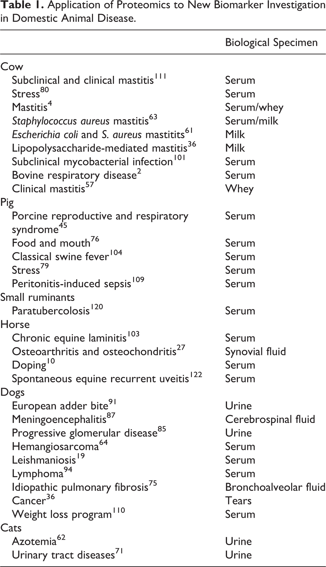
Mastitis is considered the most frequent and most costly production disease in dairy cows. It is therefore not surprising that proteomics has been widely applied to investigate mastitis pathogenesis. Intramammary infections with Staphylococcus aureus and Escherichia coli have received a lot of attention due to their economic impact. In the classical experiment of Boehmer and coworkers, 13 acute mastitis was induced by infection with E. coli in a group of dairy cows, whereas another group was treated with sterile physiologic solution. 2DE gel electrophoresis was carried out before and 18 hours after the infection, and differentially expressed proteins were identified in milk by peptide sequencing with MALDI time of flight (TOF). Intramammary infection caused by S. aureus was also investigated. 63 Three strains were used, and proteomic analysis revealed that while acute phase protein expression was almost identical, differential regulation of strain-specific host IFN-γ and antimicrobial peptides were observed. A similar experimental design was applied after challenging the mammary gland with bacteria-derived pathogen-associated molecules, such as E. coli lipopolysaccharide and S. aureus lipoteichoic acid. 56 Proteomic analysis was carried out on milk, revealing in both groups of animals an almost complete hydrolysis of casein, supposedly caused by an overexpression of proteases. The results obtained during these experiments also identified useful candidates for biomarker identification (Table 1). The importance of proteomics (and peptidomics in particular) in new biomarker search has been clearly evidenced in a recent publication, focused on peptidomics investigation during mastitis. 77 Another remarkable study demonstrated how α- and β-caseins modulate biofilm formation during mastitis of Streptococcus uberis. 112 This activity is increased by casein degradation, which can be induced by bacterial proteases.
Application of Proteomics to New Biomarker Investigation in Domestic Animal Disease.
Prerequisite of these “pathogenesis-focused” studies are preliminary experiments aimed at elucidating the mammary gland immunoproteome, identifying the proteins that are responsible, in physiologic status, for immune defense of the mammary gland in milk 55 and in mammary parenchyma. 11
Proteomic techniques were useful to explore other bovine major disease groups, such as those involving respiratory apparatus. Bronco alveolar lavage fluid represents an ideal specimen for proteomic analysis. 2DE proteomics confirmed the importance of stressors, such as transportation and weaning, in respiratory disease development. 83 Broncho alveolar fluid was also studied by LC-MS/MS following experimental induction of pneumonia with Mannheimia haemolytica. 14 Antimicrobial peptides and acute phase proteins were identified by proteomics as being upregulated in bovine mucosal defense.
Proteomic Approaches to Parasitosis in Small Ruminants
Most proteomic research on small ruminant species has been focused on sheep parasites. Pathogenesis of gastrointestinal nematode infection was recently studied by quantitatively investigating the expression of proteins by abomasal mucosa of resistant and susceptible sheep breed after experimental Haemoncus contortus infection. 86 A number of proteins (4468 in total) were identified and several of them (n = 158) were found to be differentially expressed between resistant and susceptible sheep. Proteins such as galectin-4, trefoil factor 2, fibrillin-2, and DAG were detected in luminal mucus of resistant sheep, where they can interfere with parasite adhesion processes. Immune response to other abomasal parasites, such as Teladorsagia circumcincta, was thoroughly explored by combining 2DE and MALDI-TOF proteomics, after in vivo 92 and in vitro 7 experimental infection, identifying galectin-15 as being involved in specific resistance against parasite infection. Both these studies focused on the abomasal mucosal surface, going in-depth through the network of proteins that directly interact with Teladorsagia. Epithelial cell lysates revealed obvious candidates, such as immunoglobulin A and sheep mast cell protease 1, and less obvious ones, such as calcium-activated chloride channel and intelectins. The latter may be involved in increased mucus secretion, which in turn may result in reduced parasite infection. In a further attempt to better define the pathogenesis of this parasite, proteomic analysis of lymphatic drainage obtained from cannulated abomasal lymph nodes identified other proteins, including hemopexin, α1-b glycoprotein, and gelsolin, as being altered after Teladorsagia infection. 47 The involvement of the immune and inflammatory defense mechanisms in Echinococcus granulosus was evidenced after proteomics investigation on hydatid cyst fluid, not only from sheep, but also from cattle and humans. 8 Out of 130 proteins identified from fertile cysts, only one third (n = 48) were of parasite origin, whereas the others (n = 82) were from host origin, clearly demonstrating how E. granulosus can absorb host proteins across its outer germinal layer and deceive the host immune system.
Proteomics Research in Pigs: Respiratory and Intestinal Diseases
The pathogenesis of several viral diseases, including porcine reproductive and respiratory syndrome, classical swine fever (CSF) and porcine circovirus diseases, has been explored by proteomics. In vitro infection of susceptible cells, such as porcine alveolar macrophages and Marc-145 cells, 37 provided valuable information about which cellular pathways are mostly activated during porcine reproductive and respiratory syndrome viral replication, assembly, and pathogenesis. In particular, stress proteins may be involved in all stages of the life cycle of the virus, and all the proteomics studies carried out so far have identified heat shock proteins (HSPs), such as HSP27 and HSP8, as well as proteins related to cytoskeleton assembly, including annexins, β-actin, and tubulin, as being incorporated into virion particles.
Another important swine viral disease is CSF. This virus targets endothelial cells, and widespread hemorrhages are the pathognomonic clinical signs of the disease. The pathogenesis of CSF was therefore explored by proteomics on primary porcine endothelial cells, 74 revealing that CSF virus induced a downregulation of proteins involved in energy metabolism and upregulation of others that inhibit endothelial cell proliferation. Upregulation of proteins involved in inflammatory reaction and oxidative stress was also demonstrated, partially elucidating the molecular bases of endothelial damage and related vascular permeability. Further progress on the knowledge of CSF pathogenesis was obtained by proteomic characterization of macrophages involved in the development of the disease. Both alveolar macrophages 121 and peripheral blood monocytes 104 were studied, revealing that HSP expression was altered, as well as proteins involved in energy metabolism, adhesion, oxidative stress, and protein translation and processing.
Postweaning multisystemic wasting syndrome, caused by porcine circovirus-2, was analyzed after macrophage infection, 26 revealing the involvement of cytoskeletal proteins. Immune system targeting of porcine circovirus was also explored by quantitative proteomics on lymph nodes from experimentally infected piglets, identifying acute inflammatory response-related proteins involved in the insurgency of the disease and others involved in oxidative stress. CD81, which has been already shown to be involved in other viral infections, was upregulated as well.
Pigs are asymptomatic carriers of Salmonella typhimurium, which is regarded as one of the most frequent food-borne pathogens, transmitted through contaminated pork meat. In vivo experimental infection, followed by DiGE and MALDI-TOF MS of mesenteric pig lymph nodes, 81 identified a complex interaction between Salmonella and the pig immune system, revealing an activation of pyroptosis, an innate immune mechanism against intracellular bacteria. By integrating high-throughput proteomics with classical immunohistochemistry and molecular biology, this study described at the molecular level the delicate equilibrium between the host immune system and bacteria invasion. Salmonella could reduce the apoptosis rate of neutrophils, allowing a better control of the disease, meanwhile modulating some cell functions, such as cytoskeleton rearrangement, for the benefit of the bacteria themselves.
Proteomics in Horses: Articular Disorders and Equine Recurrent Uveitis
The application of proteomics in equine disease is not extensive at present. However, 2 major horse diseases have been studied by proteomics: osteoarthritis/osteochondrosis and equine recurrent uveitis.
Proteomic studies were carried out on articular cartilage inflammation in an ex vivo experimental model, providing an in-depth perspective on the molecular pathogenesis of equine arthritis and demonstrating how treatment with IL-1 modulates the expression of matrix metalloproteinase-1 and -3 but decreases, for example, that of clusterin. 29
An excellent example of how classical histochemistry can be integrated with proteomic techniques is observed in a study focused on equine recurrent uveitis. In a label-free LC-MS/MS analysis of vitreus from uveitis, beside an increase in complement cascade-involved proteins, a significant decrease of proteins involved in Wnt signaling (ie, DKK3 and SFRP2) was recently detected. 52 Western blotting analysis allowed the quantification of the different expression of the 2 proteins, whereas immunohistochemistry integrated proteomics results by locating the expression of the 2 proteins on retinal specimens. Major immunohistochemical findings include a specific downregulation of SFRP2 in retinal outer limiting membrane and a general decrease of DKK3, which was shown to colocalize with Müller glial cells. Therefore, by coupling proteomics, quantitative Western blotting, and immunohistochemistry, this study not only demonstrated the involvement of the Wnt pathway in equine uveitis but also localized it at tissue levels where the expression pattern of these proteins is mostly modified.
Proteomics in Companion Animal Disease Pathogenesis
While there have been applications in veterinary medicine where proteomic approaches have been employed for investigations in small animals and in studies of infectious and neoplastic disease in particular, 41,53 the use of proteomics in the study of pathogenesis in companion animals is less extensive than that for farm animals.
Proteomics has been used in an attempt to identify novel antigens from Leishmania chagasi for vaccine production and diagnostic development. 32 Based on DiGE and Western blotting, 25 proteins from L. chagasi have been identified for use in serum diagnostics and for consideration of vaccine development targets. Others important dog parasites that were investigated though proteomics include Ancylostoma canum 84 and Echinococcus multicularis. 68
Proteomics has also been used to investigate bacterial pathogens of dogs and their interaction with the host. Surface proteomic analysis has been used to identify proteins of Staphylococcus pseudintermedius involved with adherence to the external matrix. 9 Lipoprotein from Ehrlichia chaffeenis, identified by LC-MS/MS, was subsequently shown to elicit an immune response in dogs. 59 The response of protein in the mitochondria of liver from dogs treated with bacterially derived lipopolysaccharide has been studied by 2DE, with 14 of the 500 identified proteins being shown to be differently expressed within 4 hours of intravenous lipopolysaccharide treatment. 33 Canine liver has also been examined in bull terriers with a genetic disorder leading to inherited lethal acrodermatitis. 49
Worth mentioning is an interesting study on rabies. Three areas of the central nervous system of dogs infected with rabies virus resulting in paralytic or furious manifestation of the disease have been compared by 2DE, followed by quadrupole TOF MS. 108 A total of 32 proteins were found to differ in the hippocampus, 49 in the brainstem, and 67 in the spinal cord. In comparing the paralytic to the furious forms of the disease, 13, 17, and 41 proteins differed in the hippocampus, brainstem, and spinal cord, respectively. The functions of the proteins were from a range of activity, including antioxidants, HSPs, metabolism, and transcription and translation proteins, with the results leading to an increase in understanding the molecular pathology and differential manifestation of the rabies in its different forms.
As in human medicine, initiatives have been taken in canine oncology to identify biomarkers of neoplastic disease with the use of proteomics. 38,54 Lymph nodes from dogs with B-cell lymphoma have been studied using 2DE and MALDI-TOF MS in comparison to lymph nodes of healthy dogs. 82 Over 90 differentially expressed protein spots were identified, among which proline dipeptidase, triosophosphate isomerase, and glutathione-S-transferase were downregulated and macrophage capping protein was upregulated in the samples from the lymphoma cases. While 2DE can identify differences in expression between diseased and healthy samples, for a diagnostic test to be based on such results requires extensive development. An approach that held promise from human medicine to identify biomarkers is surface-enhanced laser desorption/ionization MS. 94 The advances made in proteomic science can be used to reexamine established methods of veterinary diagnostics, and in the clinical investigation of lymphoma, SPE is a regularly used tool in which the proteins of serum are separated into well-known fractions, namely, albumin α1, α2, β1, β2, and γ. The protein bands of agarose SPE have been identified as a source of protein for trypsin digestion and tandem mass fingerprinting. Thus the main SPE protein bands in serum of cats with and without lymphoma, have been subjected to proteomic analysis. 45 The study confirmed established understanding of the content of the main bands but also revealed the presence of lower-abundance protein in particular fractions, such as inter-α (globulin) inhibitor 4 in the α2 band of cats with lymphoma.
Other neoplastic diseases that have been investigated by proteomics include prostate and bladder carcinomas, which have been examined by DiGE in comparison to healthy tissues. 72 Analysis by DiGE has also been used to investigate the proteins of metastatic canine mammary carcinomas 65 and identified 21 proteins that were either up- or downregulated in the carcinomas. Proteomics was also very useful in mammary carcinoma 66 staging, identifying individual proteins as markers of each stage. In the first stage, named adenoma pattern, phosphoglycerate mutase 1 was increased and calumenin decreased; in the next, carcinoma pattern, 14-3-3-zeta was increased and gelsolin decreased; and in a metastasis pattern, bomapin was increased and maspin decreased. DiGE has also been used to compare high-grade canine cutaneous mast cell tumors that have poor prognosis with low-grade tumors with better prognosis. 99
Canine brain tissue has been subjected to 2DE and MALDI-TOF MS in a model of Alzheimer disease where environmental enrichment and antioxidant-fortified food were compared to controls. 90 The group with both treatments showed the best neuronal function and cognition and, upon proteomic analysis, was shown to differ from the control group in reduced expression of parietal cortex proteins, including glutamate dehydrogenase, enolase, and glutathione-S-transferase, while creatine kinase and glyceraldehyde-3 phosphate dehydrogenase, among other proteins, were upregulated.
Using iTRAQ and LC-MS/MS technology, the proteins in early- and late-stage myxomatous mitral valve disease in dogs have been examined, 69 with 117 proteins identified in mitral valves that were differentially expressed as compared to tissue from healthy dogs. Based on hierarchical sample clustering, it was evident that the altered expression of the mitral proteins occurred in the early stage of disease progression, thereby providing a valuable model of the equivalent human disease.
Finally, canine liver has been examined in bull terriers with a genetic disorder leading to inherited lethal acrodermatitis, 49 which may be particularly interesting on the background of the possible utilization of dogs as model for equivalent human disease. In this study, 13 differently expressed proteins were identified, involved in processes such as chaperone action, calcium binding, and energy metabolism.
Proteomics and Posttranslational Modifications in Veterinary Diagnostics
Changes in posttranslational modification readily occur in inflammation but also in cancer and other disease states. Based on recent advances in the development of analytic techniques and instrumentation, MS in particular, several blood-derived glycan-based biomarkers using glycomics strategies have been identified. 1,5,98 Due to the high extent of its carbohydrate moiety (more than 40% of the weight of the protein), the acute-phase protein alpha1 acid glycoprotein (AGP) has been rightly regarded as a model to study posttranslational modifications during diseases 23 and an ideal model to implement these findings as a biomarker source. In fact, AGP glycan pattern has been found to be modified in animal diseases, as well in cats 24,93 and goats. 25 Furthermore, a proteomic approach has been utilized to determine the differences between AGP glycoforms from different tissue origins, which is one of the first steps to utilize AGP as specific biomarker for diseases. 24 Structural modifications of other proteins, such as transthyretin, in animals have been described as well, including horses 54 and apes. 100 A proteome-wide glycan analysis was carried out in dog serum though a lectin-enrichment-based approach coupled with MS, which allowed high-throughput investigation of protein glycosylation signatures. 28 While application of glycomics is still quite far from the veterinary field, posttranslational modification of proteins occurin in neoplastic disease may provide further opportunities to identify biomarkers of the disease in animals. Glycoproteomic profile of serum peptides has been examined by a combination of lectin affinity selection, global internal standard technology, high-performance LC, and MALDI-TOF to investigate canine lymphoma and transitional cell carcinoma. 115 Two peptides were identified that allowed discrimination between these conditions.
Immunoproteomics: Identifying Antigens Involved in Immune Response Against Pathogens
The immunoproteome defines the subset of proteins that induce immune response. Circulating serum antibodies represent important biomarkers per se, since their half-life varies between 7 and 20 days and they are highly stable compared to other serum proteins. The aim of immunoproteomics is to identify the antigens that these circulating antibodies have been raised against during a given infectious disease. Immunoproteomic-based available techniques include gel-based and gel-free immunoproteomics.
Gel-based immunoproteomics relies on the separation of a complex mixture of proteins derived from pathogens by 2DE and electrotransfer onto a solid support, usually nitrocellulose. Immunoreactive proteins are then identified after incubation of the membrane with serum from infected animals. Gel-based immunoproteomic techniques have been widely utilized in veterinary immunoproteomics to identify, for example, specific antigens involved in paraTBC 73 in both cattle and sheep, 60 Streptococcus suis in pig, 119 and Staphylococcus aureus isolated from bovine mastitis. 107 Immunoreactants against parasites were characterized as well, such as those from Fasciola hepatica and Schistosoma mansoni 16 in cattle and Haemoncus contortus in sheep. 118
Gel-free immunoproteomics takes advantage of different physicochemical characteristics of antigens, such as isoelectric point, hydrophobicity, and/or affinity with immune sera to sort complex mixtures in smaller groups of proteins. Protein subfractions are then identified by MS. Immunoproteomics of Brucella abortus identified differential immunoreactivity of Triton X-114-soluble antigens between naturally infected vaccinated and nonvaccinated seronegative cows, identifying antigens potentially useful for immunoassays. In a very elegant experiment, Neospora caninum antigens were identified after their interaction with CD4+ T-cell lines. 95 In a first step N. caninum tachyzoite water-soluble antigens were grossly fractionated by size exclusion chromatography. Their immune-stimulating capability was then assessed after challenging CD14+ T-cell lines with sorted protein groups, and the immunologic selected fractions were eventually analyzed by LC-ESI-MS/MS.
Clinical Proteomics: Biomarker Discovery
While single-protein concentration assessed by antibody-based affinity is still the gold standard of diagnostics for clinical practice, there is now a general agreement that a panel of independent disease-related proteins could substantially improve the diagnosis of animal diseases. High-throughput proteomics has emerged as the most powerful technique for identifying protein profiles in animal biological fluids, as shown in Table 1. The application of proteomics as a diagnostic tool has been further implemented by its capability to analyze new and “neglected” biological fluids, such as saliva
70
or tears,
102
to complement the classic fluids (ie, blood serum and milk). The identification of markers related to diseases has wide practical implications, since they provide diagnostic tools as well as potential targets for novel approaches to monitor the therapies. Notwithstanding its progress, the translation of biomarkers from the discovery field to their clinical use is still hampered by several pitfalls, which are particularly evident in veterinary medicine, including
Sample collection—since differences among age, breeds, but also sample collection and storage (ie, the amount of time between sample draw and analysis) may profoundly alter serum protein profiles.
30
Dynamic range—serum/plasma is the most important clinical specimen, but no more than 10 proteins make up 90% of the total protein serum content, more than half being made of albumin. There are therefore at least 10 orders of magnitude difference between the highest- and lowest-abundant protein. These potential obstacles can be overcome by using depletion of most abundant proteins by beads (Proteo Miner)
78
or by immunodepletion.
44
However, several potentially important proteins may be lost in the process due to their bonding to removed high-abundant proteins.
Cost—which is unaffordable for veterinary clinical practice at this stage, though it may be reduced in the future.
Therefore, while proteomics has been extremely useful so far for new potential biomarker discovery, further work is required to standardize routine utilization in veterinary practice.
Histoproteomics: The Application of Proteomics to Tissues Processed for Immunohistochemistry
Standard techniques for preserving biological material for storage and histologic processing include snap freezing in liquid nitrogen and fixation in formalin and embedding in paraffin (FFPE). Proteomic techniques have been developed to investigate the localization and expression profiles of proteins on freshly frozen pathologic specimens, meanwhile preserving the histolopathologic integrity of the tissues. LC-MS workflows have been optimized to determine laser-captured microdissected pathologic tissues and have been applied to specimens from human breast cancer tissues, 17 fixed with ice-cold ethanol (70%), and then stained with hematoxylin. Laser-capture dissection was then carried out on stained tissues, and more than 400 proteins were identified by LC, followed by MS. General application of such optimized protocols to animal disease diagnosis has been prevented, at least so far, by economic reasons. Nonetheless, as such protocols are now available, its future application in veterinary clinical practice or research can be envisaged. Cryopreserved samples provide the first choice for proteomic analyses, since proteins are not modified. It must also be said that morphologic details are not optimally preserved. Moreover, longtime storage is expensive and may not be accurate.
Embedding of samples in paraffin after formalin fixation represents a serious challenge for protein extraction. Formalin reaction with tissues results in the cross-linking between proteins, causing a significant reduction of recovery. New formalin-free fixatives that may enable both histologic and molecular analyses are now available, and their potential value for proteomic investigation has been assessed. 43,67 These new methods have proven very effective in proteomics on archived cancer tissues. 13 While providing useful protocols for future applications of proteomic techniques to novel processed pathologic specimens, protocols to extract and analyze proteins from FFPE are mostly required to take advantage from the huge histologic and pathologic archives available, which represent the most abundant and already classified pathologic specimens, allowing retrospective analysis, with extended and well-documented clinicopathological follow-up. FFPE tissues still provide major challenges to proteomics. Protein extraction issues from FFPE tissues has been recently reviewed. 113 Application of 2-dimensional DiGE on FFPE specimens was successfully carried out on dog and sheep tissues, 105,106 after deparaffination with xylene. Current protocols have to be improved: only low molecular weight peptides were successfully removed, and the yield was low. High molecular weight proteins were poorly resolved due to high background, making their identification difficult. Nonetheless, these results provide a valuable starting point and demonstrated for the first time how proteomic techniques can be effectively applied on stored tissues of veterinary interest.
Final Remarks and Future Perspectives
Better biomarkers are urgently needed in veterinary medicine for diagnosis and prognosis of diseases. The research world is entering a postgenomic era, which provides great opportunities in the pursuit of new biomarkers. Proteomics in veterinary science is still lagging, if compared with proteomics in humans and mice. It is clear that valuable information on the molecular mechanism of diseases of animals of veterinary interest is being and will be generated in the future as the technology becomes more applicable in studies designed to explore and explain the pathology of veterinary disease. Initial proteomic studies, when applied to novel areas, have tended to focus at first on describing the proteome of a particular tissue or fluid. Then the power of the techniques is recognized, and experiments to compare and quantify protein changes in experimental procedure or in comparison of disease to healthy samples become more common. The application of proteomics in the study of veterinary pathology is entering this second stage.
One of the reasons why proteomics has played a limited role in veterinary medicine and diagnostics, beside the economic one, is the scarce genomic and proteomic data available as compared with rodents and humans. The recent publication of genomes from pig and cow as well as the growing availability of proteomic reference maps of companion animal tissues and biological fluids will probably overcome these technical barriers. The cost of proteomics experiments is decreasing as well. Given these premises, the still-limited number of proteomic maps is expected to increase, providing new opportunities to utilize proteomic information for diagnosis of animal diseases. Technological advances in proteomics, such as those briefly described in the present review, have expanded the dynamic range of detection for low-abundance proteins, allowing the detection of disease-specific proteins to be used as potential biomarkers in veterinary medicine as well. The ultimate goal should be to develop diagnostic protocols on clinical samples at multiple levels, including transcriptome (RNA) and proteome (proteins). The development of techniques allowing extraction of molecular information from FFPE tissue, coupled with bioinformatics tools, will provide further advances in the identification of reliable biomarkers through accessing the extensive archives of annotated tissue samples, which can be linked to clinical information and disease outcome.
Footnotes
Acknowledgements
We are members of the COST Action FA1002—Farm Animal Proteomics, financed by the European Science Foundation.
Declaration of Conflicting Interests
The author(s) declared no potential conflicts of interest with respect to the research, authorship, and/or publication of this article.
Funding
The author(s) received no financial support for the research, authorship, and/or publication of this article.