
Abstract
This report reviews diseases of 1546 elasmobranchs representing at least 60 species submitted to Northwest ZooPath from 1994 to 2010. Cownose rays (Rhinoptera bonasus) (78), southern rays (Dasyatis americana) (75), dusky smooth-hounds (Mustelus canis) (74), bonnethead sharks (Sphyrna tiburo) (66), and bamboo sharks (Hemiscylliidae) (56) were the most commonly submitted species. Infectious/inflammatory disease was most common (33.5%) followed by nutritional (11.9%, mostly emaciation), traumatic (11.3%), cardiovascular (5.5%, mostly shock), and toxin-associated disease (3.7%). Bacterial infections (518/1546, 15%) included sepsis (136/518, 26%), dermatitis (7%), branchitis (6%), and enteritis (4%). Fungal infections (10/1546, 0.6%) included dermatitis (30%), hepatitis (30%), and branchitis (20%). Viral or suspected viral infections or disease processes (15/1546, 1%) included papillomatosis (47%), herpesvirus (20%), and adenovirus (7%). Parasitic infections (137/1546, 9%) included nematodiasis (36/137, 26%), ciliate infections (23%), trematodiasis (20%), coccidiosis (6%), myxozoanosis (5%), amoebiasis (4%), cestodiasis (1%), and flagellate infections (1%). Inflammation of unknown cause (401/1546, 26%) included enteritis (55/401, 14%), branchitis (9%), encephalitis (9%), and dermatitis (7%). Traumatic diseases (174/1546, 11.3%) included skin trauma (103/174, 60%), stress/maladaptation (9%), and gut trauma (7%). Toxicoses (57/1546, 4%) included toxic gill disease (16/57, 26%), gas bubble disease (19%), fenbendazole (7%), ammonia (7%), chlorine (5%), and chloramine (3%). Species trends included visceral nematodiasis in black-nosed sharks (Carcharhinus acronotus) (55%); sepsis in dusky smooth-hounds (41%), blue-spotted stingrays (36%), southern rays (36%), and wobeggong sharks (Orectolobus spp) (69%); emaciation in bamboo (33%) and bonnethead (32%) sharks and freshwater stingrays (Potamotrygon motoro) (32%); and trauma in bonnethead sharks (30%).
Elasmobranchs are a group of cartilaginous fish represented by approximately 400 species of sharks and 500 species of rays and skates. 7 Elasmobranchs have been exhibited in public aquariums since the 1860s. 18 These fish have become popular attractions in aquariums and zoos and frequently are housed in elaborate, mixed-species exhibits. 9 A 2008 international census estimated that there were 10 14 000 elasmobranchs representing 213 species in captivity. The most commonly exhibited fish are sand tiger sharks (Carcharias taurus), bamboo sharks (Chiloscyllium spp), catsharks (Scyliorhinus spp), nurse sharks (Ginglymostoma cirratum), cownose rays (Rhinoptera bonasus), southern rays (Dasyatis americana), thornback skates (Raja clavata), and oscillated freshwater rays (Potamotrygon motoro) (American Elasmobranch Society, Census Data, 2008 http://elasmo.org/).
In contrast to teleost (boney) fish, elasmobranchs have a cartilaginous skeleton and no bone, lack a swim bladder, and have a modification of the gills, termed the corpus carvernosum, which may function as a hydraulic skeleton or aid in osmoregulation and thermoregulation. Elasmobranchs do not have bone marrow, and hematopoiesis occurs primarily in the epigonal organ or in Leydig’s organ. Sharks, but not skates or rays, have modified scales termed dermal denticles. Elasmobranchs apparently do not have cavitary adipose tissue and store lipid in hepatocytes. 33
Despite their popularity as exhibit animals, compared with teleost fishes, there is scant literature describing the spontaneous diseases of elasmobranchs, and the author is aware of no large retrospective studies. This manuscript reports on the spontaneous diseases of elasmobranchs submitted to a private diagnostic service.
Materials and Methods
A search was conducted of the case files at Northwest ZooPath for elasmobranch submissions between 1994 and 2010. Cases were read by 1 of 4 pathologists and then reread by the author if needed for code clarification. Based on an internal coding system, cases were categorized by species and then recategorized by disease processes and affected systems to optimize detection of any subtle trends within and across species. Case materials included formalin-fixed biopsies and necropsy tissue sets submitted by clinicians from zoological, rehabilitation, or aquarium facilities or from private practices. Tissues were routinely processed, sectioned at 5 microns, and stained with hematoxylin and eosin (HE). Select tissues were stained with Gomori methenamine silver (GMS), Fite’s acid fast (AF), periodic acid–Schiff (PAS), Brown and Brenn (B&B), Giemsa, Gimenez, or Warthin-Starry (WS) techniques. When necessary, transmission electron microscopic examination was performed on wet formalin-fixed tissue or tissue retrieved from paraffin blocks. Polymerase chain reaction testing was occasionally performed when attempting to identify unknown infectious agents.
Results
From 1994 to 2010, 1546 elasmobranch submissions representing at least 60 species were received at Northwest ZooPath. The fish originated from 67 different facilities in the United States and its territories. In decreasing order, the most commonly submitted species were cownose rays (Rhinoptera bonasus) (78), southern rays (Dasyatis americana) (75), dusky smooth-hounds (Mustelus canis) (74), bonnethead sharks (Sphyrna tiburo) (66), and bamboo sharks (Hemiscylliidae) (56).
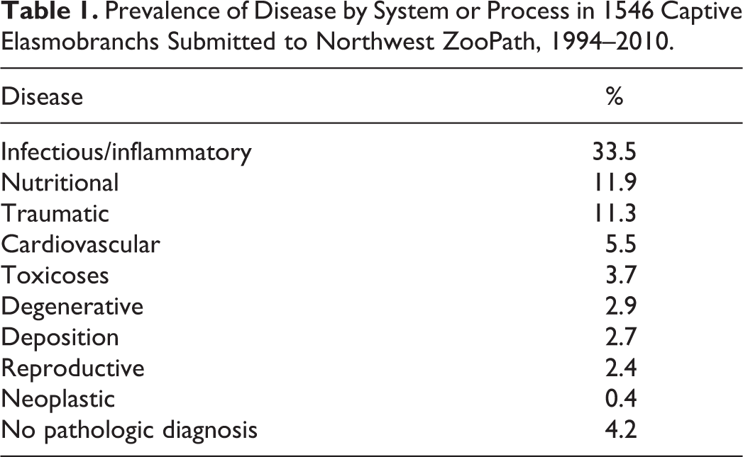
Table 1 lists the disease processes in the study group. In decreasing order, these included infectious/inflammatory (33.5%), nutritional (11.9%), traumatic (11.3%), cardiovascular (5.5%, mostly shock), possible toxin-associated disease (3.7%), degenerative (2.9%), deposition (2.7%), reproductive (2.4%), and neoplastic (0.4%). In addition, no pathologic diagnosis was established for 4.2%. Table 2 lists trends for specific disease processes recognized in 30% or more of the animals within a specific species.
Prevalence of Disease by System or Process in 1546 Captive Elasmobranchs Submitted to Northwest ZooPath, 1994–2010.
Species Related Trends in Disease Processes in Elasmobranchs Submitted to Northwest ZooPath, 1994–2010.a
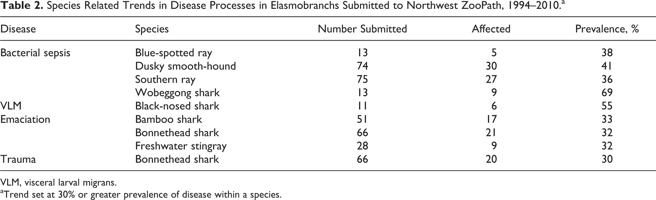
VLM, visceral larval migrans.
aTrend set at 30% or greater prevalence of disease within a species.
Infectious/Inflammatory Disease
Of the infectious diseases, bacterial infections (518/1546, 15%) were most common, based on the observation of bacteria in histologic lesions and usually associated with other disease processes, especially traumatic skin injuries or suboptimal water quality. Bacterial conditions included sepsis (136/518, 26%), dermatitis (7%), branchitis (6%), and enteritis (4%). Bacterial cultures were only rarely obtained or included in the submission information, and no trends in fatal disease related to a specific bacterial agent were identified by histologic, staining, or culture techniques. Species-related trends for sepsis were seen in dusky smooth-hounds (30/74, 41%), southern rays (27/45, 36%), blue-spotted rays (5/13, 38%), and wobeggong sharks (9/13, 69%). An epitheliocystis-like organism was associated with mild branchitis in 3 hammerhead sharks (3/7, 43%) that died following a tank pump failure, and these organisms were considered a probable incidental finding.
Parasitic infections (137/1546, 9%) included nematodiasis (36/137, 26%), ciliate infections (23%), trematodiasis (20%), coccidiosis (6%), myxozoanosis (5%), amoebiasis (4%), cestodiasis (1%), and flagellate infections (1%). A characteristic black serpigenous pattern of tract formation was grossly apparent due to Hufmanella sp nematode egg deposition in the skin of a sandbar shark, as previously reported (Figs. 1, 2). 21 No other typical gross lesions of nematodiasis were recorded. Larvae generally appeared viable and were observed encysted with little host reaction, within microgranulomas, or free within blood vessels and capillaries. The most common sites in which intravascular nematode larvae were encountered histologically were brain, skin, gut wall, liver, mesentery, and gill (Fig. 3). A species trend for nematodiasis was observed in black-nosed sharks (6/11, 55%). Intravascular nematode larvae often were associated with occlusion of capillaries, necrosis, and interstitial inflammation in the gills. In the brain, vessels containing or adjacent to nematode larvae sometimes had fibrinoid necrosis and were surrounded by zones of hemorrhage and mixed inflammation. Ciliate infections in swell sharks, zebra sharks, and horn sharks generally involved the gills, skin, brain, and liver, and the organisms were sometimes difficult to find in tissue section. These ciliates were pyriform shaped and resembled those of the subclass Scuticociliatida. These were often invasive and associated with edema, mixed inflammation, vascular necrosis, and thrombosis, and the vessel lesions were especially prominent in the meninges and neuropil of the brain. Twelve cases of cutaneous ciliate protozoan infection were seen in dusky smooth-hounds, usually in conjunction with bacterial infection, flagellated protozoa, and herpesvirus or adenovirus infections (Fig. 4). These parasites were confined to the skin and connective tissues and resembled Uronema sp. Rarely, larger ciliates that resembled Cryptocaryon sp were detected in the gill interstitium. Flatworm infections were diverse: monogeneans on or encysted in the gills were associated with significant morbidity due to associated proliferative and inflammatory gill lesions (Fig. 5). Digenic trematodes often were encysted in muscle or free in the intestinal lumen and had little if any associated tissue reaction. Occasionally, digenic trematodes and their eggs were identified in the brain and associated with severe hemorrhage, vascular necrosis, and vasculitis (Fig. 6). Coccidiosis was limited to sexual and asexual stages of Eimeria spp developing in the cytoplasm of mesothelial cells of the coelomic viscera in cownose rays. 31 The condition was associated with hypertrophy and hyperplasia of the mesothelium and usually a mild infiltrate of lymphocytes and histiocytes beneath the mesothelial layer, with occasional inflammatory cell exocytosis and mesothelial cell necrosis or exfoliation. Although the lesion was characteristic and generally diffuse on coelomic surfaces, the organisms were difficult to find histologically, usually located on the splenic capsule, epicardium, or capsular surface of the epigonal organ (Fig. 7). A common clinical observation associated with this infection was generalized turbid ascites. Myxozoan spores were most commonly encountered in the gallbladder or bile ducts, glomeruli, or meninges and generally were associated with little tissue reaction or inflammation. Protozoa resembling large amoeba were detected in a few cases, primarily associated with necrosis and hemorrhage in the liver and brain. Encysted cestode larvae were rarely encountered in the mesentery or viscera. Few cases of flagellated protozoan infections were rarely identified in the intestine and bile ducts. An outbreak of fatal microsporidiosis in leopard sharks (Triakis semifasciata) at one facility was characterized by histiocytic inflammation in the gills, spleen, pancreas, brain, and blood vessels, and typical spores were easily identifiable in the cytoplasm of histiocytes and melanomacrophages (Figs. 8, 9). 11
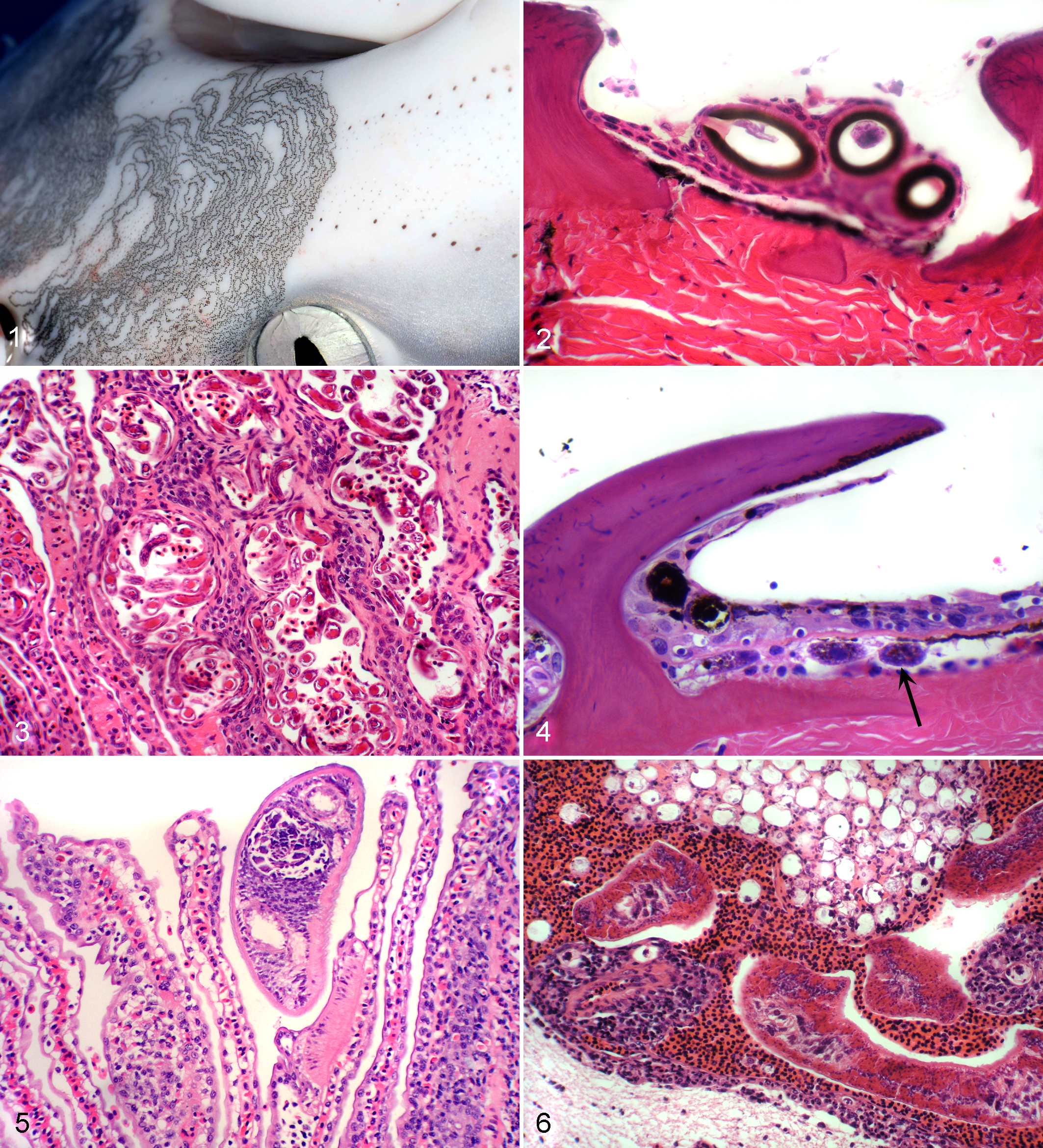
Skin; sandbar shark. Serpigenous migratory tracts associated with Huffmanella sp nematode infection. Photo courtesy of Dr S. Bullard.
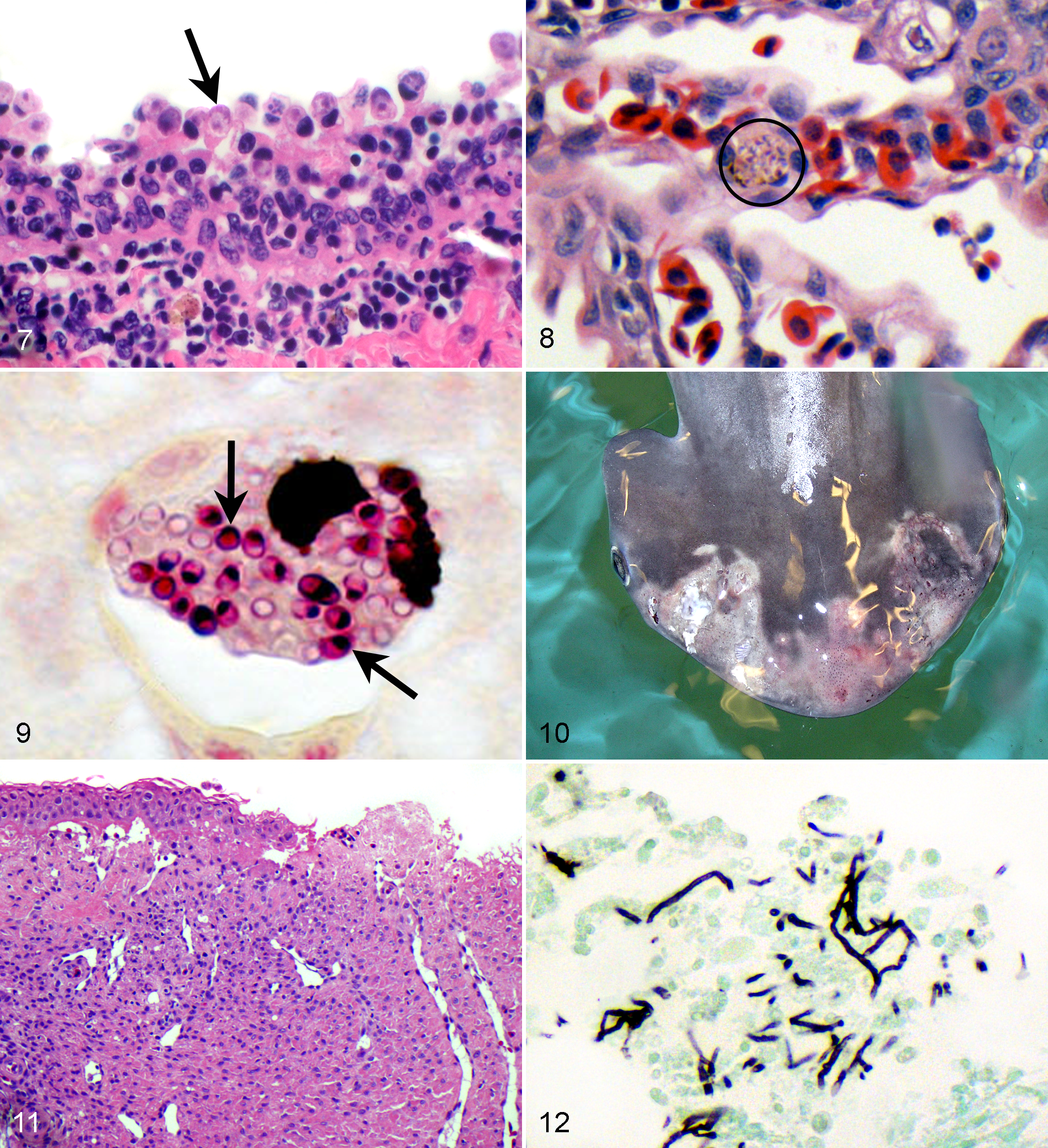
Splenic capsule; cownose ray. Intracytoplasmic coccidia in various stages of development within hypertrophied mesothelial cells, with associated inflammation within the capsule and mesothelial layer. Hematoxylin and eosin (HE).
Mycotic infections were uncommonly encountered (10/1546, 0.6%) and included dermatitis (30%) in 2 cownose rays and one each of sandbar, tiger (Galeocerdo cuvier), bonnethead, and white-tipped reef sharks. Mycotic hepatitis (25%) was seen in southern stingrays and a sandbar shark. Mycotic branchitis (25%) was seen in a tiger shark, a cownose ray, and a yellow stingray (Urobatis jamaicensis). All were hyphae in tissue, and none were further identified by culture or polymerase chain reaction (PCR), although the gross and histologic presentation in the bonnethead was typical of the Fusarium-related disease described in this species (Figs. 10 –12). 30 It is noteworthy that fusariosis in the bonnethead shark invoked necrosis and granulation tissue formation but was not granulomatous.
Viral infections (15/1546, 1%) included adenovirus (40%), herpesvirus (13%), and presumed viral papillomatosis (47%). Six smooth-hounds also had adenovirus-associated inclusion body branchitis and dermatitis detected by histologic and electron microscopic examination, but culture was unsuccessful (Figs. 13, 14). 2 Flagellates and ciliates commonly colonized ulcerated adenovirus-associated skin lesions but were not seen in the gill lesions. Herpesvirus-associated epidermitis and ulcerative dermatitis were in a spiny dogfish (Squalus acanthias) and in a dusky smooth-hound, as has been reported in these fish (Figs. 15, 16). 19
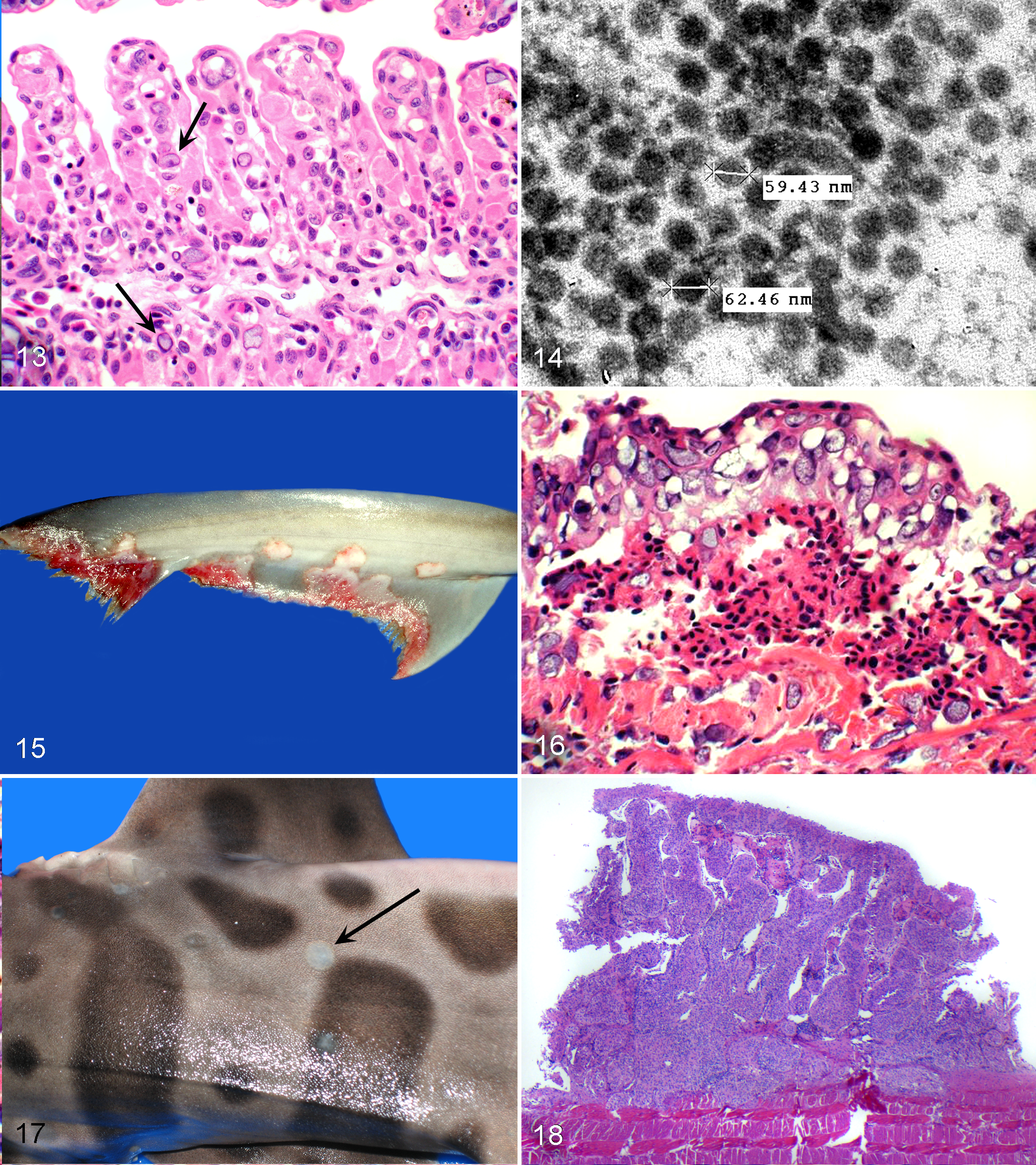
Gill; dusky smooth-hound, adenovirus infection. Note atrophy of secondary lamellae, collapse of the interlamellar space, and intranuclear inclusions in the endothelial cells of the lamellar interstitium (arrows). Hematoxylin and eosin (HE).
Papillomatosis was identified in bamboo, sand tiger, leopard, and white-tipped reef sharks (Triaenodon obesus) from 5 different facilities. This disease was generally multicentric over the trunk, and lesions were slightly raised, slightly pale plaques approximately 1 cm in greatest dimension (Fig. 17). These lesions comprised hyperplastic epidermis with orderly maturation and had no koilocytes or inclusions. Epidermis was supported by fibrovascular stroma, and vessels sometimes were congested or contained thrombi (Figs. 18, 19). These lesions would resolve spontaneously, and no malignant transformation was noted. Transmission electron microscopy did not reveal viral particles in these lesions. Immunohistochemistry did not identify herpesvirus or papillomavirus antigen, and PCR did not identify amplicons using generic herpesvirus or papillomavirus DNA probes.

Skin; leopard shark. Higher magnification of Fig. 18 showing orderly maturation of epidermal layers, absence of koilocytes, and no inclusions. Hematoxylin and eosin (HE).
One catshark had epidermitis with epithelial intranuclear inclusions, and 1 bat ray (Myliobatis californica) had vasculitis with endothelial intranuclear inclusions; these conditions were presumed to be viral, but further diagnostic procedures were not performed. An inclusion forming disorder not encountered in other species on file was identified in histiocytic or myeloid cells in the spleen and epigonal organ of several dusky smooth-hounds from one facility. This condition was presumed to be viral, although no particles were detected in the inclusions by electron microscopic examination, with the inclusions resembling degenerative cell organelles and phagocytized cell debris (Fig. 20).
Inflammation of unknown cause was identified in at least one organ system and subsequently coded for 419 submissions (27%). These included enteritis (55/419, 13%), branchitis (38, 9%), encephalitis (35, 8%) dermatitis (27, 6%), pancreatitis (8, 2%), and splenitis (6, 1%). Inflammation in these cases was usually mild and lymphocytic, and infectious agents were not apparent in histologic section; however, most of these lesions were attributed to parasite migration because lesion morphology and distribution overlapped what was seen in those cases in which parasites were identified in tissues. Three horn sharks from one facility in the same year had fatal necrohemorrhagic pancreatitis. This lesion was characterized by random foci of acute acinar necrosis and hemorrhage, sometimes delineated by small foci of granulocytic inflammation, edema, and thrombosis (Figs. 21, 22). No infectious agents were seen in HE-stained sections or sections stained with B&B, WS, GMS, AF, Giemsa, or Gimenez techniques, and the cause was not determined.
Noninfectious Disease
Cardiovascular disease was diagnosed in 69 of 1546 cases (5.5%) and included systemic shock (27/69, 39%), hemorrhage (9, 13%), osmoregulatory disturbance (6, 9%), myocardial necrosis (6, 9%), and myocardial fibrosis (4, 6%).
Degenerative disease was diagnosed in 45 of 1546 cases (2.9%) and included acute renal tubular necrosis (12/45, 27%), neuronal or neuropil necrosis (9, 20%), chronic renal disease (7, 16%), rhabdomyolysis (4, 9%), and biliary hyperplasia and fibrosis (2, 4%).
Deposition disorders included mineralization (26), miscellaneous crystals or pigments (10), tattoo ink (3), and melanosis (2). Mineralization disorders included renal tubular mineralization (11/26, 42%), metastatic mineralization (8/26, 31%), and calcinosis circumscripta (7/26, 27%). Calcinosis circumscripta was seen focally in skin and associated connective tissues of the wings or body wall of 4 rays, in the skin or connective tissues of swell shark and a coral catshark, and in the endocardium and myocardium of a bowmouth ray (Rhina ancylostoma) (Figs. 23, 24). Fatal tattoo ink emboli occurred in the gills of 3 freshwater stingrays shortly after receiving cutaneous identification tattoos.
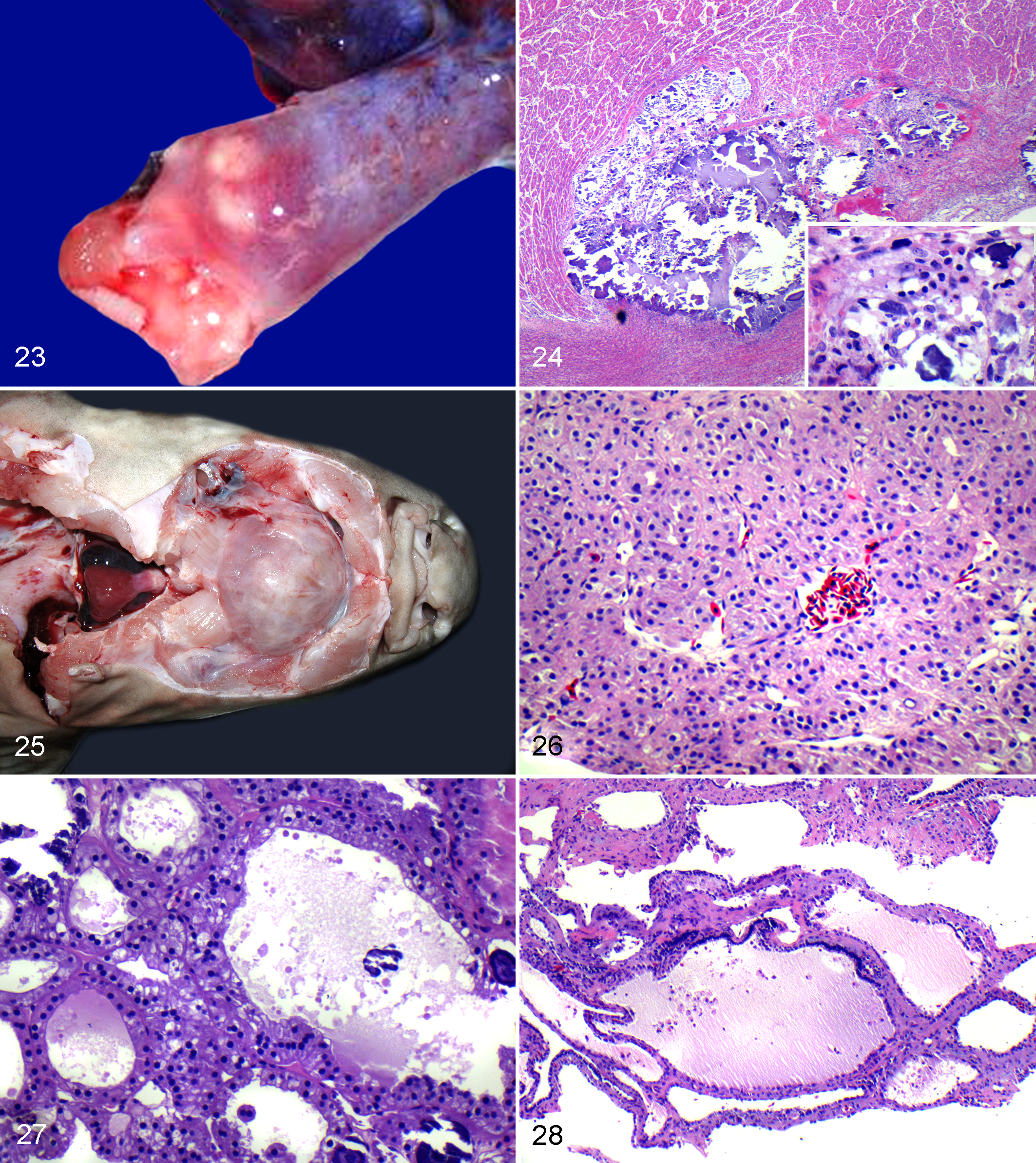
Heart; cownose ray. Note nodular foci of calcinosis circumscripta in the myocardium and great vessel at outflow tract. Photo courtesy of Dr C. Fields.
Metabolic/endocrine disease was limited to 10 of 1546 cases (0.6%) of goiter or suspected goiter in 4 swell sharks, a catshark, a nurse shark, a zebra shark, a cownose ray, and a southern stingray. The goiter was hyperplastic in the cownose ray, a nurse shark, and the zebra shark. The goiter was colloid type in the catshark, the southern ray, and a swell shark and was mixed in a swell shark (Figs. 25 –28). One swell shark biopsy was too small to fully characterize the type of goiter.
Nutritional disease was diagnosed in 184 of 1546 cases (12%) but was the sole problem in only 24 of these cases. All cases related to emaciation or suboptimal nutritional status based on depletion or absence of lipid stores from hepatocytes (Figs. 29 –32). A trend in emaciation was noted in bamboo sharks (33%), bonnethead sharks (32%), and freshwater stingrays (32%).
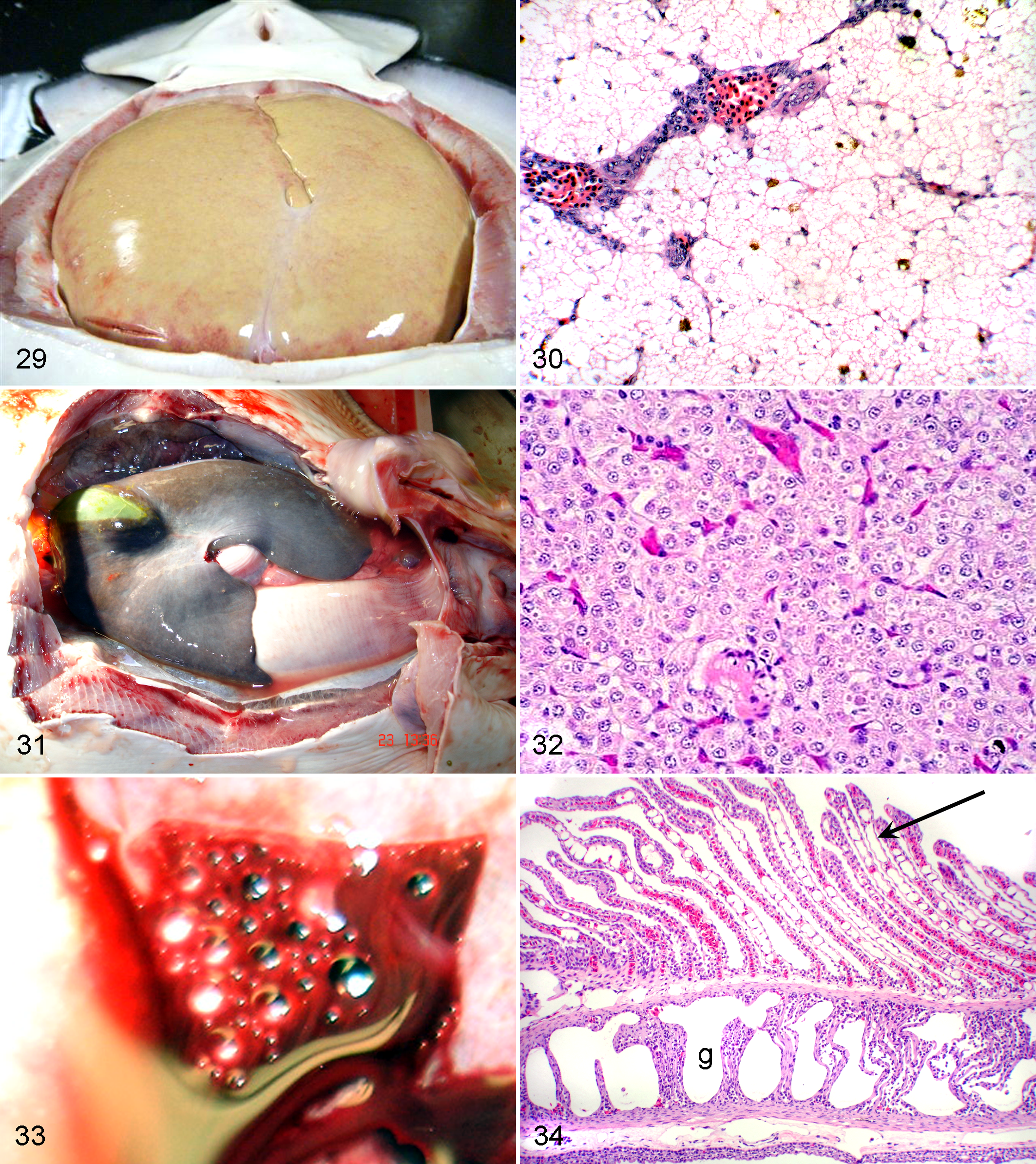
Liver; cownose ray. The bulging tan fatty appearance indicates normal lipid storage and good to perhaps corpulent nutritional status. Photo courtesy of Dr B. Doescher.
Neoplasia was diagnosed in 6 of 1546 cases (4%) and included multicentric dermal fibromas in a black-tipped reef shark, a renal fibroma in an Atlantic stingray, a pancreatic adenocarcinoma in a leopard shark, a melanoma in a nurse shark, 34 an adenoma of the corpuscle of Stannius in a stingray, and a pituitary adenoma in a zebra shark.
Various forms of trauma were encountered in 174 of 1546 cases (11%), and affected systems included skin (103), alimentary tract (12), and stress-related lesions (epidermal necrosis, lymphoid depletion, and/or rhabdomyolysis) (16). A trend in lesions related to trauma was noted in the bonnethead shark (30%).
Toxicoses were coded for 57 of 1546 cases (4%) and included embolic gas disease (11, 19%) (Figs. 33, 34), ammonia toxicosis (4, 7%), fenbendazole toxicosis (4, 7%), and chloramine toxicosis (3, 5%). “Toxic gill disease” for which the agent was not known was diagnosed in 21 of 57 cases (37%). Five cases of neuronal or glial cell necrosis were also included as possible toxicosis-related events.
Reproductive disease was coded for 30 of 1546 (2%) cases, was limited to females, and included neonatal death (20, 66%), follicular degeneration or oophoritis (5, 17%), pyometra (3, 10%), and yolk embolism (2, 7%).
Discussion
Cownose rays, southern rays, dusky smooth-hounds, bonnethead sharks, and bamboo sharks were the most commonly submitted species, and these trends correspond well with the recent census results for captive elasmobranch species (American Elasmobranch Society, Census Data, 2008 http://elasmo.org/). Inflammatory/infectious diseases were the most commonly encountered diseases. This was an expected result based on unpublished data obtained at Northwest ZooPath from zoo submissions in teleosts and other animal classes from US zoological and aquarium facilities over several years, in which inflammatory and infectious disease has consistently been the most common disease category in annual reports and retrospective studies.
There has been little documentation on the pathogenic bacteria of elasmobranchs beyond the flora in the shark mouth that may be pathogenic to humans. Grimes et al 14 described natural and experimental shark meningitis caused by infection with Vibrio carchariae and also documented the existence of bacteria in normal tissues of asymptomatic sharks. Hemorrhagic septicemia caused by Aeromonas salmonicida has recently been described in a black-tipped reef shark. 3 In the fish of this report, bacterial infections primarily involved skin wounds and compromised gills, which then served as a source of sepsis. Cultures and special stains were rarely done because these “opportunist” patterns are easily recognized as indicators of penmate aggression, suboptimal water quality or housing, or environmental maladaptation. It is noteworthy that no cases of mycobacteriosis were diagnosed in this elasmobranch study; although some species can seroconvert following exposure to mycobacteria, 17 it would appear that elasmobranchs are not particularly susceptible to mycobacteriosis, whereas the disease is common in teleosts. 15
Fungal infections are reportedly sporadic to common in teleosts, the most common agents being oomycetes such as Saprolegnia spp or dematiaceous (pigmented) fungi such as Exophiala spp (noga, bruno). 4,26 Few fungal diseases have been reported in elasmobranchs, although fusariosis is a distinct entity in the bonnethead shark. 22,30 It is noteworthy that the bonnethead shark reaction to fusariosis is ulceration and granulation tissue formation in the skin of the head and fins. Granuloma formation is not characteristic. In this regard, it is possible that fusariosis may be more common than is diagnosed histologically, since the histologic lesion does not seem to suggest infection in the HE-stained sections. It is unclear if this is true for all elasmobranchs.
Viral diseases are common in teleosts and can have a profound economic impact in the aquaculture industry. 5 Only 2 viral diseases have been described in elasmobranchs: a herpesvirus has been documented to cause ulceration and necrosis of the epidermis of wild and captive dusky smooth-hounds. 19 Grossly, these lesions present as small gray slightly raised plaques that eventually ulcerate. Histologically, these lesions comprise epithelial cells with intracellular edema or balloon degeneration, intranuclear inclusions, exfoliation, and necrosis. The lesions in the smooth-hounds reported herein were essentially identical to those of the original report. The other viral disease from the literature is viral erythrocytic necrosis, caused by an iridovirus that forms intracytoplasmic inclusions in circulating erythrocytes, and this condition was not seen in the study animals. 16
Some additional viral or suspected viral diseases were encountered in our study. An adenovirus- like infection associated with marked branchitis and dermatitis was diagnosed histologically and electron microscopically in dusky smooth-hounds, but attempts to culture this virus were unsuccessful and molecular studies of this virus were not performed. Cutaneous papillomas were diagnosed in several different species of sharks from different facilities, and these lesions had consistent morphology and behavior that suggested that they were viral-induced lesions. Although they did not have inclusions or koilocytes, they appeared to be contagious and spontaneously regressed over time. To date, electron microscopy, immunohistochemistry, and in situ hybridization have been unsuccessful in demonstrating the presence of virus in these lesions. Several dusky smooth-hounds at one facility had an unusual form of inclusion forming disorder in the myeloid and histiocytic cells of the epigonal gland and spleen. These fish usually were septic and clinically appeared stressed and maladapted to their exhibit. An immunosuppressive viral agent was considered possible but could not be demonstrated by electron microscopy in the inclusions, which contained membranous whorls of degenerative cell organelles and debris and occasional bacteria. Other cases in which inclusions were present or lesions were suspicious for a viral etiology were not further investigated.
An interesting form of necrotizing pancreatitis was encountered in horn sharks during 1999. Pancreatitis was commonly seen in elasmobranchs as a mild condition in conjunction with generalized lymphocytic inflammation due to parasite migration; however, it is not typically granulocytic, associated with necrosis and hemorrhage, or severe enough to cause clinical morbidity or death. The necrotizing lesion in horn sharks was severe and likely caused death in these fish. Its morphologic appearance was suggestive of an infectious event, and it resembled the lesion of infectious pancreatic necrosis of salmonids and other teleosts.
Various forms of parasitism are the best documented of the infectious processes of elasmobranchs. A full review is beyond the scope of this article, but some good summaries exist. 1,6,13 Most of the infections seen in this study have been previously described, and some are known pathogens such as the branchial monogeneans. Some have more vague interpretations regarding pathogenicity, such as nematodiasis or coccidiosis in cownose rays. 31 The author is of the opinion that intravascular nematode migration can be a pathologic event in elasmobranchs, especially those cases that involve the brain and gills, where there is considerable inflammation and occlusion of capillaries. Larvae generally were viable and did not have features to suggest that tissues were reactions were to dead larvae in treated animals. Also, the inflammation, proliferative mesothelial changes, and coelomic effusion that accompany coccidiosis in cownose rays likely contribute substantially to the wasting seen in affected fish. 31 A previously undescribed fatal systemic ciliate protozoan infection attributed to Philasterides dicentrarchi was identified in sharks, and a more complete review of this disease is covered elsewhere. 32 Systemic microsporidiosis was encountered in a collection of leopard sharks early in the study; although microsporidiosis is common in teleosts, this condition appears to be rare in elasmobranchs, with only 2 reports in the literature to date. 10,11 In contrast to the systemic infection seen in leopard sharks, microsporidiosis of Caribbean stingrays (Dasyatis pastinaca) caused by Dasyatispora levantinae gen. et sp. nov is limited to the disc. Infection causes development of disfiguring nodules containing yellowish-white caseous substance consisting of degraded host tissue and microsporidian sporophorous vesicles, which in turn contain developing sporonts, sporoblasts, and spores. 10 Genetic analysis of the microsporidia from the leopard sharks was not performed.
Elasmobranchs have an interesting form of soft tissue mineralization: the nodular pattern of mineral deposition surrounded by a narrow zone of macrophages and fibroblasts resembles calcinosis circumscripta (tumoral calcinosis, pseudogout), but the distribution of the lesion is not limited to the peripheral connective tissues as seen in mammals and reptiles. 12 Although the lesion may involve skin and associated tissues of the trunk in sharks and the wings of rays, it also may be seen on serosal surfaces of the viscera and on the epicardial surface of the heart, a distribution pattern reminiscent of metastatic mineralization. A few of the cases in our files have been associated with chronic renal disease, suggesting an association, although more investigation is needed to further characterize the pathogenesis of this disorder in elasmobranchs. The author has not encountered similar lesions in teleosts.
Goiter is well documented in elasmobranchs and has been described as hyperplastic, colloid, or mixed. 8 Historically, this condition has been attributed to derangements in iodine metabolism, either too much or too little iodine in the diet, or exposure to goitrogenic substances. Recent experimental studies have shown that high environmental nitrate concentrations interfere with thyroid utilization of available iodine, resulting in overstimulation of the thyroid by thyroid-stimulating hormone and subsequent hyperplastic goiter. 24 In the fish of our study, swell sharks appeared to be overrepresented for this condition; however, this is a conservative interpretation because thyroid was not always included in submitted tissue sets. Swell sharks may be overrepresented due to increased species susceptibility to environmental nitrates or perhaps have more specific dietary iodine requirements than other elasmobranchs.
The only nutritional problem coded in the study group was suboptimal nutritional status or emaciation. Elasmobranchs may have adipocytes as a component of the connective tissues of the body wall or fins 20 but apparently do not have cavitary adipose tissue; the author has never encountered it histologically and could find no references pertaining to cavitary adipose tissue. Elasmobranchs primarily store lipid in hepatocytes, which makes the liver a sensitive indicator of nutritional status. 28 In contrast to other animal classes and even teleost fishes, a lipid-laden liver in elasmobranchs indicates good nutritional status, whereas depleted lipid stores correspond to suboptimal or emaciated nutritional status.
As expected, trauma was frequently encountered in various forms. Elasmobranchs are predatory fish, are easily stressed, can be aggressive, and sometimes are in high-density mixed exhibits. Conspecific or tank mate aggression and wall collisions are common. 9 Although not proven experimentally in elasmobranchs, it is possible that stress-induced epidermal necrosis may also occur, leading to ulcerative skin and fin lesions as seen in teleosts, predisposing to opportunistic bacterial infection. 27
Fenbendazole overdoses occurred at 2 different facilities, with associated radiomimetic lesions in the gut and epigonal organs. 25 Mortality associated with exposure to volatile organic compounds occurred at one facility when elasmobranchs and teleosts were introduced to a new exhibit in which paint fumes had apparently settled in the tank. 29 Gas embolism (gas bubble disease) was commonly encountered, associated with oxygen or nitrogen supersaturation. This event can occur due to equipment malfunction, poor exhibit design (waterfalls into deep pools), use of supersaturated well water, and passage of severe low-pressure storm fronts. 23 To maintain water quality for elasmobranchs, large quantities of water must be processed; not all elasmobranchs have the same water quality parameters, and it appears that many of these fish are very sensitive to fluctuations in water quality and environmental toxins. Thus, many problems associated with water quality were encountered in elasmobranches. As with teleosts, the gills are a target tissue for this form of disease process and after acute exposure develop congestion or venous aneurysms and necrosis of the lamellar epithelium, so-called toxic gill disease. Those that live a few days will develop blunting or fusion of the secondary lamellae and hypertrophy or exfoliation of the remaining lamellar epithelial cells. Acute epidermal necrosis and ulceration may also develop, and visceral lesions that suggest hypoxic change due to gill damage may be seen in the liver, such as hepatocellular centrilobular necrosis and renal tubular necrosis. The offending toxin was known in some cases based on water evaluation (ie, chlorine, ammonia) or was suspected based on history or histologic lesions. Stress-related skin lesions have morphology that overlap those seen with lesions of environmental toxicoses, and these disease processes can be difficult or impossible to distinguish histologically without adequate information regarding history and water quality, especially in suboptimally preserved specimens.
Footnotes
Acknowledgements
I am immeasurably grateful to Drs Jim Raymond and John Trupkiewicz for slide reading during the study intervals and to Dr Nancy Stedman for angelic backup support during manuscript preparation. I also thank Roy Brown of Histology Consulting Service for slide preparation, Liz Post and Sue Hutton of Northwest ZooPath for slide and data retrieval, and the omnipresent Christie Buie of Northwest ZooPath for reference retrievals, image layout, and manuscript submission.
I also thank Robert Nordhausen of the California Animal Health and Food Safety Laboratory and Dr Dalen Agnew of the Diagnostic Center for Population and Animal Health, Michigan State University for electron microscopy support; Dr Matti Kiupel of the Diagnostic Center for Population and Animal Health, Michigan State University for molecular diagnostic support; and the veterinarians and staff at the following institutions for case submissions: Akron Zoo, Animal Medical Center/Oregon Coast Aquarium, Audubon Park Zoo, Aquarium of the Americas, Aquarium at Moody Gardens, Aquarium of the Bay, Aquarium of the Pacific, Bristol Veterinary Service, Buttonwood Park Zoo, Cabrillo High School Aquarium, California Science Center, Clearwater Marine Aquarium, Cleveland Metroparks Zoo, Columbus Zoo, Rosamond-Gifford Zoo, Dallas World Aquarium, Dallas Zoo, Disney s Animal Programs, El Paso Zoo, Exotic and Companion Animal Services, Florida Aquarium, Folly Road Animal Hospital, Fresno Chaffee Zoo, Fort Worth Zoo, Galveston Veterinary Clinic, Gentle Doctor Animal Hospital, Hecker Animal Clinic, Hendricks County Animal Hospital, Henry Doorly Zoo, Houston Zoo, Indianapolis Zoo, John Ball Zoo, Jones Animal Hospital, Kansas City Zoo, Living Exhibits, Marathon Veterinary Hospital, Marine Animal Medical Conservancy, Medfield Veterinary Clinic, Miami Metrozoo, Miami Seaquarium, Minnesota Zoo, Monterey Bay Aquarium, National Aquarium in Baltimore, National Aquarium in Washington DC, Oklahoma City Zoo, Paradise Islands, Peaceful Passing, Phoenix Zoo, Pittsburg Zoo, Pt. Defiance Zoo and Aquarium, San Francisco Zoo, San Antonio Zoo, Santa Barbara Zoo, Sea Life Aquarium, Sea Life Park Hawaii, Six Flags World of Adventure (Ohio), South Carolina Aquarium, South Wilton Veterinary Group, The Mirage, The Oregon Zoo, The Seattle Aquarium, The Toledo Zoo, Theater By The Sea, Tulsa Zoo, University of Maryland Medical School, Veterinary Aquarium Group, West Flamingo Animal Hospital, Wildlife World Zoo, Woods Hole Oceanographic Institute, and Woods Hole Science Aquarium.
Declaration of Conflicting Interests
The author(s) declared no potential conflicts of interest with respect to the research, authorship, and/or publication of this article.
Funding
The author(s) received no financial support for the research, authorship, and/or publication of this article.