
Abstract
A 23-year-old Anglo-Arabian mare was presented with tachypnea, dyspnea, and pitting edema of the ventral thoracic subcutis. On necropsy, a tan to red, friable, irregularly shaped mass (23 × 20 × 18 cm) occupied the cranial mediastinum. Histologically, the mass was classified as a liposarcoma and was composed of short interlacing bundles of spindle-shaped to irregularly rounded cells with discrete, variably sized, clear cytoplasmic vacuoles, which were stained with oil red O in frozen sections of formalin-fixed tissue.
Neoplasms of adipocytes are classified as lipomas, infiltrative lipomas, angiolipomas, and liposarcomas. Liposarcomas are found mainly in the subcutis and soft tissues, 4 less commonly in the bone marrow or alimentary tract, 7,12 but rarely in other internal organs in domestic animals. The prevalence of thoracic neoplasia is low in horses. 10,11 The more common primary neoplasms in the equine thorax are pulmonary granular cell tumor, mesothelioma, and thymoma. 2,10 Multicentric lymphoma and various metastatic neoplasms have also been reported in the equine thorax. 10,11 Although mediastinal liposarcoma is documented in humans, 5 it apparently has not been reported in domestic animals. The purpose of this report is to describe the histologic and histochemical findings in a case of cranial mediastinal liposarcoma in a horse.
Case history
A 23-year-old, 452-kg, Anglo-Arabian mare was presented with acute tachypnea, dyspnea, and pitting edema of the ventral thoracic subcutis. On thoracic auscultation, lung sounds were decreased in the right hemithorax and ventral aspect of the left hemithorax. Pleural effusion was detected by ultrasonography, and approximately 20 liters of serosanguineous fluid was drained by thoracocentesis. No evidence of infection or neoplastic cells was evident by cytologic examination of the pleural effusion. When a cranial mediastinal mass was detected radiographically after removal of the pleural effusion, euthanasia was elected. At necropsy, a 23 × 20 × 18 cm, tan to red, friable, irregularly shaped mass filled the cranial mediastinum and was focally adhered to the pericardium (Fig. 1). The cut surface of the mass was pale tan to yellow with cystic cavities containing sanguineous fluid. The mass displaced the lungs caudally but did not invade thoracic organs. The only other pertinent gross finding was a polypoid, pedunculated mass (4 × 3 × 2 cm) in the pyloric mucosa. Samples of major organs were fixed in 10% neutral buffered formalin. Representative trimmed tissues were routinely processed, paraffin embedded, sectioned at 5 μm, and stained with hematoxylin and eosin. Sections of the mediastinal mass were also evaluated with Masson’s trichrome stain, Grimelius, Alcian blue (pH 2.5), and periodic acid–Schiff (PAS). Frozen sections from the formalin-fixed samples of the mass were stained with oil red O.
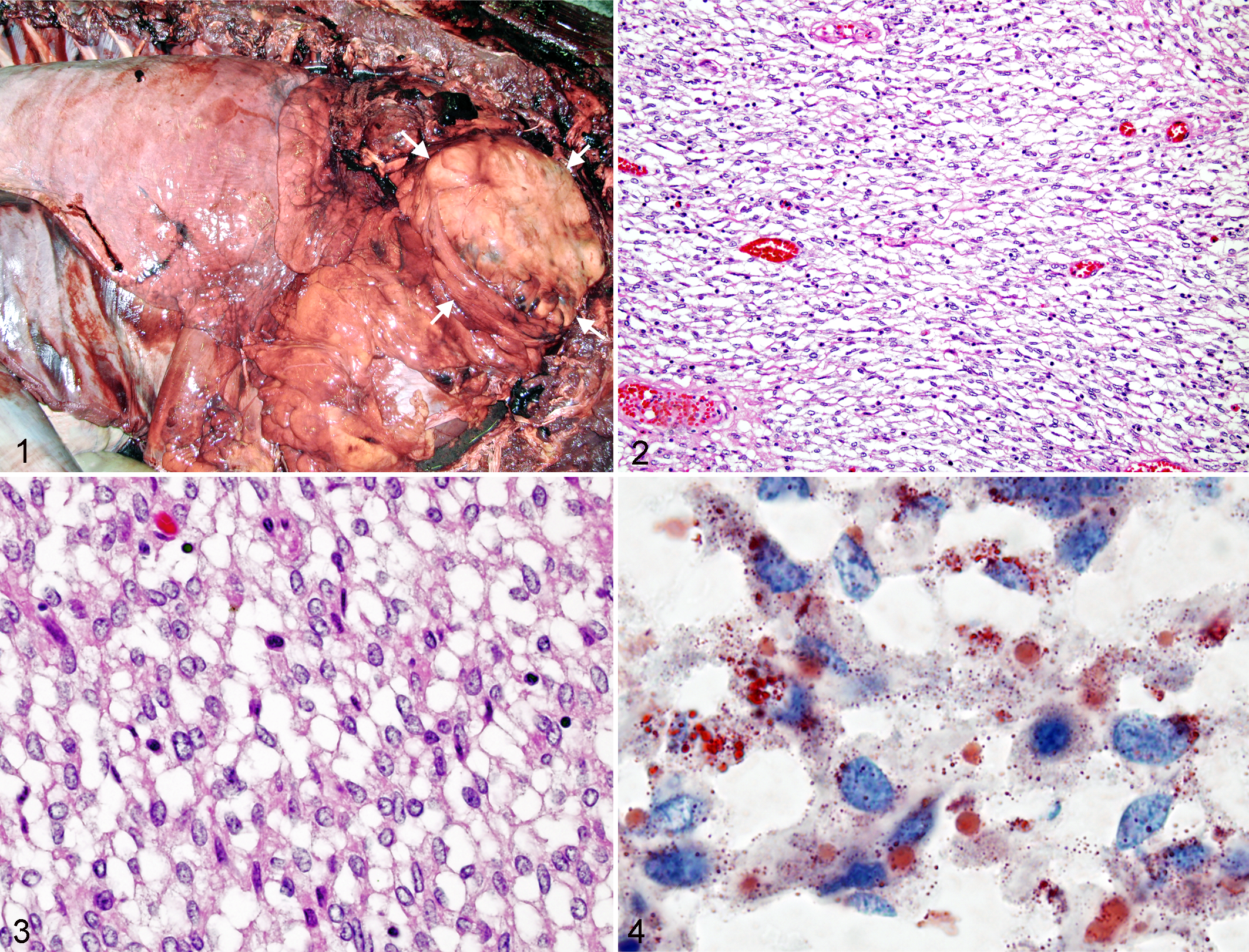
Cranial mediastinal mass; horse. The tan lobulated mass (arrows) fills the cranial mediastinum.
Histologically, the cranial mediastinal mass was composed of densely cellular, short interlacing bundles of spindle-shaped to irregularly rounded cells supported by scanty to moderate amounts of delicate fibrous stroma with multifocal hemorrhage and necrosis and few scattered eosinophils, lymphocytes, and plasma cells (Fig. 2). The stroma stained deep blue with Masson’s trichrome. The neoplastic cells had a round to elongated central to eccentric nucleus, moderate variation in nuclear and cellular diameter, coarse chromatin, distinct nucleolus, 5 mitotic figures per ten 400× fields, indistinct cell borders, and variably sized unstained cytoplasmic vacuoles with sharply delineated borders (Fig. 3). The cytoplasmic vacuoles were neither Alcian blue nor PAS positive but were stained with oil red O (Fig. 4). No argyrophilic granules were observed with Grimelius technique. The pyloric mucosal mass was classified as an adenoma and considered an incidental finding, unrelated to the mediastinal liposarcoma.
Discussion
The diagnosis of liposarcoma in this horse was based on the histologic finding of spindle-shaped neoplastic cells with cytoplasmic vacuoles stained by oil red O. Liposarcomas are classified as well differentiated, pleomorphic, or myxoid. 3,8 In this case, because most neoplastic cells resembled nonneoplastic adipocytes and contained clear lipid vacuoles, the neoplasm was classified as a well-differentiated liposarcoma. The differential diagnosis at necropsy included thymoma, lymphoma, mesothelioma and neuroendocrine tumors, such as aortic body chemodectoma. Lymphoma and mesothelioma are relatively common among equine thoracic neoplasms. 9 A lipid-rich peritoneal mesothelioma has been reported in a horse with disseminated masses that were composed of epithelioid cells, some with cytoplasmic lipid. 3 Each of these entities was eliminated from the differential diagnosis, because the mass in the horse of this report was composed of spindle-shaped cells without round cell or epithelial/epithelioid cell populations. Furthermore, the presence of a solitary mediastinal mass is not typical of equine mesothelioma. 2,3 The lymphoid cell population typically observed in thymomas was absent in this case. No cytoplasmic argyrophilic granules were detected in the neoplastic cells, as would be expected with a neuroendocrine tumor.
Intrapericardial lipoma and infiltrative lipoma in the heart have been reported in horses. 1,6 In the present case, the mass was not associated with the heart or other thoracic organs. Although focally adhered to the pericardium, pericardial origin was considered unlikely, and the cytologic features were not consistent with a benign lipoma. 6 It is difficult to define the origin of the mass in the present case, but thoracic adipose tissue would be most likely. To our knowledge, this is the first report of an anterior mediastinal liposarcoma in the horse.
Footnotes
Declaration of Conflicting Interests
The authors declared no potential conflicts of interest with respect to the research, authorship, and/or publication of this article.
Funding
The authors received no financial support for the research, authorship, and/or publication of this article.