
Abstract
The present study was performed to determine the morphologic change and selected molecular features of spontaneous lung tumors in cats examined at the North Carolina State University Veterinary Teaching Hospital. Thirty-nine primary lung carcinomas represented 0.69% of all feline cases admitted to the hospital. Most lung tumors were observed in aged cats (P < .0001), and no sex predilection was found (P < .4241). Persian cats with pulmonary carcinoma were overrepresented in the data set, at least 4 times more frequently than other breeds. The histologic tumor types included adenocarcinoma (64.1%), bronchioloalveolar carcinoma (20.5%), and adenosquamous carcinoma (15.4%). Metastasis was observed in about 80% of 39 cases, with decreasing order of intrapulmonary metastasis, intrathoracic carcinomatosis, regional lymph nodes, and distant organs, including digits. The size of the largest tumor mass was significantly associated with metastatic potential (P < .001). Based on immunohistochemistry, more than 80% (20 of 24) of feline lung tumors were positively labeled with either surfactant protein A or thyroid transcription factor 1. Epidermal growth factor receptor mutant and p53 proteins were detected in approximately 20% (5 of 24) and 25% (6 of 24) of the feline lung tumor cases, respectively. Limited sequencing analysis of K-ras and p53 genes in 3 selected normal and neoplastic lung tissues did not reveal any alteration. Results indicate that primary lung carcinomas are rare but aggressive tumors in cats, thereby warranting further studies on molecular carcinogenesis.
Keywords
Lung cancer is exceptionally deadly. It has the highest mortality rate among all types of cancer in both men and women and is second in new cancer cases. In the United States, human lung cancer accounts for 30% of cancer deaths every year, 16 and new cases and deaths from lung cancer in 2008 were 215 000 and 160 000, respectively (http://www.cancer.gov/cancertopics/types/lung). Lung cancer incidence rates stabilized in men from 1995 through 2001 but continued to increase by 0.3% per year from 1987 through 2001 in women. 15 Lung cancer now kills more women than breast cancer and more men than prostate and colon cancer combined. 16 Nearly 75% of patients who present with lung cancer have advanced local or metastatic disease that is not amenable to cure. 13
Unlike humans, animals rarely suffer from primary lung cancers. In domestic animals, spontaneous lung tumors are more frequently observed in dogs, cats, and sheep. 26 Ovine lung adenocarcinoma (AC), also known as jaagsiekte, is an enzootic contagious tumor caused by retroviral infection. 7 In contrast, lung tumors in domestic dogs and cats occur as a sporadic geriatric disease not related to any infectious cause. 26 Cats affected with lung tumors can have a variety of clinical signs, including lethargy, weight loss, wheezing, coughing, dyspnea, anorexia, vomiting, diarrhea, tachypnea, ataxia, hemoptysis, and lameness. The feline lung tumors have been treated by pneumonectomy and adjuvant chemotherapy. In cats with moderately differentiated tumors, the median survival time with complete surgical resection is 698 days; in cats with poorly differentiated tumors, the median survival time is 75 days. 11 The median survival time in cats with primary lung tumors has been statistically related only to histologic morphology of the tumor. Currently, there is no molecular biomarker for the prognosis of feline lung tumors.
Neoplasia can arise from every component of the lung. In animals, important lung cancers mostly originate from epithelium of the conducting airways or alveolar parenchyma. According to the World Health Organization classification, 26 malignant pulmonary epithelial tumors in domestic animals are classified on the basis of morphology and cell of origin: adenocarcinoma (AC), bronchioloalveolar carcinoma (BA), squamous cell carcinoma (SCC), adenosquamous carcinoma (AS), and so on. In contrast to canine pulmonary carcinomas, feline tumors of airway origin with histologic pattern of AC are more common than tumors arising from the bronchioloalveolar junction. Because of the aggressive growth and overlapping histologic pattern of tumors at different stages of tumor progression within the lung, it is often difficult to delineate the cell of origin based solely on gross and light microscopic examinations.
Localization of molecular markers by immunohistochemistry has been widely adopted to elucidate the histogenetic origin and shed light on potential mechanisms involved in the development of human and veterinary tumors. Surfactant proteins (SPs) and thyroid transcription factor 1 (TTF-1) frequently serve as diagnostic markers for lung AC, with alveolar type II pneumocytes and bronchiolar Clara cells thought to be the cells of origin. It is well known that multiple signaling pathways associated with p53, K-ras, and epidermal growth factor receptor (EGFR) genes play an important role in the genesis and progression of human and rodent lung cancers. Grossmann et al reported the comparative molecular and biological properties of a feline AC cell line established from a 12-year-old Persian cat. 8 This cell line has phenotypic characteristics of type II pneumocytes, such as intracytoplasmic lamellar bodies and immunoreactivities to surfactant protein and TTF-1. Sequencing analyses of this cell line demonstrated that the p53 gene had a homozygous G to T transversion at codon 167, while H-ras and K-ras were not altered.
To provide further insight into the histogenetic cell origin and carcinogenic mechanisms of feline lung cancers, we evaluated spontaneous pulmonary carcinomas, using light microscopy, immunohistochemistry, and sequencing methods.
Materials and Methods
Feline Lung Tumors
Thirty-nine cat lung tumor cases were included in this study after a review of the pathology reports of the feline cases admitted to the Veterinary Teaching Hospital, College of Veterinary Medicine, North Carolina State University, between January 2006 and August 2010. Clinical data, pathology reports, and paraffin-embedded tissue blocks and sections were obtained from the selected cases. The lung tumor slides were reexamined microscopically and reclassified on the basis of published guidelines that categorize these neoplasms by histologic pattern without reference to their site of origin. 26 The clinical history of the cats with primary lung cancers and the incidence of their subtypes are summarized in Table 1.
Distribution of Sex, Age, Breed, and Histologic Subtypes of Feline Lung Tumors, No. (%)a
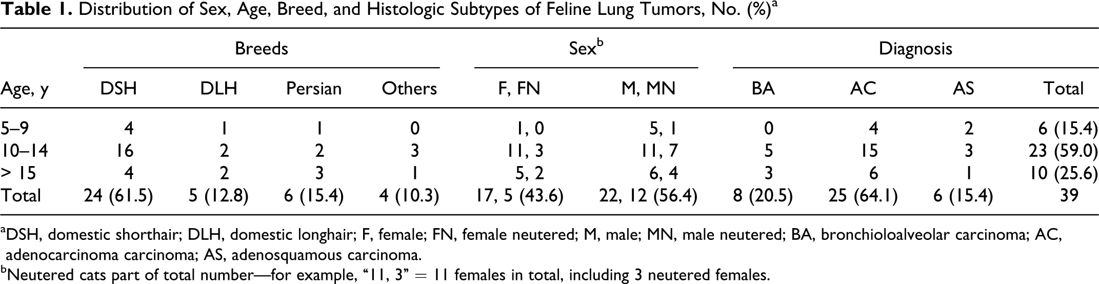
aDSH, domestic shorthair; DLH, domestic longhair; F, female; FN, female neutered; M, male; MN, male neutered; BA, bronchioloalveolar carcinoma; AC, adenocarcinoma carcinoma; AS, adenosquamous carcinoma.
bNeutered cats part of total number—for example, “11, 3” = 11 females in total, including 3 neutered females.
Immunohistochemistry and Evaluation
Formalin-fixed, paraffin-embedded lung tumor tissues were sectioned at 3 to 5 μm of thickness, mounted on glass slides, baked in an oven at 60°C for 1 hour, deparaffinized in xylene, and rehydrated through a graded series of ethanol concentrations. Antigen retrieval was performed with the target retrieval buffer in the DakoCytomation Pascal pressure chamber according to manufacturer’s recommendations. Following antigen retrieval, slides were placed in running tap water and used for immunohistochemistry. Immunohistochemistry was performed with the Vectastain ABC Kit (Vector Laboratories) based on manufacturer’s recommendations. Endogenous peroxidase activity was quenched by incubation in hydrogen peroxide (0.3% or 3%) solution in water for 30 minutes. Primary antibodies included surfactant protein A (SP-A; Biocare Medical, Clone 32E12, 1:200), TTF-1 (Dako, Clone 8G7G3/1, 1:100 dilution), p53 (Vector Laboratories, CM5, 1:200), and EGFR (Cell Signaling Technology, E746-A750 del Specific, 6B6, 1:200). As a negative control, the primary antibody was either replaced with normal serum or omitted from the staining protocol. As a positive control, we used normal cat lung tissue (for SP-A and TTF-1) and a nasal squamous cell carcinoma (for p53 and EGFR). Labeling was visualized with application of DAB substrate (Vector Labs) to the sections for 2 to 10 minutes until color developed. Sections were rinsed and counterstained with Mayer’s hematoxylin and coverslipped with Aquamount (BioGenex) or Permount (Sigma).
Slides were independently subjected to blind scoring by 2 independent researchers (S.D. and D.-Y.K.). Any discordant interpretation was resolved among the reviewing researchers at a multiheaded microscope. Immunoreactivity was evaluated as a product of the percentage of cells staining positively and the relative staining intensity. Labeling in more than 10% of neoplastic cells with moderate or strong intensity was interpreted as positive. The distribution of staining in individual cells was noted as membranous, cytoplasmic, or nuclear.
Sequencing of K-ras and p53
For the K-ras gene, total RNA was extracted from the normal and tumor areas of 3 lung samples with the RNeasy Mini Kit (Qiagen). After treatment with TURBO DNase, cDNA was transcribed with Superscript III Reverse Transcriptase and oligo dT primers. For the p53 gene, genomic DNA was extracted from the normal and tumor areas of 3 feline lung ACs with the DNeasy Blood and Tissue kit (Qiagen). Polymerase chain reaction (PCR) reactions for K-ras and p53 genes were performed on 50-μl volumes containing 1X PCR buffer, 2mM MgC12, 0.2mM dNTPs, 5μM primers, 5 U of Platinum Taq polymerase (Invitrogen), and cDNA or genomic DNA. Primers 8 included K-ras sense: 5′-GACTGAATATAAACTTGTGG-3′, K-ras antisense: 5′-CTATAATGGTGAATATCTTC-3′, p53 sense: 5′-GGCGCCTATGGTTTCCATTTAG-3′, and p53 antisense: 5′-CATCCAGTGGCTTCTTCTTTTG-3′. PCR conditions consisted of 35 cycles of denaturation at 94°C (1 minute), annealing at 50°C (1.5 minutes), and extension at 72°C (2 minutes). The PCR product was run on a gel, and DNA was purified from a gel slice using the QIAquick Gel Extraction Kit (Qiagen) according to the manufacturer’s recommendations. The extracted DNA was sequenced (Eton Biosciences) using the aforementioned K-ras or p53 primers. The sequences were compared to published sequences for feline p53 20 and K-ras 25 genes, which are registered under National Center for Biotechnology Information accession Nos. D26608 and U62089, respectively.
Statistical Analysis
To compare differences in categorical variables within the 39 samples collected, Fisher exact tests were performed (since small expected values in some contingency table cells made parametric analysis inappropriate). One-proportion t tests were performed to test for deviations in the proportions of demographic variables in the cases evaluated (eg, sex, breed, and age distribution) from the expected proportions from the full cohort data. To correct for multiple comparisons, a Bonferroni corrected α of 0.05 / 12 = 0.004 was used for ascribing significance to the results. This correction keeps the family-wise error rate of the overall study to 0.05. Analyses were performed using Stata 11 (http://www.stata.org).
Results
Feline Lung Tumors
The records of cases submitted for clinical evaluation and pathology at North Carolina State University revealed that 39 pulmonary carcinomas represented 0.69% of all feline cases admitted to the Veterinary Teaching Hospital from January 2006 to August 2010 (Table 2). The age of the affected cats ranged from 6 to 18 years, with an average age of 12.3 (Table 1). Approximately 85% of the feline lung tumors were observed in cats 10 years of age or older. This was significantly different from the expected proportion; based on hospital admission records, the expected proportion of age 10 or above is 12.01%, P < .0001. No sex predilection was found in cats with pulmonary carcinomas (P < .424). Persian cats with pulmonary carcinomas were proportionally overrepresented, with prevalence approximately 4 times that of other breeds (Table 2). This was highly significantly different from the expected proportions (P < .0001). No other breeds were significantly over- or underrepresented.
Incidence Rate of Feline Lung Tumors in Catsa
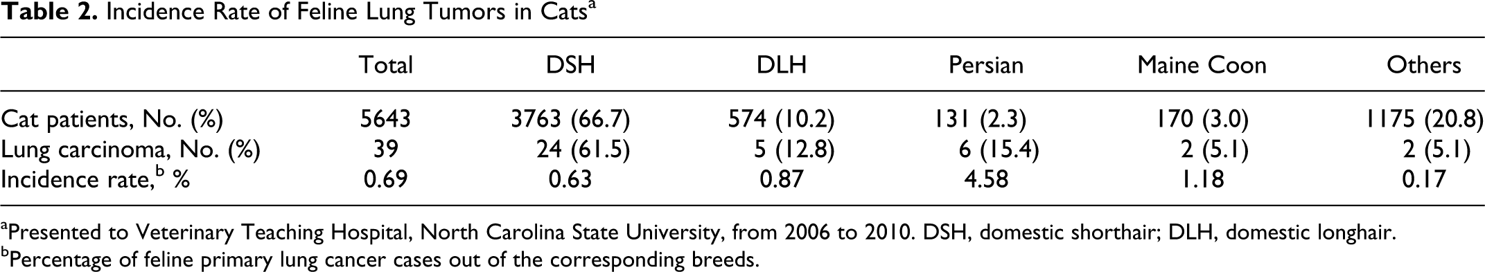
aPresented to Veterinary Teaching Hospital, North Carolina State University, from 2006 to 2010. DSH, domestic shorthair; DLH, domestic longhair.
bPercentage of feline primary lung cancer cases out of the corresponding breeds.
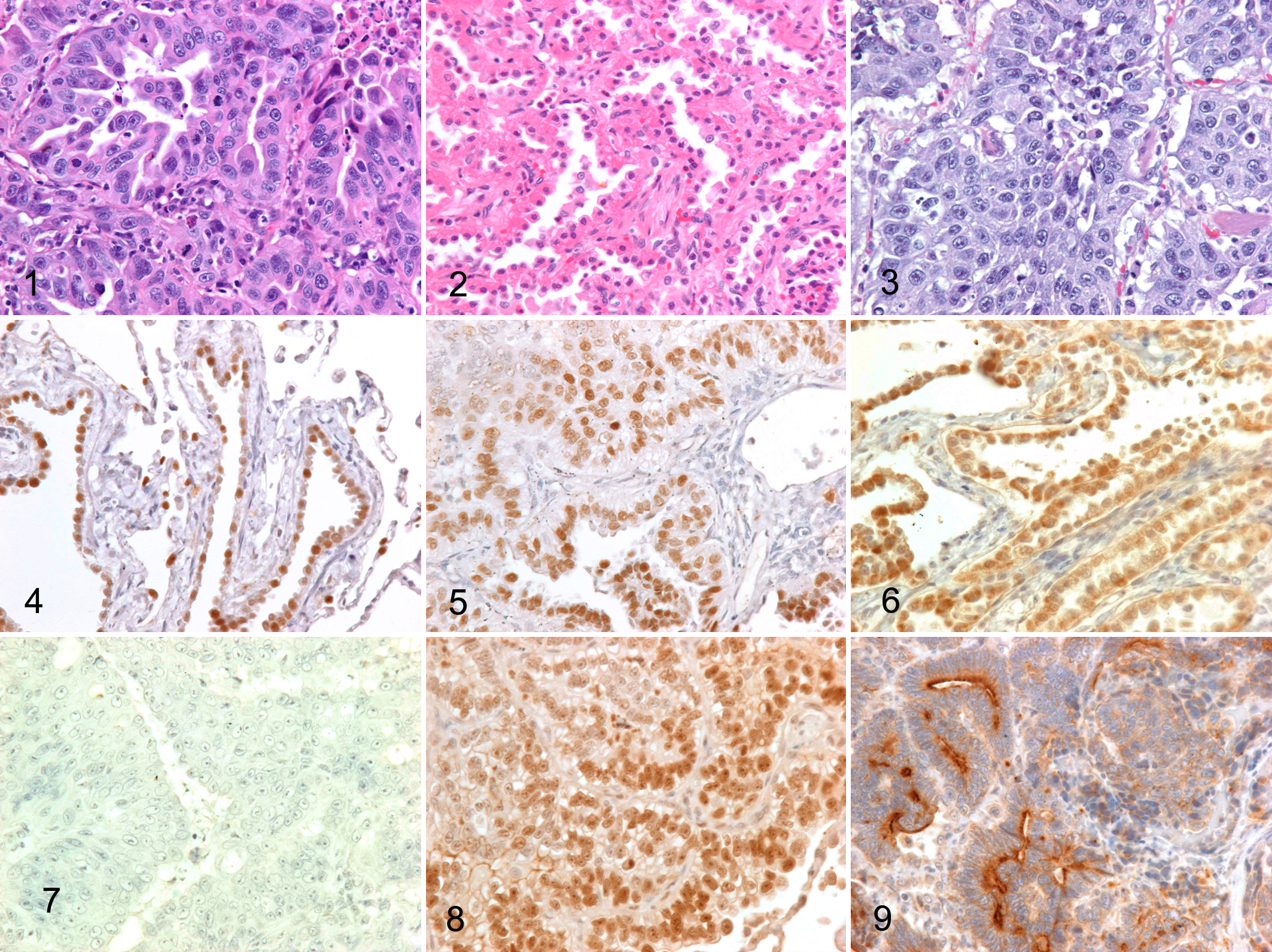
The primary pulmonary carcinomas were divided into 3 subtypes based on the published criteria. 26 Twenty-five cases (64.1%) of feline lung tumors exhibiting papillary, acinar, solid, or mixed glandular structures were classified as pulmonary AC (Fig. 1). The diagnosis of BA was made for 8 cases (20.5%) based on the presence of single-layered cuboidal cells lining the surface of fibrovascular stroma (Fig. 2). Six cases (15.4%) were diagnosed as AS with the neoplastic cells arranged predominantly as solid sheets with evidence of keratinization and intercellular bridges and as glandular structures (Fig. 3). There was no difference in the age of the subjects between each histologic type of the feline pulmonary carcinomas (P < .584).
Regardless of histologic types, approximately 80% of the feline pulmonary carcinomas metastasized (Table 3). The most frequent type of metastasis was intrapulmonary metastasis (66.7%), characterized by multiple tumor masses with similar histologic features within the lungs. Carcinomatosis within the pleural cavity (38.5%) and metastases to regional lymph nodes (33.3%) and distant organs (25.6%) were also frequently detected. Metastasis to digits was observed in 7 of 39 cases (17.9%). The size of the largest tumor mass was significantly associated with metastatic potential (P < .001); no metastasis was observed in cats with lung tumors less than 1 cm in diameter.
Metastatic Pattern of Feline Lung Tumors in Relation to the Size of the Largest Lung Mass, No. (%)a
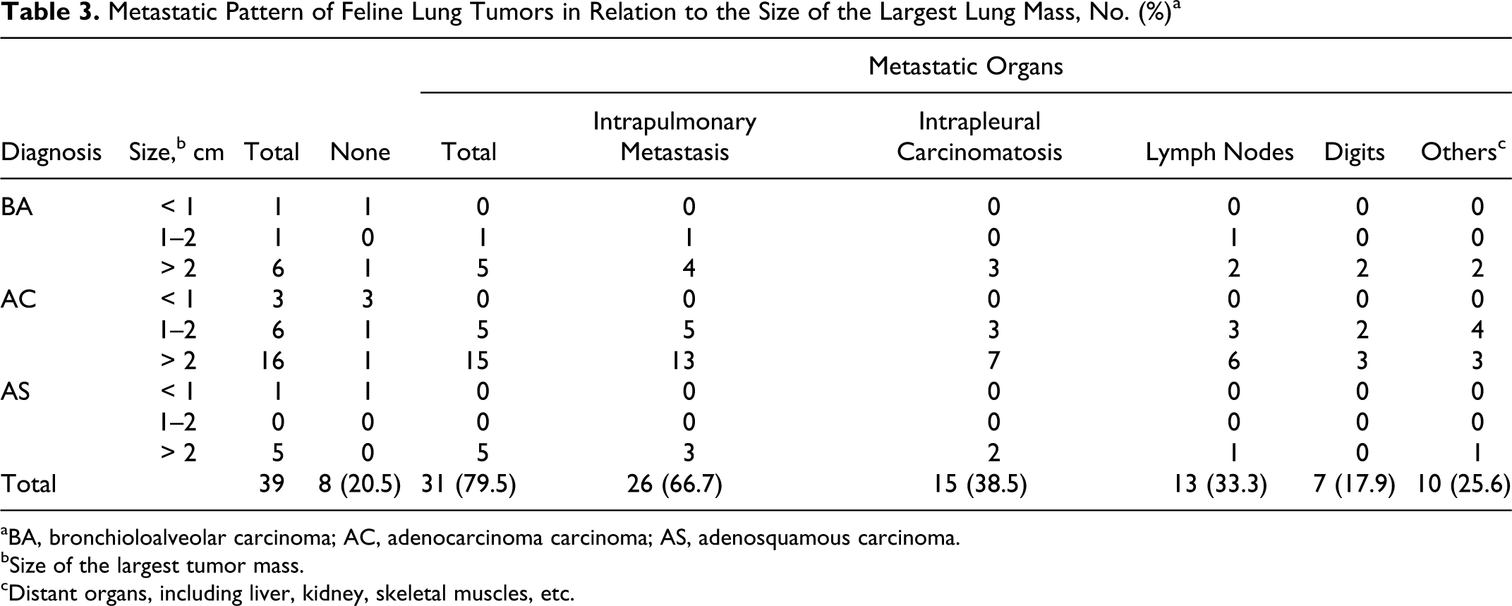
aBA, bronchioloalveolar carcinoma; AC, adenocarcinoma carcinoma; AS, adenosquamous carcinoma.
bSize of the largest tumor mass.
cDistant organs, including liver, kidney, skeletal muscles, etc.
Immunohistochemistry
Based on the tissue size and quality, 24 pulmonary carcinomas, including 6 BA, 16 AC, and 2 AS, were selected for immunohistochemistry (Table 4). TTF-1 labeled the nuclei of bronchiolar epithelial cells in normal lung adjacent to the tumors (Fig. 4). In sum, 66.7% (16 of 24) of feline lung carcinomas had positive nuclear labeling for TTF-1 in the neoplastic cells (Fig. 5). In normal lung, SP-A positively labeled intraluminal secretory material, the luminal surface, and cytoplasm of bronchiolar epithelial cells and type II pneumocytes. Positive reactivity to SP-A antibody was detected in 50% (12 of 24) of lung tumors examined (Fig. 6). TTF-1 reactivity was more intense in AC and AS than in BA. The combined results of SP-A and TTF-1 immunohistochemistry indicated that more than 80% (20 of 24) of feline lung tumors stained with at least one of these markers.
Immunohistochemical Staining Results in Cat Lung Tumors, No. (%)
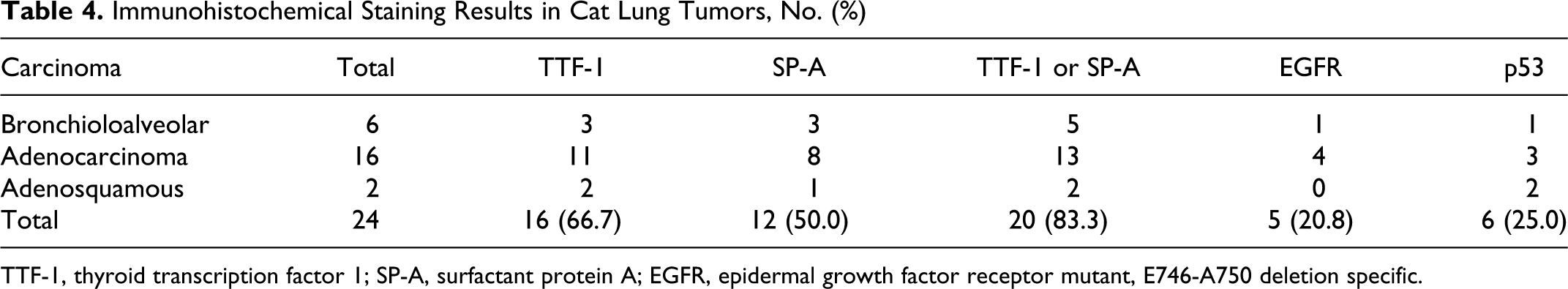
TTF-1, thyroid transcription factor 1; SP-A, surfactant protein A; EGFR, epidermal growth factor receptor mutant, E746-A750 deletion specific.
Immunohistochemistry for p53 and EGFR proteins was performed to investigate the abundance and distribution of proteins known to be involved in carcinogenesis. Normal lung was not positively labeled with either p53 or mutant EGFR. Accumulation of p53 protein in the nuclei of the neoplastic cells was detected in 25% (6 of 24) of the feline lung tumors examined. Distribution of the positive staining for p53 protein in the neoplastic tissue was variable between the areas but predominated in the areas of solid cellular growth (Figs. 7 and 8). All the AS cases had labeling of p53 protein. EGFR (E746-A750del) mutant protein was strongly detected on the luminal surface of the neoplastic cells in approximately 20% (5 of 24) of the feline lung tumors examined (Fig. 9). In contrast to p53 expression, the EGFR was detected predominantly in the AC subtype and not in the AS subtype. Only a single case of feline lung tumors had detectable expression of both p53 and EGFR mutant proteins.
Sequencing of K-ras and p53
In both the normal and neoplastic lung tissues, the feline p53 primers amplified a region larger than 1 kb that contained the mutational hot spots. The K-ras primers amplified 289 base pairs of exons I and II of feline K-ras gene, which contained the mutational hot spot codons 12, 13, 59, and 61. Sequences of p53 and K-ras genes obtained from normal and neoplastic lung tissues were identical to the published sequences of National Center for Biotechnology Information accession Nos. D26608 and U62089, respectively (data not shown).
Discussion
The incidence rate of primary lung carcinomas representing 0.69% of all feline cases admitted to the Veterinary Teaching Hospital is similar to that (0.75%) reported in a survey performed at the University of California for all feline accessions. 26 The distribution of the histologic subtypes of the feline pulmonary carcinomas is also comparable to previous reports. 10,26 In our study, pulmonary AC accounted for 64% of all feline lung carcinomas, while BA and AS accounted for 21% and 15% of all primary lung carcinomas examined, respectively. The average age of the cats with pulmonary carcinomas in our study was 12.3 years, which is consistent with previous surveys. 26 In accordance with published studies, our study showed no sex predilection of pulmonary carcinomas in cats. In contrast to the previous studies indicating no breed predisposition, 10,26 our data showed that Persian cats with pulmonary carcinomas were proportionally overrepresented among the total cases included in our analysis. However, this observation should be further validated on the basis of investigations using larger sample sizes.
Many studies have reported that feline pulmonary carcinomas frequently metastasize to distant organs. In one survey, 76% of feline pulmonary tumors had metastases. 10 Sites of metastasis included the regional lymph nodes (30%), intrathoracic organs (30%), and a variety of extrathoracic organs (16%). In the present study, approximately 80% of feline pulmonary carcinomas examined metastasized, with a decreasing order of intrathoracic carcinomatosis (38.5%), regional lymph nodes (33.3%), and distant visceral organs, including liver, kidney, and skeletal muscle. Approximately 18% of our feline carcinoma cases metastasized to the digits, validating the previously reported syndrome of feline pulmonary carcinomas. 6
Immunohistochemistry has greatly enhanced diagnostic precision in many areas of pathology, particularly in the assessment of tumors. It is a valuable diagnostic tool that should be used with clinical and morphologic findings rather than in isolation. When an AC is encountered in a lung, the principal question is often whether the lesion is a primary lung tumor or is metastatic from another site. Specific markers of cell origin, such as TTF-1 and various surfactant proteins, may be of great utility in this regard. 2 TTF-1 is a tissue-specific transcription factor that is known to play a crucial role in differentiation of lung epithelial cells. In human adult lung, TTF-1 is expressed in type II pneumocytes, nonciliated bronchiolar epithelial cells (Clara cells), and bronchiolar basal cells. TTF-1 expression identified human lung AC with a predominant Clara cell and/or type II alveolar pneumocyte component and poorly differentiated tumors. 24 In our study, TTF-1 labeled the nuclei of the normal bronchiolar epithelial cells and scattered type II pneumocytes. More than 80% of feline lung tumor cases were labeled with either TTF-1 or SP-A, suggesting the importance of these proteins as potential markers for identification of primary lung tumors. The sensitivity and specificity of these markers for the primary lung tumors should be further evaluated by examining tumors that originate from other organs. Moreover, a large portion of the primary lung tumors analyzed in this study were labeled with both TTF-1 and SP-A, indicating that the delineation of cell origin may require an additional molecular marker.
Proto-oncogenes, along with tumor suppressor genes, play regulatory roles in normal cell growth. Activation of proto-oncogenes and inactivation of tumor suppressor genes can lead to abnormal cell growth that contributes to cellular transformation and carcinogenesis. Mutations in the K-ras proto-oncogene and p53 tumor suppressor gene have been detected in a large proportion of human lung cancers. 14 Point mutations in the K-ras proto-oncogene occur in 20% to 30% of non–small cell lung carcinoma (NSCLC). 12 In humans, K-ras mutations were detected in 25% to 40% of atypical adenomatous hyperplasia, suggesting that K-ras mutation may be an early event in human lung cancer development. 22 The p53 tumor suppressor functions in a network of pathways, playing critical roles in cell cycle check points, apoptosis, DNA repair, and recombination. 9 Inactivation of the p53 gene plays an important role in the pathogenesis of human lung cancer. 12 In humans, mutation of p53 has been frequently reported in small cell lung carcinoma (75%) and NSCLC (50%), but BA exhibits p53 mutations less frequently than squamous cell carcinoma or other histologic subtypes. 3,5 In the present study, p53 protein was immunohistochemically detected in 25% of feline lung tumors examined, predominantly in AS subtypes. Physiologically, wild-type p53 protein has a very short half-life in cells; thus, its expression in normal cells is not detected by immunohistochemistry. 1 In contrast, mutant p53 protein resists the normal degradation process and accumulates in the nucleus, which is detectable by immunohistochemistry. Although our sequencing analysis did not reveal any mutation in K-ras and p53 genes, lack of mutations in the present study does not completely rule out the involvement of these genes in the feline lung carcinogenesis. Because this study examines a small fragment of the genes only in 3 samples, further studies are warranted on mutations outside the targeted areas in more tumor samples. Alternatively, mechanisms (other than somatic mutation) that regulate the activity of these genes may be involved in the carcinogenic process of feline lung cancer development.
In humans, the EGFR pathway is shown to be activated in the majority of NSCLC, providing an important target for NSCLC treatment. 18 Upon ligand binding, the EGFRs homo- or heterodimerize and subsequently activate the receptors’ intrinsic tyrosine kinase activity and broad downstream signaling cascades, mainly including Ras-Raf-MAP-kinase, PI3K-Akt, and STAT pathways. 18 All these pathways have strong stimulatory effect on cell proliferation, differentiation, survival, angiogenesis, and migration. Alterations of the EGFR gene reported for human NSCLC include point mutation, deletion, insertion, and amplification. The in-frame deletion has been associated with AC histology, in nonsmokers or light smokers, females, and East Asian ethnic minorities. 23 Overexpression of EGFR protein is detected in 40% to 80% of NSCLC patients, which is reportedly associated with aggressive clinical behaviors and poor prognosis. 21 EGFR tyrosine kinase inhibitors have been developed and used clinically in the treatment of advanced NSCLC. 18 In our study, 20% of the feline lung tumor cases showed expression of mutant EGFR protein. The antibody used in the study specifically detects EGFR (E746-A750del) mutant protein. 27 The presence of EGFR mutant protein in our feline lung tumor samples suggests similarities between human and feline lung carcinogenesis. Further studies on EGFR sequence and its clinical importance in feline lung tumors are warranted on a larger sample size.
In the present study, we could not pursue the clinical outcomes including survival date, because most of feline lung tumor cases were diagnosed at the time of necropsy. Lobectomy for extirpation of pulmonary tumors has been the treatment of choice in cats, but the prognosis is generally poor. 26 A greater number of feline lung tumors are classified as inoperable at diagnosis, either as a result of extensive disease or metastasis or due to concurrent decompensated cardiomyopathy. 19 In humans, standard systemic therapy in advanced lung cancer involves a combination of cytotoxic agents, angiogenesis inhibitors, and small molecule tyrosine kinase inhibitors. Despite recent advances in treatment, the prognosis for human lung cancer patients remains poor, with overall 5-year survival rates (10%–15%) for lung cancer among the lowest of all cancers. 16 Published reports point to the existence of cancer stem cells in each lung cancer type. 4 Squamous cell carcinomas, for example, tend to occur close to larger bronchi where the basal cells expressing keratin 5 are enriched. Bronchioloalveolar stem cells are likely the cell origin of mouse lung tumor that closely recapitulates the features of human lung AC, 17 proposing an important connection between normal lung stem cell biology and lung cancer development. Further studies regarding the role of endogenous stem cells in the lung carcinogenesis of veterinary species may be necessary to provide a new perspective on the mechanisms of this malignancy, which may lead to the development of a more efficient therapeutic regimen.
In summary, this study reports the incidence rate, histologic subtypes, and expression profiles of potential histogenetic and carcinogenetic markers of feline spontaneous lung cancers. Further evaluation of the genetic and/or biochemical aspects of feline lung carcinogenesis would enable us to identify useful markers for diagnosis, malignant progression, and prognosis, which may promote the development of efficacious preventive or therapeutic strategies for feline patients.
Footnotes
Acknowledgements
We thank Drs Ronald Herbert (National Institute of Environmental Health Sciences, Research Triangle Park, North Carolina), Mac Law (North Carolina State University), and Malcom Roberts (North Carolina State University) for proofreading the manuscript and Sandra Horton (North Carolina State University) for her technical expertise.
Declaration of Conflicting Interests
The authors declared no potential conflicts of interest with respect to the research, authorship, and/or publication of this article.
Funding
The authors received no financial support for the research, authorship, and/or publication of this article.