
Abstract
The aim of this study was to evaluate the vasculature in porcine circovirus type 2–infected (PCV2-infected) lungs and to identify the PCV2 subtypes involved in porcine pneumonia. Pulmonary samples from 140 pigs, 2 weeks to 7 months of age, from 36 Hungarian commercial herds with clinical signs of respiratory disease were examined for the presence of respiratory pathogens, with bacterial culture, pathologic evaluation, and immunohistochemistry for PCV2, porcine reproductive respiratory syndrome virus, and swine influenza virus. PCV2 was the most commonly identified pathogen (49 cases) among the 74 of 140 cases (53%) with respiratory pathogens. PCV2 was detected immunohistochemically in the wall of 13% to 100% of pulmonary vessels (mean, 89%) in 38 of 49 cases (78%). Detection of PCV2 antigen was positively correlated with the presence of vascular lesions (P < .001, odds ratio [OR]: 159.54). Other pathogens capable of vascular injury in swine were found in 29 of 49 of the PCV2-positive cases (59%). The probability of detecting vascular lesions in PCV2-infected lung was higher than in infection with porcine reproductive respiratory syndrome virus (P < .002, OR: 14.63), Pasteurella multocida infection (P < .001, OR: 5.75), or Streptococcus spp. infection (not significant, OR: 1.45). Sequence analysis of open reading frame 2 amplicons was possible in 6 PCV2-positive cases, from which 5 cases proved to be PCV2b subtype and 1 case, PCV2a subtype. In conclusion, PCV2 antigen was commonly colocalized with pulmonary vascular lesions in pneumonia in Hungarian swine, and PCV2b was the dominant subtype.
Porcine circovirus type 2 (PCV2) is highly prevalent in swine herds and has a significant economic impact on swine production. 25 The infection has been associated with various clinical syndromes, including the porcine respiratory disease complex. 10,12,18 PCV2-associated pneumonia was the second-most common PCV2-associated disease (PCVAD) in the United States 18 and is important in European swine herds as well. 25
The pathogenesis of PCV2-associated disease is not completely understood, although damage of the lymphoid system after PCV2 infection is an important feature. The outcome of the infection is influenced by host factors, coinfections, and immune modulation of the host. 18 Involvement of the vasculature in PCV2 infection has been reported. 2,4,13,17,22,23 In vitro endothelial activation and diminished coagulation time in swine naturally infected with PCV2 were demonstrated. These changes and the presence of PCV2 antigen in endothelial cells of naturally infected pigs pointed to PCV2 infection as the cause of vascular lesions. 14 Although PCV2 antigen had been found in endothelial cells in several porcine tissues, 11,15,21 severe vascular lesions have been reported only since 2004 in North American, South American, and European pigs. 2,4,13,17,22,23 A marked increase in the incidence and severity of PCVAD was observed in eastern Canada in 2004 and was attributed to the emergence of the PCV2b virus cluster. 2 PCV2b has become the most prevalent subtype in PCVAD outbreaks in North America 3,8 and Europe. 7,9,26 The aim of this study was to investigate the hypothesis that the vasculature is involved in PCV2 infection and to identify which PCV2 subtype (a or b) was more common.
Material and Methods
Samples
One hundred and forty swine, aged 2 weeks to 7 months (33 suckling, 45 nursery, 40 growing, and 22 finishing pigs), with signs of respiratory disease from 36 Hungarian commercial herds were examined for gross and histologic lung lesions and respiratory pathogens. In 29 cases, tissue samples were collected only from 2 sites of the cranial lung lobes for histologic and immunohistochemical examination. In the remaining 111 cases, samples were collected from 5 locations (cranial and middle lobes on both sides and the accessory lobe). Lung tissue samples (2 or 5) from each case were embedded into a single paraffin block. Formalin-fixed and paraffin-embedded tissues were sectioned at 4-μm thickness and stained with hematoxylin and eosin, using standard protocol. Histologic lesions were recorded, and the cases were categorized according to the presence of fibrinonecrotizing bronchopneumonia, suppurative bronchopneumonia, interstitial pneumonia, or proliferative and necrotizing pneumonia. Tissue alterations characteristic of Mycoplasma hyopneumoniae were also recorded (hyperplasia of the bronchus-associated lymphoid tissue, peribronchiolar and perivascular lymphohistiocytic infiltration), as were vascular lesions (fibrinoid necrosis, intramural edema, vasculitis, and perivasculitis) and pulmonary changes associated with vascular injury (thrombi, interlobular edema, hemorrhages, or hyaline membranes).
Immunohistochemistry
Immunohistochemistry (IHC) was used in serial tissue sections to detect PCV2, Pasteurella multocida, porcine reproductive and respiratory syndrome virus (PRRSV), and swine influenza virus (SIV). Briefly, after digestion with 0.1% protease XIV (Sigma Aldrich Co., St Louis, MO) for 10 minutes at 37°C for influenza A virus and PCV2 or heat antigen retrieval (in a microwave oven at 750 W, for 20 min in citrate buffer, pH 6.0) for P. multocida and PRRSV, the sections were incubated overnight with the appropriate primary antibodies at room temperature (influenza A virus: HYB 340-05, Statens Serum Institut, Copenhagen, Denmark, dilution 1:6000; P. multocida: rabbit polyclonal hyperimmune serum, in-house, dilution 1:2000; PCV2: 36A9, Ingenasa, Madrid, Spain, dilution 1:30 000; PRRSV: SDOW-17-A, Rural Technologies, Inc., Brookings, SD, dilution 1:200). Antibody binding was detected by a horseradish peroxidase–labeled polymer (ENVISION+, Dako, Glostrup, Denmark). Tissue sections with the relevant pathogen served as positive controls. Negative controls were prepared by replacing the primary antibody with phosphate buffer solution on serial tissue sections. Severe PCV2 infection was diagnosed when, on average, at least 10% of the cells were immunostained in the 10 fields examined at 200× magnification. Mild PCV2 infection was the diagnosis when less than 10% of the cells were immunostained. The prevalence of PCV2-positive blood vessels was evaluated by examining 1 PCV2-positive tissue sample per case.
Bacteriology
Swabs for bacteriologic examinations were collected from lungs with macroscopic lesions (n = 97 cases), whereas the spleen was sampled in all cases. These were cultured on 5% sheep blood and Drigalsky lactose agar plates under aerobic conditions for 48 hours. If Actinobacillus or Haemophilus infection was suspected, additional blood agar plates with Staphylococcus aureus streaks were incubated in an atmosphere containing 10% CO2 for 48 hours. Microbial pathogens were identified as published. 19
Polymerase Chain Reaction and Sequencing
Total DNA was extracted from formalin-fixed, paraffin-embedded lung tissue samples of 49 cases that were positive for PCV2 by IHC, using a guanidine hydrochloride–based method. 6 The presence of PCV2 genomes was tested by polymerase chain reaction (PCR) using a primer pair specific to open reading frame 1 (ORF1) (PCIIF: 5′-CTC GAT CTC AAG GAC AAC G-3′, PCIIR: 5′-ACA GCA GTT GAG GAG TAC C-3′). 16 Positive samples were amplified in 2 overlapping fragments using ORF2-specific primer pairs (ci2Ef: 5′-AGA ATT CAA CCT TAA CCT TTC TTA TTC-3′, ci2SLr: 5′-AGC GCA CTT CTT TCG TTT TC-3′, CBB2: 5′-CGC ACC TTC GGA TAT AC-3′, CORF2B: 5′-TAC ATA CAT GGT TAC ACG GAT ATT-3′) 5,6 to provide 680 base pair sequences for analysis. Purification and sequencing of the PCR products were done at Macrogen, Inc. (Seoul, Korea). 5
Statistical Analysis
The Fisher exact test was used to evaluate the independence of vascular wall lesions and associated tissue changes from the presence of PCV2 and other pathogens. 1 The association of lesions and pathogen presence was quantified by odds ratio (OR). The reported P values and 95% confidence intervals (CIs) were 2-tailed. Statistical analysis was performed using the R statistical software. 20
Results
Pathologic and Microbiologic Findings
Pneumonia was grossly evident in 97 of 140 cases (69%) and histologically evident in 135 of 140 cases (96%). Respiratory pathogens were detected in 74 of 140 cases (53%). Single pathogens were found in 42 of 140 cases (30%), whereas 2, 3, or 4 concomitant pathogens were detected in 19 of 140 (14%), 11 of 140 (8%), and 2 of 140 (1%) cases, respectively. Histologic lesions typical of M. hyopneumoniae infection were observed in 17 of 140 cases (12%).
PCV2 was the most common respiratory pathogen (49 of 140, 35%). PCV2 antigen was found in 1 of 33 suckling (aged 3 weeks) pigs (3%), 20 of 45 nursery pigs (44%), 20 of 40 growing pigs (50%), and 8 of 22 finishing pigs (36%). PCV2 infection was the sole pathogen found in 20 of 49 cases (41%) and coexisted with an additional 1 to 3 pathogens in the remaining 29 cases (59%) (Actinobacillus pleuropneumoniae, 2 cases; Actinobacillus suis, 1 case; P. multocida, 21 cases; PRRSV, 8 cases; Streptococcus spp., 5 cases; and SIV, 1 case). At gross examination, the lesions were similar in both mild and severe PCV2 cases. The PCV2-infected lungs were not collapsed but were heavy, diffusely mottled red to purple, and rubbery in 43 of 49 cases (88%). Interlobular edema was marked in 35 of 49 cases (71%); mediastinal lymph nodes were enlarged in 15 of 49 cases (31%). The histologic diagnosis was proliferative and necrotizing pneumonia in 24 of 49 cases (49%), interstitial pneumonia associated with suppurative bronchopneumonia in 16 of 49 cases (33%), fibrinonecrotizing bronchopneumonia in 8 of 49 cases (16%) and lesions suggestive for M. hyopneumoniae infection in 1 of 49 cases (2%). Lesions typical of PCV2-associated pneumonia included necrotic bronchiolitis (31 of 49 cases, 63%), granulomatous pneumonia (18 of 49 cases, 37%), and mild to marked peribronchiolar fibrosis (15 of 49 cases, 31%). Severe PCV2 infection was diagnosed in 36 of 49 cases (73%) (Figs. 1, 2). Infected cells were most commonly detected in the peribronchiolar and perivascular connective tissue where leukocytic infiltration was most prominent. PCV2 infection was considered mild in the remaining 13 cases (27%) (Fig. 3), in which immunohistochemically labeled cells (<10%) occurred multifocally. In 10 of 13 cases, PCV2 antigen was not detected in every sample. PCV2 was the only pathogen detected in 17 of 36 of the severe PCV2 cases (47%) and in 3 of 13 of the mild PCV2 cases (23%).
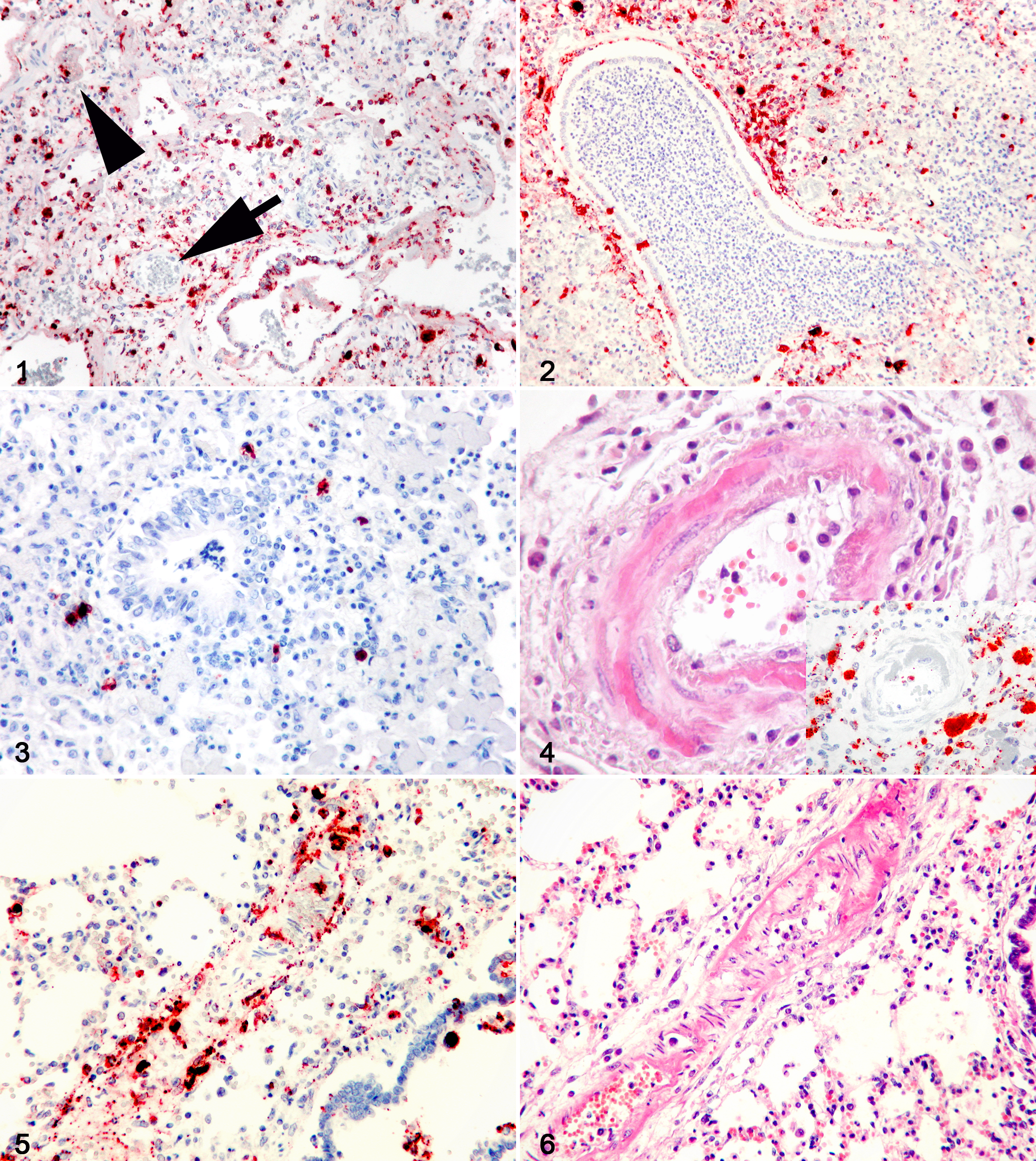
Lung; pig. Numerous porcine circovirus 2–positive (PCV2-positive) cells in a case with severe PCV2 infection. Immunolabeling in blood vessel wall (arrow) and thrombus (arrowhead) is evident. Immunohistochemistry (IHC) for PCV2, Mayer’s hematoxylin counterstain.
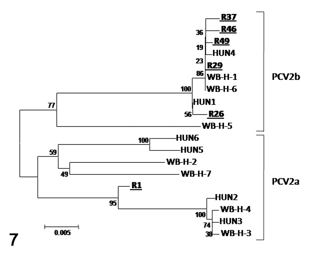
Comparison of PCV2 ORF2 sequences. The newly identified viruses are underlined and in bold. GenBank accession numbers of PCV2 from domestic pigs (HUN1-6): AY256460, AY256456, AY256458, AY256457, AY256459, AY256455, and from wild boars (WBH-1-7): AY874163, AY874164, AY874165, AY874166, AY874167, AY874168, AY874169.
PCV2 and Vascular Lesions
The association of vascular lesions with pulmonary alterations in PCV2-positive and PCV2-negative cases is indicated in Table 1. Such lesions were found in all PCV2-positive cases but in only 24 of the 91 PCV2-negative cases (26%). PCV2 was detected in the vascular wall in 38 of 49 cases (78%); the average proportion of infected blood vessels was 89% in these cases (range, 13% to 100%). Of the 13 cases with mild PCV2 infection, only a few PCV2-positive cells were found in vascular walls and only in 2 cases. In contrast, PCV2-positive cells were few to numerous in vascular walls in all 36 cases with severe PCV2 infection (Fig. 1). Fibrinoid necrosis (Figs. 4–6) was the most severe vascular lesion and had higher prevalence in severe PCV2 infection (24 of 36, 67%) compared to mild infection (2 of 13, 15%) or PCV2-negative cases (5 of 91, 6%).
Correlation of Vascular Lesions and Associated Pulmonary Changes With Porcine Circovirus 2 (PCV2) Immunoreactivity
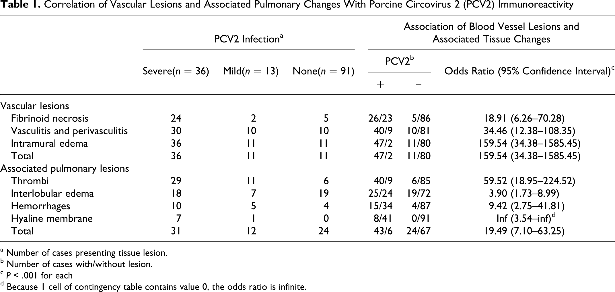
a Number of cases presenting tissue lesion.
b Number of cases with/without lesion.
c P < .001 for each
d Because 1 cell of contingency table contains value 0, the odds ratio is infinite.
Immunolabeling was detected in vessels with and without lesions. The number of labeled cells in vessels varied even within the same case, and no correlation was found between the number of infected cells and the presence or severity of the vascular lesions. Several vessels with severe circumferential fibrinoid necrosis had only a few PCV2-positive cells (Fig. 4). PCV2 antigen was found in all layers of the blood vessels in different cell types, including endothelial cells, smooth muscle cells, infiltrating leukocytes, and intravascular monocytes (free or entrapped in thrombi).
Contribution of Other Pathogens to Vascular Lesions
Infectious agents other than PCV2 that can induce vascular lesions were detected in PCV2-positive and PCV2-negative cases. A. suis, P. multocida, PRRSV, or Streptococcus spp. were detected in 16 of 36 cases with severe PCV2 infection (44%). A. pleuropneumoniae, β-hemolytic Escherichia coli (septicemia), P. multocida, PRRSV, Salmonella spp. (septicemia), or Streptococcus spp. were detected in 10 of 13 cases with mild PCV2 infection (77%). In PCV2-negative cases, A. pleuropneumoniae, A. suis, and P. multocida were detected in 9 of 24 cases with vascular lesions or associated tissue changes (38%). No vascular lesions were found in 13 of 91 PCV2-negative cases (14%) despite the detection of P. multocida, PRRSV, and Streptococcus spp.
PCR and Sequencing
PCV2 DNA was detected with the ORF1-specific primer pair in all 49 IHC-positive cases, but sequence analysis of the larger ORF2 amplicons was possible in only 6 cases, due to the poor quality of the DNA extracted from the formalin-fixed, paraffin-embedded tissues. The nucleotide sequences of the 6 PCV2 amplicons were similar to those published for Hungarian domestic pigs and wild boars, 5,6 with only minor differences (Fig. 7). Five sequences belonged to the PCV2b subtype and 1 sequence to the PCV2a subtype.
Statistical Analysis
Every type of vascular lesion with the associated tissue changes was positively correlated (P < .001) with the presence of PCV2 in the lung (Table 1). The vascular lesions and associated tissue changes also were positively correlated with the presence of PRRSV (OR: 14.63, 95% CI: 1.93–657.74, P = .002; OR: 11.02, 95% CI: 1.45–495.14, P = .007 respectively) and P. multocida (OR: 5.75, 95% CI: 2.45–14.21, P < .001; OR: 3.84, 95% CI: 1.66–9.33, P < .001). Positive association with the presence of Streptococcus spp. (OR: 1.45, 95% CI: 0.32–6.63, P = .741; OR: 1.69, 95% CI: 0.38–8.54, P = .520) was not statistically significant. Parallel occurrence of PCV2 was found in 80% of PRRSV-positive cases (8 of 10), 51% of P. multocida–positive cases (21 of 41), and 50% of Streptococcus spp.–positive cases (5 of 10).
Discussion
PCV2 was the most common porcine respiratory pathogen in this study, with viral antigen detected in the lungs of 35% of the pigs with a clinical history of pneumonia. This supports previous findings and indicates that PCV2 is an important respiratory pathogen in swine. 12,18,25 Furthermore, PCV2 was the sole pathogen detected in 41% of the PCV2-positive cases. Histologic lesions in bronchi, bronchioles, and alveoli were similar to those reported in PCV2-associated pneumonia of swine. 10,12,18 The occurrence of porcine parvovirus and M. hyopneumoniae, which were frequently found in PCV2-associated pneumonia, 12,18 was not examined in this study. The presence of PRRSV and SIV was examined by IHC, for the parallel demonstration of tissue lesions and intralesional pathogens, but IHC usually is less sensitive than PCR for these 2 viruses, which may explain the higher prevalence of single PCV2 infections in the present study compared to the results of earlier publications. 12,18
PCV2b was the dominant subtype in the current study and in previous studies in Hungary. 5,6 Based on a short 137-base amplicon sequence, a distinct subtype within PCV2b was described in Switzerland (PCV-2b-CH). 26 Our study, which used a much longer PCV2 sequence, could not confirm the existence of such subgroup. Although minor differences at the nucleotide level among the newly described PCV2 ORF2 sequences of this study were detected, these differences did not justify the establishment of a novel subtype. The sequencing results indicated that the PCV2 in this study did not differ from the widespread PCV2 present in both clinically ill and asymptomatic animals.
Coinfections occurred in most of the PCV2-positive cases, as reported previously, and are thought to be the consequence of virus-induced injury of the host immune system. 12,18 In addition, involvement of the vasculature in multiple organs was recently implicated in the pathogenesis of PCVAD in selected cases of naturally or experimentally infected pigs. 2,13,14,17,22,23 The most prominent vascular lesions were lymphohistiocytic vasculitis, 14,17 necrotizing vasculitis, 22 vasculitis with fibrinoid necrosis, 2,4,13,23 intravascular thrombi, 13,23 acute edema, 13,22,23 hyaline membranes, 23 and acute hemorrhages. 4,13,22,23 PCV2 antigen was detected in endothelial cells, smooth muscle cells of the tunica media, infiltrating leukocytes, intravascular and perivascular macrophages, and intravascular monocytes in the present study and in previous reports. 4,11,13 –15,17,21 –23 Furthermore, intimal spindle cell proliferation was found in a few cases (data not shown) and was recently reported in a cesarean-derived/colostrum-deprived pig inoculated with PCV2b. 13 No correlation was found between the number of infected cells and the presence or severity of the vascular lesions, suggesting a host immune system–induced alteration rather than a direct PCV2-induced lesion.
In a study of 456 porcine multisystemic wasting syndrome–affected pigs, the authors reported a link between vasculitis and PCV2 immunolabeling in the brain in 73 of the 107 cases with lymphohistiocytic vasculitis. 4 However, no statistical analysis was performed in that study. The data from the current study support the hypothesis that blood vessels play a role in the pathogenesis of PCV2 infection of swine. A significant positive relationship (P < .001) was found between the presence of PCV2 in the lungs and all types of vascular lesions and associated tissue changes. Furthermore, PCV2 was the sole pathogen detected in 47% of the severe PCV2 cases. Vascular lesions were colocalized with PCV2 antigen in 100% of those cases, and 90% of the blood vessels were PCV2 positive. Finally, the most severe vascular lesion (fibrinoid necrosis) was more common in cases with severe PCV2 infection (67%) compared to mild PCV2 infection (15%) or PCV2-negative cases (6%).
In pigs, vasculitis can be caused by different viruses. 13 Aujeszky disease, African swine fever, and classical swine fever viruses typically cause vascular lesions, but these viruses are not present in domestic swine in Hungary. Bovine viral diarrhea virus can induce vascular changes in cattle and can infect pigs, but in a recent study, vascular changes were not observed after experimental bovine viral diarrhea virus infection of swine. 13 Sporadically, ovine herpesvirus 2 infection can cause vascular lesions in swine; however, the pigs of the current study originated from herds with no contact with sheep. Thus, only PRRSV was examined among the viruses capable of inducing vascular lesions. Vascular lesions were found in 9 of 10 PRRSV-positive cases (90%), and a significant relationship was found between the presence of PRRSV and vascular lesions and associated tissue changes. However, in 8 of these 9 cases, PCV2 was also detected. As for bacteria, A. pleuropneumoniae, A. suis, β-hemolytic E. coli, P. multocida, Salmonella spp., and Streptococcus spp. were found in the present study and can induce vascular lesions in swine. 13,24 These bacteria could have been responsible for vascular lesions in 16 of 36 cases with severe PCV2 infection (44%), in 10 of 13 cases with mild PCV2 infection (77%), and in 9 of 24 cases without PCV2 infection (38%). Vascular lesions and associated tissue changes were positively associated with only the presence of P. multocida and Streptococcus spp. (not significant), but about half those cases were also PCV2 positive. The probability of detecting vascular lesions was significantly higher in cases with PCV2 infection (OR: 159.54) compared to infections with other pathogens capable of inducing vascular alterations (OR: 1.45–14.63). The results of the present study support the hypothesis that infection of the vasculature contributes to the pathogenesis of PCV2-induced swine diseases and that PCV2b is the dominant subtype in PCV2-associated pneumonia in Hungarian swine.
Footnotes
Acknowledgement
The excellent technical assistance of Mrs Ágnes Mészáros is gratefully acknowledged.
The authors declared no potential conflicts of interest with respect to the research, authorship, and/or publication of this article.
The authors received no financial support for the research, authorship, and/or publication of this article.