
Abstract
Vascular lesions and their association with porcine circovirus type 2 (PCV2) were evaluated in multiple organs from 10 pigs affected with PCV2–systemic disease (PCV2-SD). Animals had vascular lesions in multiple organs, consisting of lymphohistiocytic lymphangitis and/or phlebitis, mild to severe necrotizing arteritis, and thrombosis within splenic arterioles and choroid plexus capillaries. Variable amounts of PCV2 nucleic acid detected by in situ hybridization were present within endothelial cells, tunica media myocytes, and perivascular and/or intralesional inflammatory cell infiltrates. PCV2 nucleic acid was detected within endothelial cells of both lymphatic and blood vessels without lesions in the associated tissues. Necrotizing arteritis was principally present in lymph nodes and kidney and consisted of degeneration, necrosis, and pyknosis of myocytes, often with intracytoplasmic, brightly eosinophilic inclusion bodies that were strongly positive for PCV2 nucleic acid. Segmental or circumferential fibrinoid necrosis was mainly present in vessels of the lymph node, spleen, and choroid plexus and was variably associated with PCV2 nucleic acid. Severe lymphangitis associated with strong intralesional PCV2 labeling was frequently detected within the mesenteric and mediastinal lymph nodes and the lamina propria of the ileum. In most tissues, medium and large lymphatics and/or veins often had disruption of the intima and mild mononuclear inflammatory cell infiltration that was variably associated with PCV2 nucleic acid. The present study indicates that vasculitis is a frequent finding in natural cases of PCV2-SD and that PCV2 may have a direct cytopathic effect on tunica media myocytes of small- and medium-sized arteries as well as endothelium.
Keywords
Porcine circovirus type 2 (PCV2) belongs to the Circoviridae family, which includes small, single-stranded, nonenveloped DNA viruses. 31 PCV2 is highly prevalent in swine herds worldwide, and infection is mostly subclinical. 21,28 PCV2 has been linked to various clinical entities, such as postweaning multisystemic wasting syndrome (PMWS), reproductive disorders, porcine respiratory disease complex, enteritis, porcine dermatitis, and nephropathy syndrome and proliferative and necrotizing pneumonia, which are together termed porcine circovirus diseases (PCVDs). 21,27 PMWS and PCV2 subclinical infection are the most economically important PCVDs. Very recently, it has been suggested to replace the term PMWS with PCV2–systemic disease (PCV2-SD). 26
PCV2-SD is characterized by wasting, dyspnea, and lymphadenopathy. The most prominent histologic findings are in lymphoid organs and consist of extensive lymphocyte depletion, infiltrates of histiocytes, occasional multinucleated giant cells, and intracytoplasmic viral inclusion bodies. 11,13 The amount of PCV2 antigen or nucleic acid within lymphoid tissues positively correlates with the severity of lymphocyte depletion and granulomatous infiltration in lymphoid organs and with clinical signs of wasting and growth retardation. 29
Lesions of necrotizing arteritis, periarteritis, and fibrinoid vasculitis have been described. 3 –5,12,14,20,25,30 These lesions are occasionally present in lung, kidney, spleen, brain, lymph nodes, liver, and reproductive organs. In some cases, infarcts and necrotic lesions are present in brain, lymph nodes, and spleen in association with thromboses. 3 –5,13,25 Only 3 recent publications have specifically focused on cardiovascular lesions associated with PCV2 infection. In one publication, myocarditis and myocardial necrosis were described in 4 natural PCV2-SD cases; one of these cases also had periarteritis and endarteritis affecting several organs. 20 In the same report, granulomatous and necrotizing lymphadenitis were associated with thrombosis in experimentally PCV2-infected pigs. The second report documented vascular lesions in pneumonic lungs. 30 In the third report, 2 pigs experimentally infected with PCV2 developed fatal PCV2-SD with acute vasculitis, and 5 additional animals developed chronic lymphohistiocytic and plasmacytic peri- and endo-arteritis without clinical signs. 14 Furthermore, a prothrombotic state characterized by increased coagulation time and thrombin plasma activity, high platelet aggregation ability, and diminished platelet count has been described in PCV2-SD–affected pigs, concomitant with vasculitis and PCV2 infection of the endothelium. 15
Existing research on PCV2 infection, based on natural and experimental studies, suggests that the cardiovascular system plays an important role in the pathogenesis of PCVDs. The objective of the present study was to systematically characterize vascular lesions in multiple tissues of a randomly selected group of pigs diagnosed with naturally occurring PCV2-SD and assess their correlation with PCV2 nucleic acid presence.
Materials and Methods
Case Selection
Study cases were selected from a database generated from a previously published study. 8 Selected animals (10) were 14.0 ± 2.1 (mean ± SD) weeks of age and were diagnosed with PCV2-SD by means of clinical, pathologic, and virologic criteria. 26 Selected cases had severe PCV2-SD lymphoid lesions and a large amount of PCV2 nucleic acid in various lymphoid tissues. A second level of selection was dictated by the availability of most of the following tissues: lung; liver; cerebellum; kidney; spleen; heart; tonsil; mediastinal, mesenteric, inguinal, and submandibular lymph nodes; thymus; ileum; jejunum; and colon. All PCV2-SD–affected pigs were real-time quantitative polymerase chain reaction (PCR) 17 positive with a mean ± SD of 3.9 × 10⁷ ± 5.7 × 10⁷ PCV2 DNA copies/ml of serum (Table 1).
Summary of PCV2 Load, Serology Against Other Pathogens, and Microscopic Lesions Affecting the Vascular System of PCV2-SD Affected (Nos. 1–10) and Healthy (Nos. 11–15) Pigs.
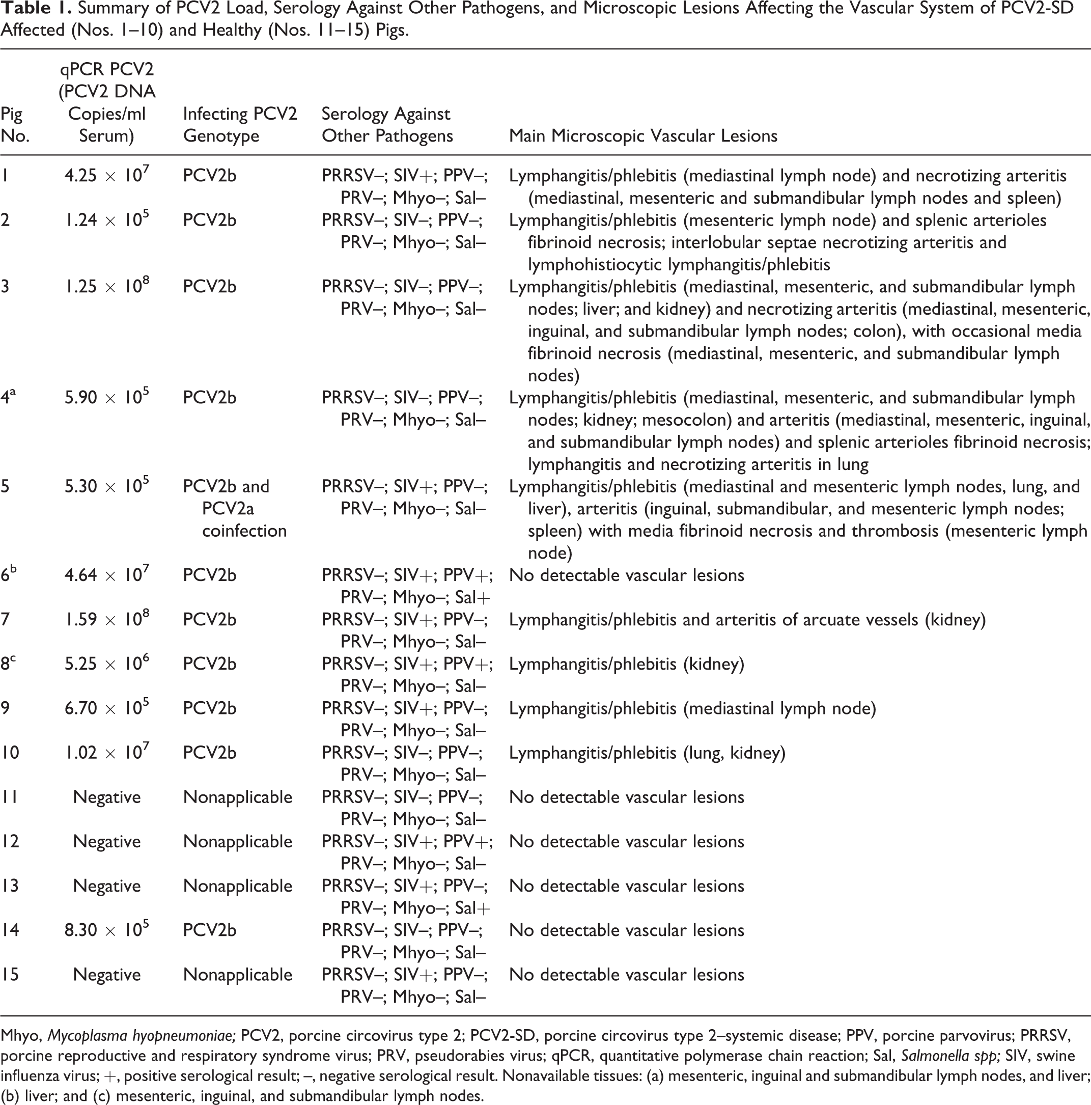
Mhyo, Mycoplasma hyopneumoniae; PCV2, porcine circovirus type 2; PCV2-SD, porcine circovirus type 2–systemic disease; PPV, porcine parvovirus; PRRSV, porcine reproductive and respiratory syndrome virus; PRV, pseudorabies virus; qPCR, quantitative polymerase chain reaction; Sal, Salmonella spp; SIV, swine influenza virus; +, positive serological result; –, negative serological result. Nonavailable tissues: (a) mesenteric, inguinal and submandibular lymph nodes, and liver; (b) liver; and (c) mesenteric, inguinal, and submandibular lymph nodes.
Five healthy pigs 14.7 ± 2.5 weeks of age from the same study 8 were negative controls. These pigs did not have clinical signs, microscopic lesions compatible with PCV2-SD, or detection of PCV2 by in situ hybridization (ISH) of lymphoid tissues. A single animal had a PCV2 load of 8.3 × 10⁵ PCV2 DNA copies/ml in serum (Table 1).
All case/control pigs were antibody tested for major swine pathogens, including porcine reproductive and respiratory syndrome virus (PRRSV), swine influenza virus (SIV), porcine parvovirus (PPV), Salmonella spp, pseudorabies virus, and Mycoplasma hyopneumoniae 8 (Table 1). Lungs and lymphoid tissues were tested for PRRSV antigen by immunohistochemistry (IHC), 8 yielding negative results. Samples from the colon of pig No. 3 were cultured due to a fibrinonecrotizing colitis; Salmonella spp were isolated.
Histopathology and PCV2 Detection in Tissues
From each pig, two 4-μm-thick sections were prepared from formalin-fixed, paraffin wax–embedded samples of lung, liver, cerebellum, kidney, spleen, heart, tonsil, thymus, ileum, jejunum, colon, and mediastinal, mesenteric, inguinal, and submandibular lymph nodes and processed for hematoxylin and eosin staining and PCV2 nucleic acid detection by in situ hybridization (ISH). 24 Paraffin-embedded material was no longer available for ISH of some tissues (mesenteric, inguinal, and submandibular lymph nodes from pig Nos. 4 and 8; the liver from pig Nos. 4 and 6; and the spleen from pig No. 5). A 41 digoxigenin-labeled DNA oligonucleotide probe corresponding to the open reading frame (ORF) 1 of PCV2 (DIG-50-CCT TCC TCA TTA CCC TCC TCG CCA ACA ATA AAA TAA TCA AA-30) was used. ISH controls included lymph node known to be negative or positive for PCV2.
Results
Vascular lesions were present in multiple organs of 9 of 10 PCV2-SD–affected pigs (Table 1). These consisted of lymphohistiocytic lymphangitis and/or phlebitis in multiple organs such as lymph nodes, lung, kidney, small and large intestine, and liver. Necrotizing arteritis was present in 5 pigs, mostly in lymph nodes (4 pigs) and less commonly in the lung (2 pigs) and kidney (2 pigs). A summary of the different types of vascular lesions per tissue examined, as well as their association with PCV2 detection by ISH, is presented in Table 2. Differentiation of lymphatic vessels from veins was not always possible; therefore, the terminology of lymphangitis/phlebitis is used when necessary. No significant histologic vascular or lymphoid lesions, or PCV2 DNA (detected by ISH), were observed in tissues of healthy control pigs.
Vascular Lesions Found in PCV2-SD–Affected Pigs and Their Association With PCV2 Nucleic Acid Detection.
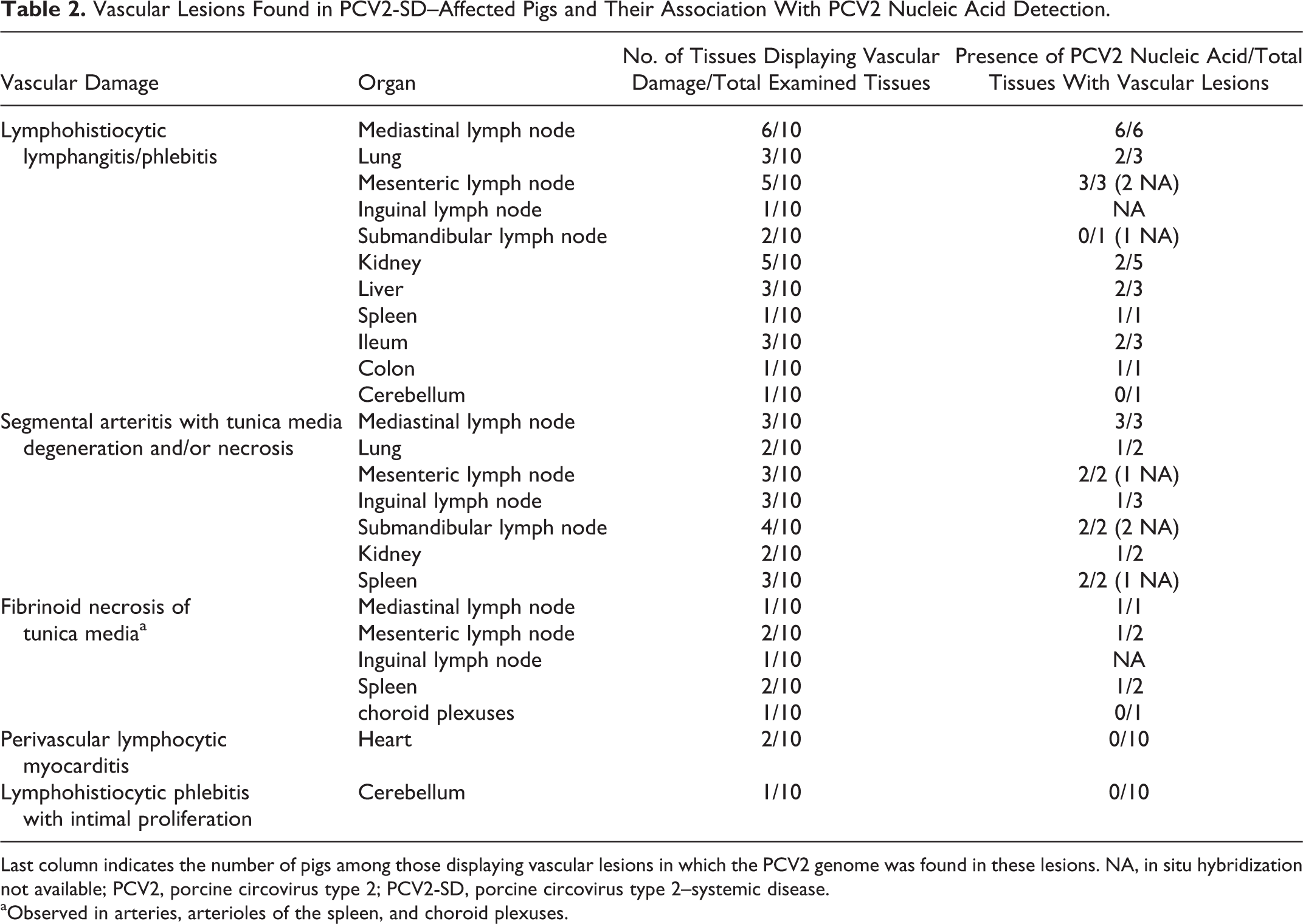
Last column indicates the number of pigs among those displaying vascular lesions in which the PCV2 genome was found in these lesions. NA, in situ hybridization not available; PCV2, porcine circovirus type 2; PCV2-SD, porcine circovirus type 2–systemic disease.
aObserved in arteries, arterioles of the spleen, and choroid plexuses.
The tunica media of medium and large arteries had multifocal areas with myocytes having cytoplasmic vacuolation and areas containing granular eosinophilic material with pyknotic and karyorrhectic nuclear debris (necrosis) (Fig. 1). This finding was specifically present in the capsule and interlobular septa of lymph nodes (4/10 pigs), spleen (3/10 pigs), pulmonary interlobular septa (2/10 pigs), and renal medullary and arcuate arteries (2/10 pigs). Variably within those foci, some myocytes had round, hypereosinophilic, 1- to 3-μm diameter inclusions (Fig. 1). These necrotic and degenerative tunica media lesions closely correlated with the presence of PCV2 nucleic acid as determined by ISH (Fig. 2), suggesting they might be viral inclusions. In these arteries, PCV2 nucleic acid was detected in high amounts within the media (Figure 2a) and in variable levels in the endothelium (Fig. 2a,b).
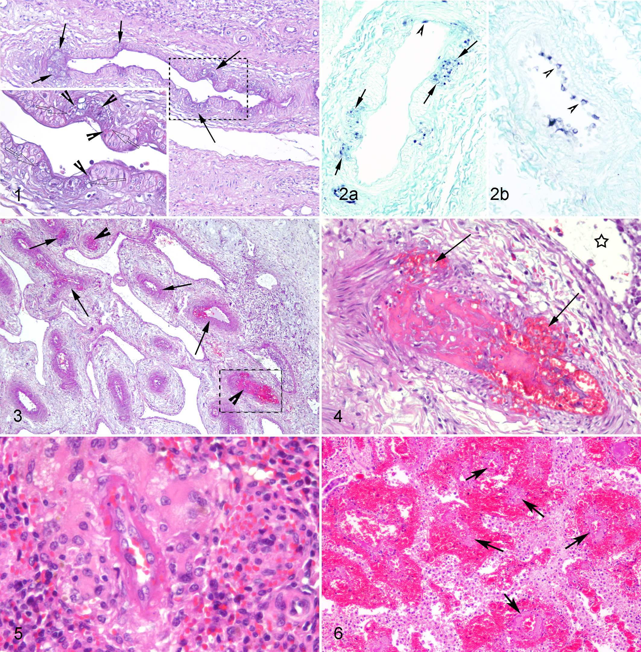
Pigs affected by porcine circovirus 2–systemic disease (PCV2-SD).
Arteries with disruption of the tunica media by edema, erythrocytes, and small numbers of inflammatory cells were in the lung of pig No. 2, in the mesenteric lymph node of pig No. 5 (Figs. 3, 4), and inguinal lymph node of pig No. 3. Fibrinoid necrosis of the tunica media was in the splenic arterioles of pig Nos. 2 and 4 (Fig. 5), the cerebellar choroid plexus arterioles in pig No. 5 (Fig. 6), and lymph node arteries in pig Nos. 3 and 5. PCV2 nucleic acid was detected in the media of the splenic arterioles with fibrinoid necrosis and also in adjacent normal arterioles in pig No. 4 (Fig. 7). PCV2 was not consistently detected in damaged arteries within the lung of pig No. 2 and the cerebellar choroid plexus arterioles of pig No. 5 (Table 2).
Lymphohistiocytic lymphangitis and/or phlebitis of medium-sized vessels were in various tissues in a total of 9 pigs (Table 2), mainly in the mediastinal and mesenteric lymph nodes and kidney. Phlebitis was characterized by disruption of the tunica intima by edema, lymphocytes, and macrophages (Fig. 8) and infiltration of the adventitia by variable numbers of inflammatory cells. PCV2 nucleic acid was present in variable amounts in some of these lesions within endothelial cells and transmurally within inflammatory cells (Fig. 9). Lymphangitis within the mesenteric lymph node hilar and capsular vessels (Fig. 10), as well as in the submucosa and serosa of the intestine, was characterized by numerous luminal macrophages and syncytial cells (Fig. 10) that were strongly positive for PCV2 nucleic acid (Fig. 11). Histocytic lymphangitis in the mesentery and intestine was associated with severe histiocytic infiltration of the mesenteric lymph nodes, histocytic enteritis and colitis (3/10 pigs), and transmural mesocolonic edema in pig No. 4. In the lung, mild to moderate lymphangitis and/or phlebitis were present within interlobular septa, along with moderate interlobular edema. Pig No. 5 had leptomeningeal phlebitis, with intra- and perivascular infiltration of lymphocytes and macrophages. There was also endothelial (intimal) proliferation (Fig. 12). This pig had PCV2 labeling in the brain, in scattered and rare endothelial cells, but not in the proliferative lesions. This pig also had marked vascular lesions within the choroid plexus vessels with severe perivascular hemorrhage (Fig. 6). The choroid vascular damage was not associated with PCV2 nucleic acid detection.
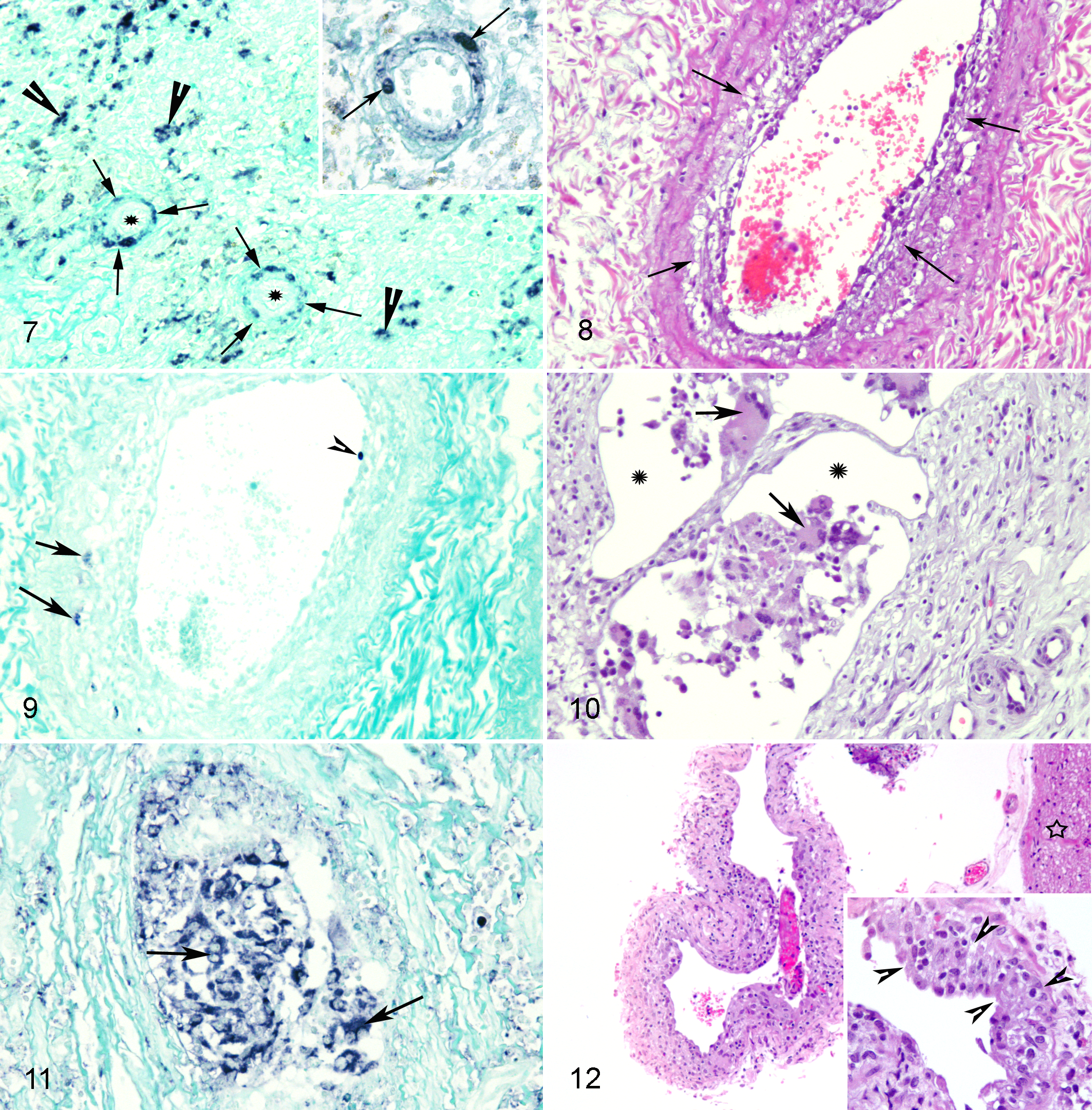
Pigs affected by porcine circovirus 2–systemic disease (PCV2-SD).
Overall, cases with less severe lymphoid depletion and less PCV2 nucleic acid in tissues tended to have less prominent and generalized vascular lesions compared with that in the more severely affected animals (Table 1). Furthermore, PCV2 nucleic acid was often detected in the endothelium of nondamaged, normal, blood, and lymphatic vessels in multiple tissues, although the number of endothelial cells labeled in each vessel was generally very low.
Discussion
Vascular lesions consisting of lymphohistiocytic lymphangitis and/or phlebitis, necrotizing arteritis, arterial and capillary fibrinoid necrosis, and vascular thromboses were detected in multiple organs of growing pigs with naturally acquired PCV2-SD. These diseased pigs were randomly selected from a wider database of affected animals with broad availability of tissues and not specifically because of the existence of vascular lesions. Therefore, this study confirms that a high percentage of pigs fulfilling the case definition of PCV2-SD have lesions in the circulatory system.
In accordance with recent reports, 12,14,20 a higher frequency of vascular lesions was present in mesenteric and mediastinal lymph nodes and kidneys. Although a high proportion of damaged vessels were ISH positive for PCV2, apparently normal vessels from PCV2-SD–affected pigs had occasional ISH endothelial labeling, which has been described in diseased pigs with high systemic viral loads. 14,20,24 Such changes were not observed in vessels of control, healthy pigs despite one pig being PCV2 viremic. This indicates that PCV2 endothelial infection is not always associated with vascular damage and further supports the theory that high viral loads may be related to endothelial/vascular infection. 10,22
In the study presented herein, lesions varied from degenerative to necrotizing arteritis, including fibrinoid necrosis of arterial and arteriolar walls, as reported in previous studies. 14,20 In contrast to published reports, this study did not detect more chronic arterial lesions, such as lymphohistiocytic arteritis and periarteritis. 14,20 It is possible that the degree of vascular damage may vary depending on the chronologic stage of PCV2 infection, as well as on putative intercurrent infections. More than half of the total arterial lesions were associated with PCV2 labeling, except when fibrinoid necrosis was present. In these cases, detection of the virus was less consistent. Similar observations have been described regarding different types of vascular damage and association with PCV2 nucleic acid or antigen detection. 5,14,30
Pathogens able to induce vasculitis in pigs include PRRSV, African swine fever virus, classical swine fever virus, bovine viral diarrhea virus (BVDV), ovine herpesvirus type 2, pseudorabies virus, Salmonella choleraesuis, Streptococcus suis, enterotoxemic Escherichia coli, and Erysipelothrix rhusiopathiae. 14 Vitamin E and selenium deficiency cause a distinctive vasculopathy mainly affecting the heart. 14 African and classical swine fever are eradicated from Spain, and ovine herpesvirus type 2 has never been detected in commercial Spanish swine herds. Moreover, study animals were seronegative for pseudorabies and PRRSV. Although PRRSV was further excluded by immunohistochemistry of lung tissue, exclusion of PRRSV infection cannot be completely ruled out without reverse transcription PCR evaluation of both serum and tissue samples. BVDV status is unknown in the Spanish swine population, but a recent sero-survey indicated a very low prevalence of this pestivirus in a farm without clinical and negative productivity parameters. 23 In addition, pigs had no macroscopic and microscopic indications of bacterial infections or vitamin E and selenium deficiency. Salmonella spp are capable of causing vascular damage and was present in pig No. 3, which had a necrotizing colitis. However, vascular lesions in this animal were associated with the presence of PCV2 nucleic acid and, therefore, most likely caused by PCV2 rather than salmonella. Synergism between the two infectious agents cannot be totally ruled out. Bacterial coinfections are common in PCV2-SD 18,26 and may contribute to the pathogenesis of vascular damage in naturally acquired disease. In a recently published study, 29 limited to the assessment of vascular damage within PCV2-associated pneumonia, concomitant pathogens capable of causing vascular injury in the lung were found in 59% of PCV2-positive pneumonia cases. These results suggest an alternative explanation for the lack of PCV2 antigen/nucleic acid detection in vascular lesions in cases of PCV2 pneumonia, such as in this study.
Apoptosis of endothelial cells has been linked to vasculitis in brain lesions of pigs naturally affected by PCV2-SD. 25 Thus, a direct cytopathic effect of PCV2 on vascular cells (endothelium, tunica media myocytes, and/or pericytes) may be possible. Another possible mechanism of vascular damage in PCV2-SD is an indirect, immune-mediated response of (1) cellular origin, with cytokine secretion by infected cells and recruited inflammatory cells, or (2) humoral origin, with deposition and/or in situ formation of immune-complexes or complement activation in the vessel walls. 1,9 PCV2 infects both endothelial cells and myocytes of the tunica media. 12,14,30 PCV2 replicates in vivo in endothelial cells. The close association of PCV2 nucleic acid with vascular damage in this study and in previous reports 14 suggests that a direct cytopathic effect of the virus in the pathogenesis of vascular lesions may be possible.
Necrotizing lymphadenitis can be found in at least one lymph node in ∼10% of pigs affected by PCV2-SD in both natural and experimental disease. 7,14,20,24,26 Some reports 7,14,20 point to an association between severe vascular damage and necrosis, suggesting that thrombosis and ischemia underlie the pathogenesis of these lesions in PCV2-SD–affected pigs. The present study supports this hypothesis.
Abundant PCV2 nucleic acid was present within lesions of mesenteric lymphangitis and the resultant meso-colonic edema in one pig, suggesting that PCV2 could underlie the pathogenesis of these lesions. In two cases, interlobular pulmonary edema was associated with lymphangitis and/or phlebitis, as well as arteritis of the interlobular pulmonary vessels. Interlobular pulmonary edema has been previously reported in association with similar vascular lesions under an unusual acute PCV2-SD presentation and in other cases of pneumonia attributed to PCV2. 3,14,30
Thromboembolic hemorrhagic lesions have been reported in the cerebellum of PCV2-SD–affected pigs and were associated with vascular fibrinoid necrosis and PCV2 endothelial infection. 5,25 In the present study, one pig had similar lesions, but an association with PCV2 was not proved. PCV2 nucleic acid was not detected in lesions of fibrinoid necrosis in choroid plexus vessels.
Two other unusual vascular findings in the present study were intimal proliferative lesions in the leptomeningeal vessels (pig No. 4) and intracytoplasmic inclusion bodies in myocytes of the tunica media. Intimal proliferation was recently reported in a pig experimentally infected with PCV2b 14 and represents a stereotypic vascular response to extensive endothelial damage to various insults, including viral injury. Reports relating intimal proliferative lesions to viral infections are scarce. 2 Intracytoplasmic inclusion bodies within myocytes of the tunica media have been rarely reported in PCV2 infections. 12
Nonsuppurative myocarditis present in two pigs was negative for PCV2 by ISH and, therefore, the etiology of this finding was undetermined. Nonsuppurative myocarditis with focal myocardial necrosis associated with cytoplasmic PCV2 antigen in cardiomyocytes can be found in aborted fetuses 16 and rarely in young pigs (4–7 weeks of age). 20
In conclusion, this report describes different types of vascular lesions and their association with PCV2 in growing pigs affected by PCV2-SD. Present results suggest that PCV2 is implicated in the pathogenesis of vascular lesions and that vascular lesions are more often found in diseased pigs than previously reported.
Footnotes
Acknowledgements
We are thankful to Mónica Peréz and Maria Jesus Navas Sanchéz, from CReSA, for their excellent technical assistance. Ana R. Resendes was supported by a postdoctoral fellowship from FCT (Fundação para a Ciência e a Tecnologia, reference SFRH/BPD/70897/2010, Portugal). We are grateful to Dr Carlos Martins (Professor of Microbiology and Virology, at Universidade Técnica de Lisboa, Faculdade de Medicina Veterinária, Portugal), for cosponsoring the fellowship application of Dr Resendes and for the critical review of the manuscript.
Declaration of Conflicting Interests
The author(s) declared no potential conflicts of interest with respect to the research, authorship, and/or publication of this article.
Funding
The author(s) received no financial support for the research, authorship, and/or publication of this article.