
Abstract
Scleral cartilaginous metaplasia was detected by routine histologic examination of globes from 5 Suffolk sheep from a scrapie pathogenesis study. The extent of the metaplasia varied among the sheep but was always posterior to the tapetal fundus. The matrix surrounding chondrocytes stained intensely with alcian blue and was immunopositive for type II collagen. Retrospective evaluation of additional eyes from Suffolk and Cheviot sheep used in various scrapie pathogenesis studies at the authors' facility revealed similar histologic changes in 40% and 12.7% of eyes examined, respectively. The clinical significance of this previously unreported finding is unknown.
The sclera constitutes the largest portion of the fibrous tunic of the eye and consists of a dense network of collagen fibrils, predominantly of collagen types I and III. 6 The inner portion of the sclera, or lamina fusca, also contains elastin fibers and scattered melanocytes. The sclera of many vertebrate species, such as birds, reptiles, and fish, normally contains cartilage and/or scleral ossicles. Osseous and, less often, cartilaginous metaplasia has been described in rodent sclera, 7 but the presence of scleral cartilage or cartilaginous metaplasia has not been reported in nonlaboratory domestic animal species.
Animals and Methods
Eyes were examined from 5 Suffolk sheep, aged 22 to 28 months, that had been part of a scrapie pathogenesis study. 2 All animals had been orally inoculated with scrapie as neonates and euthanized by barbiturate overdose when they showed advanced clinical signs of scrapie. Eyes from an additional 3 Suffolk ewes (> 24 months of age) from the National Animal Disease Center (NADC) scrapie-free flock were also examined. One globe with a segment of optic nerve of approximately 1 to 2 cm was extracted from each sheep at necropsy and immersed in 10% neutral buffered formalin. The contralateral globe was frozen for later molecular-based assays. Tissues were allowed to fix for at least 1 week, at which time a 5-mm-thick vertical section from the caudal aspect of the globe, containing retina and optic nerve, was embedded in paraffin, sectioned at 4 μm, and stained with hematoxylin and eosin (HE). For histologic examination of the contralateral eye, the globe was thawed slightly, and a similar section was trimmed, immersed in 10% neutral buffered formalin for 24 hours, and processed and stained with HE. Additional sections were stained with alcian blue at pH 1.0. Briefly, sections were stained with a 1% alcian blue solution in 0.1 N hydrochloric acid (HCl) for 30 minutes at room temperature, rinsed in 0.1 N HCl, blotted dry, and counterstained with nuclear fast red. Immunohistochemistry was performed with a monoclonal antibody against type II collagen (Millipore, Billerica, MA) at a dilution of 1:100. Briefly, sections were deparaffinized, incubated in a 0.5% pepsin solution in 5 mM HCl for 20 minutes at 37°C, incubated in 3% hydrogen peroxide, blocked in a 1.5% goat serum solution, and incubated in primary antibody for 2 hours at room temperature. Sections were then washed and primary antibody labeling was detected with DakoCytomation’s LSAB2-HRP kit and 3,3′-diaminobenzidine. Sections were lightly counterstained with hematoxylin. This study was carried out in accordance with guidelines established by the NADC Animal Care and Use Committee.
Histologic Findings
Histologically, the sclera of all 5 scrapie-inoculated sheep contained putative chondrocytes in chondroid matrix. In sheep No. 1, rare round to ovoid cells had a moderate amount of basophilic to amphophilic cytoplasm and were surrounded by variably thick, homogeneous basophilic matrix (Fig. 1). The sclera of sheep No. 2 also had scattered individual cells, but these cells were mostly aggregated in amphophilic to basophilic matrix. In sheep Nos. 3 and 4, locally extensive islands of scleral cartilaginous metaplasia could be appreciated at low magnification. In a few areas, the chondroid cells appeared to be in lacunae. The sclera of sheep No. 5 was most notably affected, with multifocal to coalescing islands of cartilaginous metaplasia up to 1 × 5 mm (Figs. 2, 3). The extracellular matrix surrounding these cells was intensely alcianophilic (Fig. 4), indicating the presence of sulfated acid mucopolysaccharides. Immunohistochemically, this matrix was positive for type II collagen, the predominant collagen type in hyaline cartilage but not in sclera (Fig. 5). Despite artifacts related to freezing, similar histologic findings were identified in the corresponding regions of the contralateral eyes. This alcianophilic matrix was conspicuous in 4 of 5 contralateral eyes. The eye without alcianophilic matrix was from the sheep with the least discernable changes on examination of HE-stained sections (No. 1). Similar scleral changes were observed in only 1 of 3 scrapie-free sheep, in which rare, mostly individual, cells in lacunae were surrounded by matrix that was positive by alcian blue staining and type II collagen immunohistochemistry.
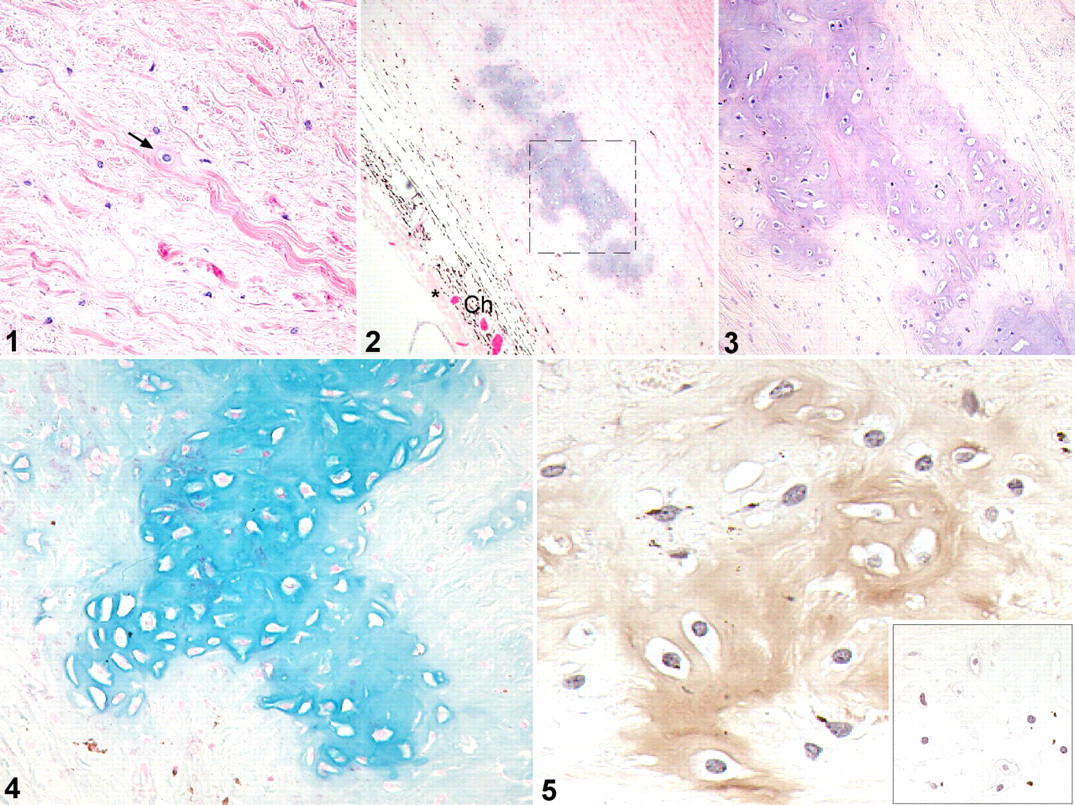
Sclera; sheep No. 1. Rare, individual, chondrocyte-like cells (arrow) surrounded by a thin basophilic matrix are scattered among collagen fibers. HE.
In all sheep, lesions were limited to the inner half of the sclera and were observed only posterior to the tapetal fundus. Evidence of underlying disease associated with these areas of cartilaginous metaplasia or other portions of sclera was not detected.
Discussion
Except in monotremes, mammalian sclera normally lacks cartilage, and reports of nonneoplastic disease with cartilage formation in the sclera are uncommon. Proteoglycan deposition was reported in the sclera of horses with degenerative suspensory ligament desmitis, 1 but cartilaginous metaplasia was not described. Osseous metaplasia is a fairly common aging change in the sclera of the Fischer 344 rat; cartilaginous metaplasia is observed infrequently. 7 In one study, metaplastic bone was detected in the sclera of the superior hemisphere in more than 95% of Fischer 344 rats. 4 Similarly, in all sheep in this study, cartilaginous metaplasia was observed only posterior to the tapetal fundus. Scrapie-infected sheep had typical neuropathologic changes of spongiform encephalopathy, but no evidence of scleral trauma, inflammation, or other abnormalities was observed histologically in the sheep.
Human sclera and cartilage have similar gene expression profiles, and chondrogenesis has been demonstrated in vitro in human scleral cells. 5 Our observations suggest that sheep sclera may have characteristics in common with cartilage and that scleral cells may possess chondrogenic potential.
The clinical significance of scleral cartilaginous metaplasia is unknown. There were no overt signs of impaired vision in any of these sheep, although subtle or subclinical visual deficits may have been present. Conditions that alter the scleral matrix in such a way that the sclera is thinned or weakened could affect vision, as in some human cases of myopia. 3
The unlikely possibility that scrapie caused the cartilaginous metaplasia was initially considered but later thought unlikely, given the observation of similar changes in the sclera of a scrapie-free sheep and the absence of this lesion in numerous cases of natural and experimental scrapie. A genetic cause is possible because these lambs and their dams were all obtained from the NADC’s scrapie-free flock. Somewhat surprising, retrospective evaluation of eyes from 70 Suffolk sheep in various scrapie pathogenesis studies at our facility revealed a prevalence rate of 40%. Similar examination of eyes from 55 Cheviot sheep revealed a prevalence of 12.7%. These findings suggest that scleral cartilaginous metaplasia may not be rare in sheep nor restricted to the Suffolk breed. The increased prevalence in the Suffolk sheep that we examined may indicate a true breed predilection or merely reflect the inbreeding of this flock.
In summary, cartilaginous metaplasia was detected in the sclera of sheep of two breeds, both with and without scrapie infection. The role of scrapie in development of this lesion could not be determined, because most specimens were from scrapie-infected sheep and no negative control eyes were available from those studies for comparison. Likewise, connective tissues other than sclera were unavailable for examination. Visual deficits were not detected in affected sheep, so the significance of this lesion is unknown.
Footnotes
Acknowledgements
We thank Virginia Montgomery, Martha Church, Judith Stasko, and James Fosse for excellent technical assistance. Mention of trade names or commercial products in this report is solely for the purpose of providing specific information and does not imply recommendation or endorsement by the U.S. Department of Agriculture.
The authors declared no conflicts of interest with respect to the authorship and/or publication of this article.
The authors received no financial support for the research and/or authorship of this article.