
Abstract
Three 4-week-old Yorkshire-Hampshire cross piglets from a litter of 9 (7 liveborn) developed convulsions the day of weaning. They were subsequently obtunded, ataxic, and hypermetric and had intention tremors. An affected male pig was presented live for necropsy on day 5 postweaning. This animal was euthanatized and necropsied. No significant grossly visible postmortem lesions were found. Histologic examination of the brain disclosed laminar necrosis of the submeningeal cerebral and cerebellar cortices with replacement by broad sheets of gitter cells. Occasional cerebral and cerebellar leptomeningeal and parenchymal vessels were surrounded by lymphocytes with fewer eosinophils. The morphologic diagnosis was severe multifocal subcortical cerebral and cerebellar laminar necrosis with moderate multifocal lymphocytic and eosinophilic cerebral and cerebellar leptomeningeal and parenchymal perivasculitis. The history and histologic findings are consistent with an etiologic diagnosis of sodium ion intoxication.
Three 4-week-old Yorkshire-Hampshire cross piglets from a litter of 9 (7 liveborn) developed convulsions the day of weaning. The owner characterized the affected animals as the “most thrifty.” The herd was positive for the PSS gene (ie, porcine stress syndrome). Nursing was replaced by hand-feeding goat milk replacer and “electrolyte water” (Milk Products, LLC, Chilton, WI). The examined male pig refused the milk replacer and electrolyte solution on the first day postweaning. It was somnolent the day of weaning and for 4 days postweaning. It was presented live for necropsy on day 5 postweaning. This animal weighed 22 pounds and had a body condition score of 3 of 5. Temperature, pulse, and respiration were within reference ranges. There were 2 recent castration wounds in the scrotum (13 days before presentation), and he was ataxic and hypermetric and had intention tremors. Euthanasia followed by necropsy disclosed no significant gross lesions. Intestine was cultured for evidence of bacterial pathogens, especially hemolytic E. coli. No significant growth resulted. Samples of most organs were fixed overnight in 10% neutral buffered formalin. Five-micron-thick sections stained with HE were prepared from paraffin-embedded tissue blocks using standard methods. Tissue sections were examined via standard light microscopy.
Differential Diagnoses
Common differential diagnoses for pigs of this age with neurologic disease include sodium ion intoxication, swine cerebral angiopathy (edema disease), streptococcal meningitis, poliomyelomalacia, and pseudorabies.
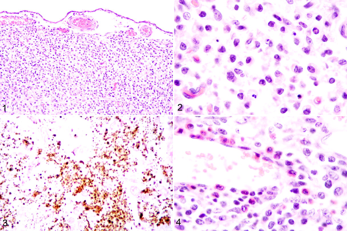
Microscopic Findings
The only tissues that contained significant histologic lesions were from the brain. There were extensive areas of submeningeal laminar cortical necrosis of the cerebrum and cerebellum with replacement with broad sheets of macrophages (Figs. 1, 2). Focal areas of necrotic neuropil were mineralized (Fig. 3). Lymphocytes and fewer eosinophils surrounded scattered cerebral and cerebellar leptomeningeal and parenchymal vessels (Fig. 4).
Diagnosis
The morphologic diagnosis was severe multifocal laminar subcortical cerebral and cerebellar necrosis with mild multifocal cerebral and cerebellar lymphocytic and eosinophilic leptomeningeal and parenchymal perivasculitis. These lesions are diagnostic of sodium ion intoxication.
Discussion
Among the differential diagnoses, swine cerebral angiopathy is generally characterized by edema, often in the eyelids, snout, greater curvature of the stomach, and mesentery of the spiral colon. Neurologic lesions may occur in the absence or presence of systemic edema and are characterized as focal symmetrical encephalomalacia. These lesions are distributed in the caudate nucleus extending to the medulla. 2 Vascular lesions accompany the malacia and are characterized as fibrinoid necrosis with karyorrhexis of vascular smooth muscle.
Streptococcal meningitis is one of a constellation of lesions characterized as fibrinosuppurative polyserositis. In this case, no clouding of the meninges was noted at gross examination, and significant meningitis was absent at histologic examination.
Focal symmetrical poliomyelomalacia in swine is a paralytic disease with flaccid tetraparesis (symptoms not noted in this case). The lesions are bilaterally symmetric and limited to the ventral gray columns of the cervical and lumbar intumescences. Wallerian degeneration accompanies these lesions. The cause is believed to be selenium toxicosis. 2
Pseudorabies in swine is generally accompanied by pyrexia and nystagmus, in addition to ataxia, recumbency, and seizures. Histologic lesions include nonsuppurative ganglioneuritis and meningoencephalomyelitis, along with neuronal necrosis, neuronophagia, and a mixed inflammatory cell influx, 2 none of which were present in the examined animal.
Dorsolateral cerebral cortical necrosis in pigs, accompanied by macrophages and scattered perivasculitis that includes eosinophils, is pathognomonic for sodium ion intoxication. 1 Also called salt poisoning, water deprivation syndrome, and water intoxication, this condition may occur in any species, although it is most common in swine. Early lesions principally consist of perivasculitis. In pigs, eosinophils often dominate the perivascular infiltrate early in the disease. Animals that survive past the initial period develop dorsolateral cerebral cortical necrosis, a lesion that is characteristic of cerebral anoxia. Although the disease may occur because of salt overload, it more commonly occurs secondary to dehydration and subsequent water overload.
The mechanism of toxicity of water deprivation and intoxication is related to electrolyte imbalance associated with dehydration. During the initial dehydration phase, there is an osmotic loss of water from the brain secondary to an increased blood sodium concentration. This is followed by an influx of ionic sodium, potassium, and chloride into the brain, which inhibits anaerobic glycolysis. When rehydration is delayed past the initial phase of dehydration, there is an influx or production of organic molecules, such as amino acids, polyols, and methylamines. These osmoles result in cell swelling upon rehydration. The osmotic differential cannot be equalized quickly enough to prevent edema of the brain. Clinical signs and typical lesions result. 3 Although a syndrome of sodium ion intoxication secondary to water deprivation–overload is thought to occur in a variety of animal species, cerebral laminar necrosis is best documented in swine. 1
In the case reported here, loss of water associated with inappetence at weaning, combined with voluminous replacement of fluids exacerbated by the use of electrolyte water, served to establish conditions ripe for the creation of sodium ion intoxication. It is also possible that PSS was at least partially responsible for electrolyte imbalance and inability to ingest feed or fluids.
Footnotes
The authors declared no conflicts of interest with respect to the authorship and/or publication of this article.
The authors received no financial support for the research and/or authorship of this article.