Simultaneous phMRI measurement of cerebral perfusion and blood-cerebrospinal fluid barrier function using interleaved echo-time ASL
C Perera1, I Harrison1, M Lythgoe1, D Thomas2,3,4 and J Wells1
1UCL Centre for Advanced Biomedical Imaging, Division of Medicine, University College London, United Kingdom
2Neuroradiological Academic Unit, Department of Brain Repair and Rehabilitation, UCL Queen Square Institute of Neurology, United Kingdom
3Leonard Wolfson Experimental Neurology Centre, UCL Queen Square Institute of Neurology, United Kingdom
4Wellcome Centre for Human Neuroimaging, UCL Queen Square Institute of Neurology, University College London, United Kingdom
Abstract
Background: We have recently developed a MRI technique that quantifies the rate of arterial blood water delivery to ventricular CSF, a non-invasive surrogate measure of Blood-Cerebrospinal Fluid Barrier (BCSFB) function.1
Aim: Here, we introduce interleaved echo-time (TE) ASL – a novel approach which simultaneously captures two distinct components of brain physiology: a) parenchymal perfusion, and b) BCSFB function, to a single dose of a drug or ‘challenge’ with high temporal resolution. This provides an efficient means to investigate the response of vessels comprising the BBB and BCSFB to pharmacological modulation in the healthy and diseased brain.
Method: Interleaved-TE ASL MRI (9.4T) was applied to investigate the effects of vasopressin, CO2, and caffeine on cortical perfusion and BCSFB function in anesthetized mice. We then investigated the mechanistic link between BCSFB function and vasopressin in ageing by capturing the response to exogenous vasopressin in aged and adult strain-matched mice (23- and 5-months).
Results/Conclusions: The commonly used vasodilatory agent, CO2, induced similar increases (∼21%) in both cortical perfusion and the BCSFB-ASL signal. Caffeine administration triggered a marked decrease in BCSFB-mediated labelled water delivery (41%), with no significant changes in cortical perfusion. Finally, we demonstrate a approx. 3-fold impairment in the BCSFB functional response to vasopressin in the aged vs adult brain (Figure 1).
Here, we highlight the value of this translational approach to non-invasively capture simultaneous and differential pharmacological modulation of vessel tone at the BBB and BCSFB, and how this relationship may be modified in the ageing brain.
Reference
EvansPG, et al.
Non-invasive MRI of blood–cerebrospinal fluid barrier function.Nat Commun2020;
11: 1–11.a-0a-2
64
Spreading depolarisations in ischaemia after subarachnoid haemorrhage (DISCHARGE-1), a diagnostic phase III study
J Dreier and J Woitzik
Charite University Medicine Berlin
Abstract
Background: Focal brain damage after aneurysmal subarachnoid haemorrhage (aSAH) results from intracerebral haemorrhage (ICH), and early (ECI) and delayed (DCI) cerebral ischaemia.
Aim: We investigated whether spreading depolarisation (SD) variables correlate to focal damage.
Method: Consecutive patients who required neurosurgery were enrolled in six university-hospitals from 09/2009 to 04/2018. Subdural electrodes were implanted.
Results/Conclusions: Longitudinal magnetic resonance images, and computed tomography studies, if clinically indicated, revealed that 162/180 patients developed focal damage during the first two weeks. During 4.5 years of cumulative recording, 6777 SDs occurred in 161/180 patients and 238 electrographic seizures in 14/180. Ten patients died early; 90/170 developed delayed infarction ipsilateral to the electrodes. Primary objective was to show that a 60-min cutoff for the peak total SD-induced depression duration of a recording day during delayed neuromonitoring (PTDDDdelayed) predicts delayed infarction with sensitivity >0.60 and specificity >0.80. Primary analysis was significant regarding sensitivity [ = 0.756 (95% confidence interval: 0.654–0.840), P = 0.0014] but specificity was 0.588 (0.472–0.696), i.e. lower than 0.80 (P < 0.0001). Nevertheless, the area under the receiver operating characteristic curve of PTDDDdelayed was 0.758 (0.685–0.831, P < 0.0001) for delayed infarction and 0.878 (0.814–0.941, P < 0.0001) for DCI (delayed neurological deficit and/or ipsilateral infarction). In secondary analysis, a new cutoff value of 180min indicated delayed ipsilateral infarction with a targeted sensitivity of 0.622 and specificity of 0.825, and the prespecified 60-min cutoff indicated DCI with a sensitivity of 0.740 and specificity of 0.815. The results suggest that PTDDDdelayed not only indicates impending infarction but also transitory neurological deficits.
73
Sex- and diabetes-specific impairment of capillary perfusion after transient middle cerebral artery occlusion in mice
W Zhu1, C Davis1, E Allen1, S Feller1, T Bah1 and N Alkayed1,2
1Department of Anesthesiology & Perioperative, Oregon Health and Science University
2Knight Cardiovascular Institute, Oregon Health and Science University
Abstract
Background: Diabetes exacerbates stroke damage, reduces efficacy of endovascular therapy and worsens long-term functional outcome after stroke. One-third to half of patients with successful recanalization of large cerebral vessels do not have good clinical outcome. This is in part due to incomplete microvascular reperfusion termed focal “no-reflow” that is more common in diabetic patients.
Aim: We determined if capillary perfusion remains impaired despite proximal reperfusion following MCA occlusion in diabetic mice.
Methods: MCAO was induced in type 2 diabetic mice using an intraluminal filament and re-perfused 60 minutes later. OCT-based imaging was used to measure deep brain capillary flux.
Results: Capillary flux was significantly reduced at 24 h after stroke compared to pre-stroke baseline in male and female diabetic mice despite proximal reperfusion. When normalized to baseline, only male diabetic mice exhibited a significant flux difference compared to non-diabetic mice (16.8% ± 3.8% vs. 47.3% ± 6.3%, p < 0.01) at 24 h post-MCAO, whereas female diabetic mice were not statistically different from non-diabetic females (57% ± 13.2% vs. 75.2% ± 12.3%, p = 0.98). Consistent with the flux data, diabetic male, but not female mice had significantly larger infarct volume in caudate-putamen compared to non-diabetic mice (69.3% ± 11.4% vs. 51.8% ± 11.8%, p < 0.05).
Conclusions: Diabetes impairs capillary perfusion in male but not female diabetic mice. The observation suggests that females are protected against diabetic capillary injury.
76
Zebrafish drug screening identifies candidate therapies for neuroprotection after spontaneous intracerebral haemorrhage
P Kasher1, A Parry-Jones1, X Wang2, J Selley1, J Cook1, V Tapia1, C Anderson2, S Allan1 and S Crilly1
1The University of Manchester
2University of New South Wales
Abstract
Background: Development of alternative pre-clinical models of spontaneous intracerebral haemorrhage (ICH) is urgently required to help address the ongoing failure to translate specific medications to patients. Our previous work indicates that spontaneous ICH in zebrafish larvae induces brain injury, locomotor deficits and neuroinflammation. Due to their high fecundity, small size and translucency, zebrafish larvae offer a unique system to visualise disease biology within the brain in real-time and allow for large-scale drug screening.
Aim: The aim of this study was to use zebrafish drug screening to identify compounds that offer neuroprotection after ICH
Method: Using a transgenic cell death reporter strain and live imaging, we applied the Spectrum Library compounds (n = 2000 drugs; 25 µM) to zebrafish larvae acutely after ICH to screen for neuroprotective agents. Sub-analysis of the INTERACT2 clinical trial dataset was performed to assess effects of angiotensin converting enzyme inhibitors (ACE-I) when administered to ICH patients acutely.
Results/Conclusions: We identified 150 compounds that were capable of inhibiting brain cell death after ICH in the zebrafish larval model, including two ACE-I. Ramipril and quinapril were further assessed to confirm a significant 55% reduction in brain cell death. Proteomic analysis revealed potential mechanisms of neuroprotection, including targeting of mitochondria. Sub-analysis of INTERACT2 clinical trial data revealed a significant association (P = 0.009) between ACE-I use after ICH and a good outcome (mRS 0–2) at 90 days. We conclude that zebrafish larvae can be used as a reliable drug screening platform, and has identified compounds which may offer neuroprotection following acute ICH, including ACE-I.
77
Expression and function of enhancer RNAs in the male and female cortex during cerebral ischemia
D Ruiz and A Dharap
University of South Florida
Abstract
Background: Enhancer RNAs (eRNAs) are a novel class of regulatory noncoding RNAs that modulate gene expression and phenotypic outcomes, but their expression and functions in cerebral ischemia are virtually unknown.
Aim: Here we mapped the genome-wide expression of eRNAs in the male and female mouse cerebral cortex during transient focal ischemia and evaluated their functional significance in post-ischemic brain damage.
Method: Adult male and female mice underwent transient ischemia via middle cerebral artery occlusion (MCAO), followed by 6-h of reperfusion. In males, ipsilateral cortices were used for genome-wide H3K27ac ChIP-seq (n = 3/group) and high-throughput RNA-seq (n = 3/group) to map genome-wide enhancers and eRNAs, respectively. In females, ipsilateral cortices were used for qPCR (n = 4/group) to evaluate eRNA expression profiles, and compared to those in males to identify commonly altered eRNAs between the sexes. One such commonly altered eRNA – eRNA_06347 – was inhibited by antisense oligo treatment via intracerebroventricular infusion, then MCAO was induced, and infarct volumes were quantified at 24-h of reperfusion using cresyl violet-stained sections (n = 6–7/group/sex).
Results/Conclusions: We identified 77 eRNAs that were significantly upregulated in the male cortex in response to stroke versus sham controls. Of these, 55 eRNAs were exclusively expressed in stroke. In females, only 20 of these 77 eRNAs were significantly induced in stroke. Knockdown of the commonly induced eRNA_06347 resulted in significant and closely matched increases in infarct volumes in the two sexes, indicating a sex-independent neuroprotective role for eRNA_06347. Overall, this study establishes eRNAs as an important class of novel regulatory noncoding RNAs in cerebral ischemia.
82
Brain microglia activation and peripheral inflammation in Parkinson’s disease: A PET study
S Liu1, H Qiao1, T Song1, O Barret2, G Tamagnan3, X Liu1, Y Yao1, J Lu1 and P Chan1
1Xuanwu Hospital
2Université Paris-Saclay
3Xingimaging LCC
Abstract
Background: Abnormal activation of immune system is an important pathogenesis of Parkinson’s disease, but the relationship between peripheral inflammation, central microglia activation and dopaminergic degeneration still remains unclear.
Objectives: To evaluate the impact of central microglia activation on disease severity and dopaminergic presynaptic function, and the associations between the central microglia activation and peripheral inflammatory biomarkers related to T lymphocytes.
Methods: In this case-control study, we recruited 23 healthy participants and 27 participants with early-middle stage Parkinson’s disease. We used 18F-PBR06 PET/MR to evaluate microglia activation, 18F-FP-DTBZ PET/MR to assess dopaminergic denervation; the percentage of T cells and subpopulations of T helper (Th1/Th2/Th17) cells and the level of serum inflammatory cytokines were assessed to measure peripheral inflammation. We used Sanger sequencing to exclude the mix-affinity binders of 18F-PBR06-PET.
Results: Compared to healthy controls, patients with Parkinson’s disease had increased 18F-PBR06-PET standardized uptake value ratio (SUVR) in the ventral putamen, which were positively associated with 18F-FP-DTBZ-PET SUVR. The relative increase was less prominent in patients with long disease duration. Patients with Parkinson’s disease had elevated Th1 cells and serum levels of IL10 and IL17A compared to healthy controls. The serum levels of TNFα, IL6 and IL17A were positively correlated with microglia activation in the putamen.
Discussion: Parkinson’s disease is associated with early putaminal microglial activation which decreases as disease progresses. An increase of serum cytokines associated with Th17 lymphocytes appears to be involved with central microglia activation in Parkinson’s disease.
88
Subclinical atherosclerosis and brain metabolism in middle-aged individuals: The PESA study
R Toribio-Fernandez1,2, M Cortes-Canteli2, J Gispert2,3, G Salvadó3, C Tristao-Pereira2, L Fernandez-Friera2,4, J Sanchez-Gonzalez5, B Ibanez1,2,4, J Molinuevo3 and V Fuster2,6
1Instituto De Investigacion Sanitaria Fundacion Jimenez Diaz (IIS-FJD)
2Centro Nacional de Investigaciones Cardiovasculares Carlos III (CNIC)
3Barcelonaβeta Brain Research Center, Pasqual Maragall Foundation
4CIBER de Enfermedades Cardiovasculares (CIBERCV)
5Philips Healthcare Iberia
6Icahn School of Medicine at Mount Sinai
Abstract
Background: Atherosclerosis has been linked to cognitive decline in late life; however, the impact of cardiovascular risk factors (CVRFs) and subclinical atherosclerosis on brain metabolism at earlier stages remains unexplored.
Aim: To determine the association between brain metabolism, subclinical atherosclerosis and CVRFs in middle-aged asymptomatic individuals.
Method: This study included 547 asymptomatic middle-aged participants (50 ± 4 years, 82% men) from the Progression of Early Subclinical Atherosclerosis study with evidence of subclinical atherosclerosis. Participants underwent 18F-fluorodeoxyglucose-positron emission tomography (FDG-PET). Global brain FDG uptake and voxel-wise analyses were used to evaluate the associations of cerebral metabolism with CVRFs and atherosclerotic plaque burden in carotids and femorals assessed by 3D-vascular ultrasound.
Results/Conclusions: Global FDG uptake showed an inverse correlation with 30-year Framingham Risk Score (30y-FRS) (β = −0.15, p < 0.001). This association was mainly driven by the presence of hypertension (d = 0.36, p < 0.001). Carotid plaque burden was inversely associated with global brain FDG uptake (β = −0.16, p < 0.001), even after adjusting for 30y-FRS. Voxel-wise approaches revealed that the brain areas most strongly affected by hypometabolism in association with 30y-FRS, hypertension, and carotid plaque burden were parietotemporal regions (angular, supramarginal, and inferior/middle temporal gyri) and the cingulate gyrus. These cerebral areas showing hypometabolism include those known to be affected in dementia. Our data reinforce the urgent need to control CVRFs and screen for subclinical atherosclerosis early in life in order to reduce the high incidence of cognitive dysfunction in late life.
95
PET imaging of synaptic density in healthy ageing mice
M Xiong1, S Roshanbin1, D Sehlin1, J Eriksson2,3, J Rokka1 and S Syvänen1
1Department of Public Health and Caring Sciences, Uppsala University
2Department of Medicinal Chemistry, Uppsala University
3PET Centre, Uppsala University Hospital
Abstract
Background: Synaptic alteration in certain brain structures is directly related to cognitive decline. Synaptic loss in many neurodegenerative diseases can be visualized by positron emission tomography (PET) imaging of synaptic vesicle glycoprotein 2A (SV2A). However, whether SV2A PET can reveal synaptic alterations during healthy ageing is still a question.
Aim: To investigate if synaptic density measured by SV2A PET decreases with age in healthy mice, and further, to correlate the PET results with biochemical measurements of synapse density.
Method: Three age groups (4–5 months: n = 7, 12–14 months: n = 11, 17–19 months: n = 8) of C57BL/6J mice were PET scanned with [18F]SynVesT-1 followed by a CT. Brain retention of [18F]SynVesT-1 was quantified through kinetic modelling based on an image-derived input function and displayed as the volume of distribution (VT). Following scanning, mouse brains were extracted after cardiac perfusion to remove blood and divided into two hemispheres. Cryosections from the right hemisphere were used for autoradiography and immunohistochemistry. The left hemisphere was divided into cortex, hippocampus, cerebellum and middle brain and then homogenized for biological assays.
Results/Conclusions: The highest VT of [18F]SynVesT-1 was observed in mice aged 12–14 months. Moreover, the two younger groups showed significantly higher VT than the oldest group. The difference in brain exposure was not likely due to blood exposure since all age groups displayed similar blood concentrations. In conclusion, synaptic density measured by [18F]SynVesT-1 did not decrease linearly with age in healthy mice, but rather suggested a drop at very old age.
97
Individualized signaling density map of the human brain
G Castrillon1,2, K Kurcyus1, L Utz1, S Epp1, A Bose1, A Ranft3, L Sundar4, J Rauschecker5, I Yakushev6, C Preibisch1 and V Riedl1
1Neuroradiology Klinikum rechts der Isar Technical University Munich
2Ayudas diagnósticas SURA
3Anesthesiology Klinikum rechts der Isar Technical University Munich
4QIMP Group Medical University of Vienna
5Neuroscience Georgetown University
6Nuclear Medicine Klinikum rechts der Isar Technical University Munich
Abstract
Background: During evolution, the human brain has continuously expanded and increased its energy demands relative to the body (Herculano-Houzel-2011), where younger brain-areas associated with higher cognitive functions (Tomasello-M-1997) have high energy demands related to neural processing (Aiello-L-1995).
Aim: Local energy metabolism is a good proxy for synaptic signaling (Li-S-2021), therefore, we hypothesized that signaling activity (CMRglc) increases with the degree of functional connectivity (DC), reflecting the underlying density of information processing.
Method: Quantitative FDG-PET and BOLD-fMRI data of 11 healthy participants were simultaneously acquired during rest. Two independent within-subject samples acquired in the same (n = 9) and an external (n = 10) (Sundar-L-2018) center were included to validate our findings.
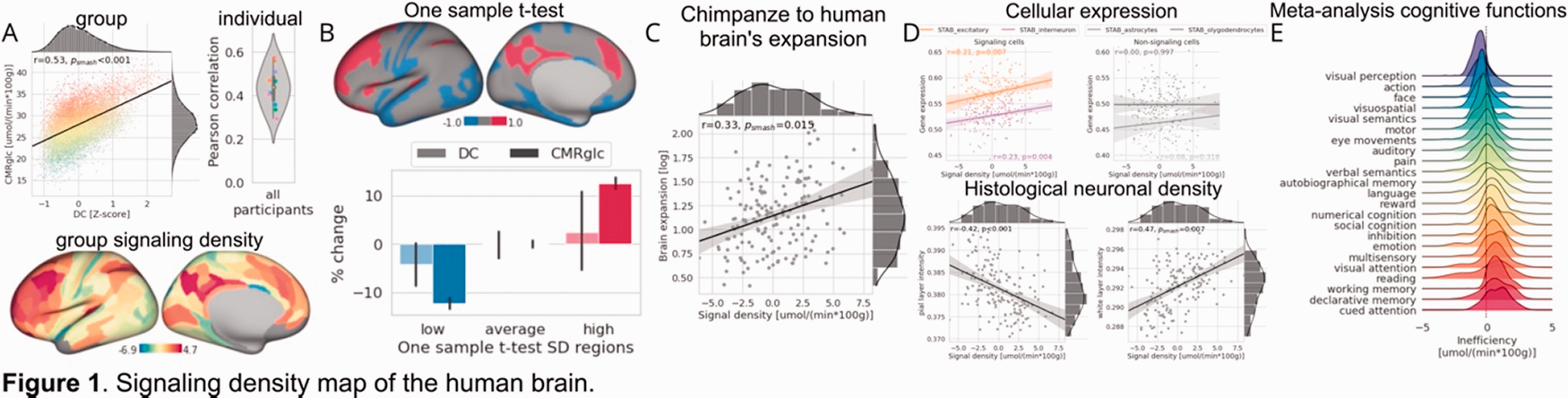
Results: The signaling-related energy demand correlated with the DC across the cortex at the group (Figure 1(a)-top-left) and individual levels (Figure 1(a)-top-right). Next, we defined an integrative parameter of signaling density (SD) as the deviation of measured from expected energy cost for a given level of connectivity (Figure 1(a)-bottom). Signaling dense regions (red-regions in Figure 1(b)) had an extra energy demand of up to 25%, but a similar DC (Figure 1(b)-barplot). On a macroscale, SD correlated with the extent of cortex expansion from chimpanzees to humans (Figure 1(c)) (Wei-Y-2019). On a microscale, SD was i) positive-correlated to the gene-expression (Markello-RD-2021) of signaling-cells but not of non-signaling-cells (Figure 1(d)-top), and ii) negative-correlated with cellular density in outer-cortical layers but positively correlated in lower-cortical layers of high-resolution histological data (Figure 1(d)-bottom) (Paquola-C-2021). Finally, signaling dense regions were predominantly involved in human-specific cognitive functions (Figure 1(e)) (Yarkoni-T-2011). Importantly, all Results were replicated in all cohorts.
98
Using an endothelial specific RiboTag mouse to understand how elevated ADMA alters cerebrovascular function
L Dowsett and J Leiper
1Institute of Cardiovascular and Medical Sciences, The University of Glasgow
Abstract
Background: Asymmetric dimethylarginine (ADMA) is a cerebrovascular risk factor with elevated plasma concentrations associated with an increased incidence of stroke and vascular dementia. ADMA is a known inhibitor of nitric oxide (NO) production however, plasma concentrations clinically associated with stroke are far below that necessary to block NO synthesis.
Aim: This study aims to understand how subpressor concentrations of ADMA alters cerebrovascular function; and to identify new targets of ADMA that contribute to cerebral endothelial cell (EC) dysfunction independent of NO blockade.
Method: We generated an endothelial specific RiboTag mouse (RPL22-HA Tie2Cre+) to isolate EC ribosomal mRNA from whole brain tissue. Mice were provided ADMA (20mg/kg/day) or L-257 (25mg/kg/day), an inhibitor of ADMA metabolism, in their drinking water for 3 days. EC ribosomes were immunoprecipitated from brain lysates followed by RNA isolation. Sequencing libraries were prepared using Ilumina Truseq Stranded mRNA sample kits and sequenced in 75 base, paired end mode on the Illumina NextSeq 500 platform. Differential expression was assessed using Bioconductor packages Tximport6 and DESeq2.
Results/Conclusions: We found a total of 350 genes regulated by ADMA and 380 genes regulated by L-257. Of these 50 genes were significantly regulated by both ADMA and L-257. Following pathway analysis using Panther ‘Go-SLIM’ we found ‘calcium ion membrane transport’ (p = 7.14E-05, FDR = 7.9E-02), ‘regulation of transmembrane receptor Ser/Thr kinase signalling’ (p = 6.65E-0.5, FDR 1.45E-01) and ‘glutamate receptor activity’ (p = 3.22E-05, FDR 8.95E-03) to be significantly enriched. These pathways suggest that excessive ADMA alters endothelial signalling pathways that are key to maintaining cerebrovascular health.
105
An automated framework for FDOPA PET imaging analysis
G Nordio1, R Easmin1, A Giacomel1, O Dipasquale1, D Martins1, S Williams1, F Turckheimer1, O Howes2,3,4 and M Veronese1,5
1King’s College London
2Department of Psychosis Studies, Institute of Psychiatry, Psychology & Neuroscience, King’s College London
3MRC London Institute of Medical Sciences, Hammersmith Hospital
4Institute of Clinical Sciences (ICS), Faculty of Medicine, Imperial College London
5Department of Information Engineering (DEI), University of Padua
Abstract
Background: FDOPA PET neuroimaging is widely used to measure pre-synaptic dopamine synthesis capacity (DSC). The method has the potential to become a non-invasive, precision medicine biomarker applicable across a range of brain disorders including Parkinson’s disease, gliomas and psychosis.1 However, technological advancements in data quantification, validation and management are needed prior to global rollout.
Aim: To present an automated analytical framework for FDOPA PET imaging to quantify dopamine function in the living human brain.
Method: The imaging platform XNAT was used to create an institutional repository including FDOPA PET scans, demographics, clinical information, structural MRI and genetics when available. A fully-automated analysis pipeline for image processing and data quantification was implemented in Python and integrated into XNAT. Reproducibility was assessed in test-retest datasets both in controls and patients with psychosis. Robustness of the method was assessed by comparing automated analysis estimates to historical results. A sensitivity analysis exploring the effect of experimental (injected radioactivity, tomograph) and demographic (weight, age, gender) variables on the dynamic FDOPA quantification was performed on baseline scans of healthy controls.
Results: The database includes the largest available repository of 892 FDOPA PET imaging scans organized in 23 studies, collected from five imaging sites. The automated analysis pipeline results are in good agreement with historical estimates (Pearson’s correlation = 0.93), confirming the good reproducibility of the biomarker (ICC controls: 0.884, ICC patients: 0.711). From the sensitivity analysis (N = 107), only gender is statistically associated with DSC in striatum (Table 1).
Conclusions: The proposed infrastructure ensures full control on the analytical process and robust and reproducible results for FDOPA brain PET imaging across sites, studies and patient populations. The Method has the potential to serve as a biomarker for early diagnosis, patient stratification and individualised treatment planning in brain disorders with altered dopamine function.
Association of demographical and experimental variables with Kicer of the whole striatum.
Dependant variable: Whole striatum Kicer
Full sample (n=108)
With Siemens biograph only (n=97)
Pearson r
t
p
Pearson r
t
p
Age
−0.064
−1.608
0.111
−0.137
−1.494
0.139
Weight
0.014
1.056
0.293
−0.009
1.161
0.249
Gender
−0.311
−3.405
0.001*
−0.332
−3.553
0.001*
Injected Radioactivity
0.042
0.013
0.989
0.021
0.208
0.836
Tomograph
0.112
1.498
0.137
0.097
1.261
0.210
Reference
VeroneseM, et al. 2021. Neuropsychopharmacology. ▪▪▪.a-1
106
Automated detection of spreading depolarizations for real-time diagnosis of delayed cerebral ischemia in aSAH patients
S Major1,2,3, C Lemâle1,2, J Dreier1,2,3,4,5
1Charité – Universitätsmedizin Berlin, Center for Stroke Research Berlin
2Charité – Universitätsmedizin Berlin, Department of Experimental Neurology
3Charité – Universitätsmedizin Berlin, Department of Neurology
4Bernstein Center for Computational Neuroscience Berlin
5Einstein Center for Neurosciences Berlin
Abstract
Background: It is now increasingly recognized that spreading depolarizations (SDs) are the mechanism of the initial, still reversible, phase of cytotoxic edema in brain gray matter. This also applies to the damage from delayed cerebral ischemia (DCI) between 3 and 14 days after aneurysmal subarachnoid hemorrhage (aSAH). Longitudinal neuroimaging in our study ’DISCHARGE-1’ in 180 patients revealed that the average patient with severe aSAH admitted to the ICU after aneurysm treatment has already lost 46 ml of brain tissue but will lose another 34 ml in the next two weeks. These 34 ml currently cannot be saved, in part because the treating intensivists recognize far too late when DCI develops in the often comatose patients.
Aim: In DISCHARGE-1, we found that SD monitoring indicates the occurrence of DCI in real-time with 0.74 sensitivity and 0.82 specificity. Because decisions on treatment should be made as early as possible, we aimed to develop a system that can automatically identify SDs in the continuous electrocorticogram (ECoG).
Method: In 1223 days of ECoG recording from 123 aSAH patients, we manually identified 3733 SDs with 8367 depressions periods. We used this data to train classification models with multiple supervised machine learning algorithms and subsequently compared their performance.
Results/Conclusions: Currently, we are able to detect 93% of SDs with a positive predictive value of 35%. This suggests that supervised machine learning on manually analyzed ECoG data allows the generation of classification models that can identify SDs and thus upcoming DCI, which could improve patient treatment.
107
The m6A epitranscriptomic reader YTHDF1 is indispensable for minimizing ischemic brain damage
R Vemuganti, A Chokkalla and S Mehta
University Of Wisconsin
Abstract
Cellular RNAs are pervasively tagged with diverse chemical moieties, collectively defined as epitranscriptomic modifications. The methylation of adenosine at N6-position generates N6-methyladenosine (m6A), which is the most abundant modification in the adult brain and is highly sensitive to various types of acute injuries, including stroke, traumatic brain injury and spinal cord injury. In the injured brain, the m6A readers dictate the fate of methylated mRNA by interacting with mRNA processing machinery. For example, the m6A reader YTH domain-containing family protein 1 (YTHDF1) binds and recruits ribosomal complex to promote translation of the methylated mRNA. We previously showed that stroke induces cerebral m6A hypermethylation of inflammatory and apoptotic transcripts along with the upregulation of the m6A reader YTHDF1. Currently, we evaluated the role of YTHDF1 in post-stroke brain damage by subjecting the adult homozygous YTHDF1 knockout male mice to transient middle cerebral artery occlusion. YTHDF1 deletion exacerbated post-ischemic motor deficits manifested by decreased time on the rotarod and increased number of foot faults, and a significantly increased infarction compared to the wild-type control. Furthermore, RNA-sequencing analysis revealed that YTHDF1 knockout mice display significantly perturbed expression of genes related to neuronal migration, synapse assembly, mRNA processing and inflammation compared with the wild-type control. Collectively, this study demonstrated that YTHDF1 deficiency worsens the post-stroke neurologic outcome by modulating multiple developmental and pathological processes.
110
Evaluating changes in neurovascular coupling as a function of cerebral autoregulation
D Acharya1, S Schmitt1,2, J Yang1, A Ruesch2, W Scammon1, E Crane1, M Smith1,2 and J Kainerstorfer1,2
1Department of Biomedical Engineering, Carnegie Mellon University
2Neuroscience Institute, Carnegie Mellon University
Abstract
Background: The ability of the brain to regulate blood flow in response to changes in cerebral perfusion pressure (CPP) is compromised in some pathologies and can lead to permanent neural damage if not treated. However, the assessment of autoregulatory state and associated neuronal health remains difficult, partially because CPP estimation is invasive. Additionally, there is a lack of understanding on how neuronal function is affected by CPP and autoregulatory changes.
Aim: To evaluate the relationship between CPP and neural function, we propose the measurement of local neural and vascular signals to evaluate neurovascular coupling (NVC) as a function of CPP. We plan to model NVC for both robust stimulus-evoked signals and spontaneous activity at rest, to provide clinical tools for varying levels of patient participation and monitoring durations.
Methods: In controlled pre-clinical studies, we altered CPP using a hydrocephalus-type animal model. We monitored NVC at both rest and to visual-stimulus evoked activity using electrophysiological (EEG) and diffuse spectroscopy (NIRS) techniques. We then studied NVC changes with CPP.
Results/Conclusion: We found changes in stimulus-evoked neural and vascular signals with CPP. We used the recorded EEG and NIRS signals to model a hemodynamic response function (HRF). The HRF shape was altered at extreme CPP levels (Figure 1). The estimated “healthy” HRF from stimulus-evoked data also well-predicted the hemodynamic response to neural activity at rest. However, this prediction became worse at CPP corresponding to impaired autoregulation (Figure 2). These preliminary results indicate that NVC can act as a non-invasive, bedside-compatible biomarker for cerebral autoregulatory assessment.
119
Protecting against cerebrovascular injury with pro-homeostatic docosanoids
N Bazan1, M Reid1, P Mukherjee1, A Obenaus2, L Khoutorova1, C Roque3, N Petasis4, R Oria3, L Belayev1, A Resano5, M Barajas5 and R Palacios-Pelaez6
1Louisiana State University Health Sciences Center
2University of California Irvine, Department of Pediatrics School of Medicine
3Federal University of Ceara School of Medicine Laboratory of the Biology of Tissue Healing, Ontogeny and Nutrition, Department of Morphology and Institute of Biomedicine
4University of Southern California, Department of Chemistry
5Department of Health Science, Public Univ
6Laboratorios Diater
Abstract
Background: This study focuses on the neuroprotective bioactivity of docosanoid mediators: Neuroprotectin D1 (NPD1), Resolvin D1 (RvD1), and their combination against experimental stroke. These lipid mediators are biosynthesized “on-demand” in response to stroke onset to resolve neuroinflammation and restore homeostasis.
Aim: To investigate the dose-response and therapeutic window after middle cerebral artery occlusion (MCAo).
Method: Male SD rats were subjected to 2h of MCAo, the behavior was evaluated on days 1, 2, 3, and 7, and ischemic core and penumbra were computed from T2WI on day 7. Dose-response study: NPD1 (111, 222, and 333 µg/kg), RvD1 (111, 222, 333 µg/kg), NPD1+RvD1 or vehicle, administered IV at 3h after MCAo. Therapeutic window study: NPD1 and RvD1 (222 µg/kg), NPD1+RvD1 were administered at 3, 4, 5, and 6 h after MCAo.
Results/Conclusion: All physiological variables showed no significant differences among groups. Treatments with NPD1 and RvD1 alone (111, 222, and 333 µg/kg) improved total neurological scores by 31–35% on day seven. In contrast, the neuroprotective effect was enhanced using NPD1+RvD1 by 41% compared to the vehicle group. Ischemic core, penumbra, and total lesion volumes (computed from T2WI) were reduced with NPD1+RvD1 by 69, 70, and 67% compared to the vehicle. Therapeutic window: NPD1 and RvD1 alone improved behavior on day seven by 35%. NPD1+RvD1 improved behavior when administered at 3, 4, 5, and 6 h by 56%, 23%, 26%, and 28%, respectively. We concluded that NPD1+RvD1 therapy is more effective than the single therapy when administered up to 6 h focal cerebral ischemia in rats.
This study was supported by NIH, NINDS grant R01NS104117, R01109221 (NGB and LB), and Brazilian CAPES (88881.311939/2018-01) (CRR)
139
Characterization of the novel mutant-huntingtin-aggregate-directed PET radioligand [11C]CHDI-009R in a mouse model of Huntington’s disease
F Zajicek1, J Verhaeghe1, S De Lombaerde1,2, A Miranda1, S Stroobants1,2, M Skinbjerg3, Y Wang3, I Munoz-Sanjuan3, C Dominguez3, V Khetarpal3, J Bard3, L Liu3, D Bertoglio1 and S Staelens1
1Molecular Imaging Center Antwerp (MICA), University of Antwerp
2Department of Nuclear Medicine, Antwerp University Hospital
3CHDI Management/CHDI Foundation
Abstract
Background: Huntington’s disease (HD) is a progressive autosomal dominant neurodegenerative disorder caused by an extended polyglutamine repeat in exon 1 of the huntingtin gene, which, in its mutated form, encodes for mutant huntingtin (mHTT) protein. Measuring mHTT aggregates with Positron Emission Tomography (PET) explores HD-related pathology during disease progression and may provide a method to evaluate the efficacy of therapeutic interventions relying on mHTT-lowering approaches as well.
Aim: We characterized the novel mHTT-aggregate-directed PET radiotracer [11C]CHDI-009R in a mouse model of HD.
Method: Ninety-minute dynamic PET imaging was performed in 9-month-old heterozygous (HET; n = 22) and wildtype (WT; n = 20) zQ175DN mice. We investigated radiometabolite profiles, the mass dose effect on volume of distribution (VT) estimations calculated with image-derived input functions (IDIF) and test-retest (T-RT) variability. Logan VT(IDIF) estimations of HET animals were compared to VT(IDIF) values of WT mice.
Results/Conclusions: Following administration of [11C]CHDI-009R, plasma or brain radiometabolite profiles displayed no reduction of intact radioligand in both genotypes (plasma40min: 95.8 ± 1.5%). Due to the correlation of injected masses and VT(IDIF) values (r = −0.5840; p = 0.0011), it is recommended to keep injected masses below 1.5 µg/kg. mHTT quantification with [11C]CHDI-009R revealed significantly higher VT(IDIF) estimations in HET animals (Striatum: 96.1 ± 6.1%; p < 0.0001) (Figure 1). T-RT variability analysis is ongoing. The available data depicts [11C]CHDI-009R as a suitable radiotracer for mHTT aggregate quantification.
142
Proteoglycan alterations after traumatic brain injury
N Siddiqui1, K Oshima1, J Orfila2, D Carter1, P Herson2, E Schmidt1,3 and J Hippensteel1
1University of Colorado Anschutz Medical Campus
2The Ohio State University
3Denver Health Medical Center
Abstract
Background: Nearly seventy million people are affected by Traumatic Brain Injury (TBI) annually worldwide resulting in immense societal burden. The glial scar, composed of many activated cell types and extensive extracellular matrix (ECM) alterations, is central to its pathophysiology. While significant attention has been paid to the cellular and general extracellular constituents of this scar, less is known about its glycobiology.
Aim: We sought to determine the spatiotemporal dynamics of glycosaminoglycans (GAGs) and proteoglycans at the injury site after TBI. Further, we sought to determine if brain-penetrating GAGs known to be released in diseases associated with systemic inflammation like TBI contribute to ECM changes at the injury site.
Method: We performed controlled cortical impact (CCI) or craniotomy-alone (CA) in mice then measured GAGs in brain and plasma at 1 and 7d via mass spectrometry (HPLC MS/MS). We compared transcriptomic profiles of CCI site to contralateral tissue. We confirmed increased expression of proteoglycans identified by transcriptomics via immunohistochemistry.
Results/Conclusions: We observed elevations in GAGs at the injury site compared to contralateral tissue after CCI and CA. We did not observe increases in circulating GAGs after CCI when compared to CA. Transcriptomics at the CCI site revealed isolated increases in the proteoglycans glypican3 and decorin. Increased expression of these proteoglycans was verified via immunohistochemistry. Our results suggest that both CCI and CA significantly impact local GAG levels. GAG alterations after TBI may be specifically driven by upregulation of glypican3 and decorin. The precise biologic consequences of these upregulated proteoglycans warrants further investigation.
160
Longitudinal simultaneous fMRI and mesoscale Ca2+ imaging in a mouse model of Alzheimer’s disease
F Mandino1, X Shen1, D O’Connor2, B Mukherjee3, K DeLuca3, A Owens3, A Hamodi4, A Qu1, J Onofrey1,2,5, M Crair4,6, X Papademetris1,2, S Strittmatter3 and E Lake1
1Department of Radiology and Biomedical Imaging, Yale University School of Medicine
2Department of Biomedical Engineering, Yale University School of Medicine
3Cellular Neuroscience, Neurodegeneration and Repair Program, Departments of Neurology and Neuroscience, Yale School of Medicine
4Kavli Institute for Neuroscience, Yale School of Medicine
5Department of Urology, Yale School of Medicine
6Department of Neuroscience, Yale School of Medicine
Abstract
Backgrounds: Alzheimer’s disease (AD) is a fatal progressive neurological disease that impacts the neurogliovascular unit (NGVU) as a whole. Preclinical models are instrumental for gaining mechanistic insights into precursors of AD which can be translated to the clinic. To this end, multimodal imaging methods have an active role to play in uncovering, and understanding, clinically accessible imaging biomarkers.
Aim: Using our simultaneous multimodal mesoscopic calcium (Ca2+) and whole-brain fMRI (fMRI) approach, we propose to examine spontaneous NGVU activity, in a double knock-in, GCaMP-positive, mouse model of AD, throughout the life span. With this approach we record cell-type specific cortical activity optically with simultaneous whole-brain BOLD fMRI.
Method: All mice (DKI = 14; WT = 11) are induced to express GCaMP6s Ca2+ fluorescence beginning at P0. Optical windows (intact skull with bone thinning) for Ca2+ imaging are implanted at 3 months, followed by four longitudinal multimodal imaging sessions (at 4, 6, 9 and 12 months). Functional connectomes are computed using the Allen Atlas. All region-to-region connectivity strengths are measured using Pearson’s correlation.
Results/Conclusions: The integrity of the optical implants (for cortico-optical access and immobilization) are preserved throughout the 9-month imaging protocol. Biologically plausible resting-state functional connectomes in both AD and WT mice are observed. Motion estimates in optical data show little-to-no motion in the X-Y field, suggesting that the major contribution to fMRI motion lies in the Z field. Encouragingly, motion in fMRI data is also low. Further investigations into disease progression and how mesoscopic-neuronal signals relate to the BOLD signal will follow.
162
PET imaging of diffuse amyloid-beta aggregates enables early confirmation of drug effects
S Meier1, D Sehlin1, S Roshanbin1, V Lim Falk1, T Saito2,3, T Saido2, U Neumann4, J Rokka1, J Eriksson1,5 and S Syvanen1
1Uppsala University
2RIKEN Center for Brain Science
3Nagoya City University
4Novartis
5Uppsala University Hospital
Abstract
Background: PET radioligands used for amyloid-beta (Aβ) imaging in Alzheimer’s disease (AD) bind to fibrillary forms of Aβ deposited as insoluble plaques. However, levels of nonfibrillar and diffuse aggregates of Aβ are likely to be affected by Aβ reducing treatments prior to alterations in plaque load. Thus, imaging of diffuse Aβ aggregates could enable an early evaluation of drug efficacy in clinical trials.
Aim: The aim of this study was to compare 11C-PiB, the “gold standard” Aβ plaque radioligand with an antibody-based PET ligand, targeting diffuse Aβ aggregates. The comparison was done before and after treatment with β-secretase inhibitor NB360 in two AD mouse models, with a study design similar to a clinical trial of a new drug candidate.
Methods: Mice (Tg-ArcSwe and APPNL-G-F) were treated with food supplemented with β-secretase inhibitor NB-360 or standard non-supplemented food for 2 months. Mice were PET scanned with [11C]PiB to measure Aβ plaque load followed by a scan with the 124I-labelled antibody-based RmAb158-scFv8D3 to investigate diffuse Aβ aggregates. Brain tissue was isolated after scanning and Aβ levels were assessed.
Results and Conclusions: There was no difference in [11C]PiB signal between NB360-treated and untreated mice, and further, both groups tended to show higher [11C]PiB signal after the 2-months long treatment compared to baseline (Figure 1(a) and (b)). In contrast, reduced [124I]RmAb158-scFv8D3 signal was observed in NB360-treated mice compared to controls (Figure 1(a) and (c)). Post mortem analysis confirmed a reduction of brain Aβ in NB360-treated mice (Figure 1(d)). Thus, imaging of diffuse Aβ, instead of plaques, may enable earlier effect confirmation in clinical studies of anti-Aβ therapies.
163
Subarachnoid hemorrhage induces sub-acute and early chronic impairment in learning and memory in mice
A Regnier-Golanov, E Golanov and G Britz
The Houston Methodist Hospital
Abstract
Background: Subarachnoid haemorrhage (SAH) leads to significant long-term cognitive deficits, the post-SAH syndrome. Existing neurological scales used to assess outcomes of SAH are prevalently focused on sensory-motor functions.
Aim: To better evaluate short term and chronic consequences of SAH we explored and validated a battery of neurobehavioral tests to gauge the functional outcomes in mice after the circle of Willis perforation-induced SAH.
Method: Behaviour of Sham and SAH mice was compared using Garcia scale, open-field, Y-maze, dyadic social interaction, Barnes maze, and novel object recognition tests during 30 days postictal period.
Results/Conclusions: The 18-point Garcia scale, applied up to 4 days, detected impairment only at 24hrs with no significant differences between the groups. A decrease in locomotion in both groups was detected at 4-days post-surgery in the open field test but recovered at 30-days. However, an anxiety-like behaviour undetected at 4-days developed at 30-days in SAH mice. At 4-days post-surgery, Y-maze revealed an impairment in working spatial memory in SAH mice. Dyadic social interactions showed decreased sociability in SAH mice, which spent less time interacting with the stimulus mouse. At 30-days, SAH mice displayed spatial learning and memory deficits in the Barnes maze committing significantly more errors and used more time to find the escape box. Novel object recognition test revealed cognitive dysfunction in the SAH mice. Data suggest dysfunction of the limbic system and hippocampus in particular. This battery of 5 basic behavioural tests allows to detect neurocognitive deficits in a sub-acute and chronic phase following the SAH.
170
In vivo staging of tau pathology in progressive supranuclear palsy with multicenter F-18-PI-2620 PET
M Rullmann1, M Brendel2, J Rumpf3, J Levin4, R Perneczky5, V Villemagne6, A Stephens7, O Sabri1 and H Barthel1
1Department of Nuclear Medicine, University of Leipzig
2Department of Nuclear Medicine, University Hospital of Munich, LMU Munich
3Department of Neurology, University of Leipzig
4Department of Neurology, University Hospital of Munich, LMU Munich
5Department of Psychiatry and Psychotherapy, University Hospital, LMU Munich
6Department of Psychiatry, University of Pittsburgh
7Life Molecular Imaging GmbH
Abstract
Background: Progressive supranuclear palsy (PSP) is a primary 4R tauopathy. Six pathological stages of tau have been identified in postmortem studies. Tau aggregates begin to accumulate in the subthalamic nucleus (STN), with subsequent involvement of the globus pallidus (GP), striatum (ST), cerebellum with dentate nucleus (DN), frontal cortex (FC), and occipital cortex (OC).
Aim: We aimed to test whether this staging system is reproducible with in vivo F-18-PI-2620 tau PET.
Method: We analyzed data from 85 patients with clinical diagnosis of PSP, including 44 patients with Richardson syndrome (RS)-PSP, 41 nonRS-PSP, and from 26 healthy controls. Dynamic F-18-PI-2620 PET imaging was performed over 60 minutes in a multicenter setting. After kinetic modeling, parametric distribution-volume-ratio images were analyzed and classified in the mentioned hierarchy. Data from controls defined the normal range for each tau-in-PSP VOI.
Results/Conclusions: Tau-in-PSP VOI positivity varied regionally for STN (16%), GP (18%), ST (33%), DN (19%), FC (1%), and OC (5%). 64% of RS-PSP patients were PET positive, with 41% classified hierarchically as stage 1, 16% as stage 2, and 7% as stage 3. The non-RS-PSP group showed lower frequencies (46%, 36%, 5%, and 5%, respectively). The non-RS-PSP patients showed a correlation between the highest PSP stage and age (r = 0.37, p = 0.02). In vivo tau PET staging was not associated with clinical disease scores (PSPRS, SEADL, or disease duration).
In summary, F-18-PI-2620-PET appears to be capable of performing PSP tau staging in vivo. However, our data question the concept of a single hierarchical system. More data are needed to also clarify the relationship between F-18-PI-2620-PET signal and the severity of the clinical phenotype.
175
Fibroblasts repair blood brain barrier damage and hemorrhagic brain injury via TIMP2
L Xu1,2, T Xu2, A Nirwane1, K Devasani1 and Y Yao1,2
1University Of South Florida
2University of Georgia
Abstract
Background: Controversial results exist on whether fibroblasts exert a beneficial or detrimental role after CNS injury. This is due to the lack of fibroblast-specific markers. Almost all fibroblast markers also label other cell populations that are known to regulate CNS injury. A recent single-cell RNAseq study identified Col1α1 as a fibroblast-specific marker in the CNS.
Aim: We aim to investigate the functions of fibroblasts in both homeostatic and intracerebral haemorrhage (ICH) conditions.
Method: We generated fibroblast-ablated mice (termed FKO) by crossing the fibroblast-specific Col1α1-Cre line with the iDTR knock-in mice, and investigated the functions of fibroblasts using these FKO mice.
Results/Conclusions: FKO mice were grossly normal under homeostatic conditions. After ICH, however, FKO mice exhibited exacerbated blood-brain barrier (BBB) damage, increased injury volume and worse neurological function at the subacute phase, indicating a beneficial role of Col1α1+ fibroblasts in ICH. Consistent with these results, fibroblasts significantly attenuated endothelial permeability in an in vitro ICH model. Next, we showed that fibroblasts promoted BBB repair in ICH mainly via up-regulating tight junction proteins without affecting transcytosis-associated proteins, suggesting a paracellular rather than transcellular mechanism. Subsequent mechanistic study revealed that the BBB-protecting effect of fibroblasts is partially mediated by TIMP2. Moreover, we found that exogenous TIMP2 decreased BBB leakage in FKO mice after ICH. These findings suggest that Col1α1+ fibroblasts repair BBB damage in ICH via the paracellular pathway in a TIMP2-dependent manner, and that fibroblasts and TIMP2 may be targeted in the treatment of ICH.
176
Targeting brain injury after intracerebral hemorrhage with lipid nanoparticles
Z Al-Ahmady1,2, B Dickie3, I Aldred2, D Jasim2, J Barrington3, M Haley3, E Lemarchand3, G Graham Coutts3, S Kaur1, J Bates1, A Parry-Jones4,5,6, K Kostarelos2 and S Allan3,6
1Pharmacology Department, School of Science and Technology, Nottingham Trent University
2Division of Pharmacy and Optometry, Faculty of Biology, Medicine and Health, AV Hill Building, The University of Manchester
3Division of Neuroscience and Experimental Psychology, School of Biological Sciences, Faculty of Biology, Medicine and Health, University of Manchester
4Division of Cardiovascular Sciences, Lydia Becker Institute of Immunology and Inflammation, School of Medical Sciences, Faculty of Biology, Medicine and Health, Manchester Academic Health Science Centre, The University of Manchester
5Manchester Centre for Clinical Neurosciences, Salford Royal NHS Foundation Trust, Manchester Academic Health Science Centre, Salford
6Geoffrey Jefferson Brain Research Centre, The Manchester Academic Health Science Centre, Northern Care Alliance NHS Group, University of Manchester
Abstract
Background: Alteration of blood-brain barrier (BBB) integrity occurring post intracerebral haemorrhage (ICH) could be utilized to effectively deliver drugs to the brain. However, to achieve this a greater understanding of the mechanisms and timeframe of BBB disruption in ICH is needed. Previously we showed that we can target ischemic-stroke lesions by selective translocation of liposomes through the site of BBB disruption.
Aim: Our aim in this study is to investigate if similar selective targeting is possible in ICH, with loss of BBB integrity being the primary mechanism by which entrance is permitted.
Method: ICH was induced in mice (C57BL/6) by intra-striatal collagenase injection. Liposomes were injected intravenously at 3h, 24h & 48h post-ICH, and accumulation in the brain studied using SPECT/CT, in-vivo optical imaging and histology. BBB integrity, brain water (oedema), and brain iron were characterised using dynamic contrast-enhanced MRI, T1 mapping and T2*-weighted imaging respectively using 7T MRI with imaging timepoints matched to the liposome administrations.
Results/Conclusions: In-vivo SPECT/CT imaging and optical imaging data show biphasic liposomes uptake into the haematoma, with an early phase of increased uptake at 3–24h post-ICH, followed by a second phase at 48–72h. MRI measurement of BBB leakage of Gd-DOTA showed a similar biphasic pattern post-ICH, suggesting a common enhanced entry into the brain through disrupted tight junctions and damaged endothelial cells. Overall, our findings suggest that selective liposomal accumulation into the haematoma is linked to biphasic BBB hyper-permeability after ICH, offering a novel route for drug delivery into the haemorrhaged brain.
179
MCAo stroke increases hippocampal soluble amyloid beta production in a TRPM2-dependent manner
J Basak1, M Falk1, D Mitchell1, B Wassermann1, N Quillinan1 and P Herson2
1The University of Colorado School of Medicine
2The Ohio State University School of Medicine
Abstract
Background: Post-stroke cognitive impairment and dementia (PSCID) is a phenomenon of increasing concern in survivors of large vessel ischemia. The molecular mechanisms underlying PSCID are poorly defined, but may involve perturbations in pathways involved in both neurodegeneration and oxidative stress.
Aim: In this study we used the middle cerebral artery occlusion (MCAo) stroke model in mice to evaluate how large vessel ischemia alters soluble amyloid beta (Aβ) levels and to determine whether the ion channel TRPM2 interacts with Aβ in the setting of focal ischemia.
Methods: MCAO was performed on adult (8–10 week) wild-type and TRPM2 KO mice and Aβ levels were measured in the tissue via ELISA 7 days post-injury. To measure BACE enzyme activity and protein levels, a FRET-based enzyme assay and immunohistochemical analysis of BACE antibody were performed.
Results/Conclusions: Soluble Aβ40 and Aβ42 levels were increased in the ipsilateral hippocampus in MCAo mice when compared to sham-treated mice (71.1% increase for Aβ40, p = 0.04; 91.7% increase for Aβ42, p = 0.03). No changes in Aβ levels were observed in TRPM2 knock-out mice. We also found that the activity of the BACE enzyme, responsible for generating Aβ, is increased in the ipsilateral hippocampus of the MCAo mice (40.7% increase in comparison to sham mice), with no changes in BACE expression levels. BACE activity was not increased in TRPM2 KO mice. Our data highlights that a transient MCAo stroke leads to increases in the production of soluble endogenous Aβ40 and Aβ42 in the hippocampus of the injured brain in a manner that requires the presence of TRPM2.
180
GLUT1 dysfunction, metabolic and neurovascular alterations sustained by cortical dysplasia post-seizure induction in brain
C Ghosh1,2, R Westcott1, C O’Connor3 and I Najm3
1Lerner Research Institute, Cleveland Clinic
2Cleveland Clinic Lerner College of Medicine of Case Western Reserve University
3Charles Shor Epilepsy Center, Neurological Institute
Abstract
Objectives: Focal cortical dysplasia (FCD) is associated with compromised blood-brain barrier (BBB) function, which has clinical relevance and presents the opportunity for therapeutic intervention in patients with difficult-to-treat epilepsy. The underlying cellular and molecular factors in cortical dysplasia (CD) associated with progressive neurovascular challenges during the pro-epileptic phase, post-seizure and during epileptogenesis remain unclear.
Methods: We studied BBB function in a rat model of congenital (in utero radiation-induced, first hit) CD and longitudinally examined the baseline and progressive neurovascular alterations, glucose transporter-1 (GLUT1) expression and glucose metabolic activity at 2, 15, and 30 days following a second-hit using pentylenetetrazole-induced seizure. In the cortex, the neurovascular target proteins associated with BBB integrity, glucose metabolism and mammalian target of rapamycin (mTOR) signaling were analyzed by immunoblotting and immunohistochemistry. Cortical brain glucose-lactate levels and ATPase activity were evaluated. We also assessed the effect of lactate supplementation during glucose deprivation in human neuronal cell culture on mTOR activation and monocarboxylate transporter-2 (MCT2) expression. In addition, the FCD brain tissues from focal epilepsies are used to corroborate the BBB-metabolic connection.
Results: Our study revealed that 1) altered vascular density and prolongation of BBB albumin leakages continued through 30 days post-seizure in CD rats, 2) CD brain tissues showed elevated matrix-metalloproteinase-9 levels through 15 days post-seizure and microglial overactivation through 30 days post-seizure, 3) BBB tight junction protein and GLUT1 levels were decreased and neuronal MCT2 and mTOR levels were increased in the CD rat brain, and 4) ATPase activity is elevated and a low glucose/high lactate imbalance exists in the brain of CD rats. Further, the mTOR pathway is found to be activated and MCT2 elevated in the presence of high lactate during glucose starvation in neuronal brain cells. Similar pattern was identified in human dysplastic epileptic brain regions that showed low glucose-high lactate, decreased GLUT1 levels and upregulated vascular endothelial growth factor levels. These findings suggest BBB metabolic disturbances and lactate compensatory mechanism interplay in the dysplastic brain.
Conclusions: In summary, the study indicates that BBB anomalies, including GLUT1 dysfunction, may contribute to epilepsy and its progression in CD rat model and human epileptic brain through multiple mechanisms. These metabolic factors at the neurovasculature could have potential implication in the diagnosis and early therapeutic intervention of FCD in medically refractory epilepsy.
Funding Support: This study is supported in part by NIH-NINDS grant R01NS095825 awarded to Dr. Chaitali Ghosh.
185
Inhibition of STAT1 improves outcome after traumatic brain injury via mitigating proinflammatory microglial/macrophage responses
Y Zhao1, C Ma1, S Li1, M Bennett2, C Dixon3,4, J Chen1,3 and Y Shi1,3
1Department of Neurology, University of Pittsburgh
2Dominick P. Purpura Department of Neuroscience, Albert Einstein College of Medicine
3Geriatric Research, Education and Clinical Center, Veterans Affairs Pittsburgh Health Care System
4Department of Neurosurgery, University of Pittsburgh
Abstract
Background: Microglial/macrophage (Mi/Mø) polarization critically influences neuroinflammation and traumatic brain injury (TBI) outcome. The transcription factor STAT1 is a strong determinant of proinflammatory M1-polarization in peripheral macrophages. Whether STAT1 can be targeted to modulate brain Mi/Mø phenotype and improve long-term TBI outcome is unknown.
Aim: We hypothesized that STAT1 drives proinflammatory Mi/Mø responses after TBI, and investigated whether STAT1 deletion or pharmacological inhibition improves TBI outcome via mitigating proinflammatory Mi/Mø responses.
Method: TBI was induced by controlled cortical impact in mice with tamoxifen-induced, Mi/Mø-targeted knockout (mKO) of STAT1 or wild-type mice. A selective STAT1 inhibitor fludarabine (5mg/kg) was administered i.p. 2h after TBI and then daily for 5d. Neuroinflammation, Mi/Mø phenotype, and various neurobehavioral deficits were assessed for 35 days post-injury (dpi).
Results/Conclusions: STAT1 was upregulated in Mi/Mø 2h after TBI and lasted for 5dpi (p < 0.005, n = 3-6). STAT1 inhibition by fludarabine reduced proinflammatory Mi/Mø (CD16/32 immunostain) and infiltration of peripheral immune cells (flow cytometry), thus ameliorating brain inflammation (ELISArray; 9 out of 14 markers) 3–5dpi (p < 0.05, n = 6). STAT1 mKO reproduced fludarabine’s effect and markedly reduced proinflammatory Mi/Mø (CD16/32+, CD86+, and TNF-α+ cells; flow cytometry; n = 6). By shifting Mi/Mø away from a proinflammatory phenotype, STAT1 mKO attenuated long-term (35dpi) neurological deficits and cortical lesion size (n = 8-12). Remarkably, fludarabine significantly improved long-term TBI outcome for 35dpi, but this effect was lost in STAT1 mKO mice (n = 8-12). Our findings suggest that STAT1 dictates post-TBI proinflammatory Mi/Mø responses, and fludarabine is a promising treatment for TBI to alleviate neuroinflammation and promote long-term functional recovery.
186
miR-98 and let-7g* protect BBB, decrease leukocyte infiltration and microglia activation in ischemic stroke
S Gajghate1, M Winfield1 and S Rom1,2
1Department of Pathology and Laboratory Medicine, Lewis Katz School of Medicine, Temple University
2Center for Substance Abuse Research, Lewis Katz School of Medicine, Temple University
Abstract
Objective: Stroke is incapacitating cardiovascular disease. Following an ischemic attack, inflammatory processes result in recruitment of immune cells and the activation of microglia, and lead to increased Blood-Brain Barrier (BBB) permeability. Inflammation plays a critical role in pathogenesis of ischemic stroke followed by reperfusion (IS/R) and mitigation of inflammatory responses, reduces infarct size and avoids neurological deficits. Inspite stroke’s high incidence, morbidity and mortality, treatment options remain limited. Lately, non-coding RNAs (microRNAs or miRs) have arose as key regulators in a variety of inflammatory conditions, however, miRNAs’ role is still understudied.
Methods: In the present study we utilized both in vivo and in vitro IS/R models, a mouse transient middle cerebral artery occlusion (tMCAO) and oxygen-glucose deprivation (OGD) followed by reperfusion.
Results: We have identified a 2 key non-coding RNAs, from lethal (let) miR family, miR-98 and let-7g* which levels were severely reduced as result IS/R conditions, respectively. Liposome-based delivery of these miRs attenuated the inflammation after stroke, reduced BBB permeability, limited brain infiltration by CD4+ T-cells, neutrophils, and proinflammatory LY6Chi monocytes, also in significant reduction of proinflammatory cytokines. All of these accompanied by diminished microglia activation and neuronal death and subsequently enhanced clinical outcomes.
Conclusion: We demonstrated that post-stroke treatment with either let-7g* or miR-98 may salvage the integrity of tight junctions within the vasculature of the cortex. This study postulates identification and functional assessment of miRNAs in brain endothelium and lays the groundwork for improving therapeutic approaches for patients suffering from ischemic attacks.
189
Role of hemodynamic impairments in white matter lesions – Healthy ageing vs. carotid artery stenosis
S Kaczmarz1,2,3, J Göttler1,2, L Schmitzer1, K Weiss3, M Hansen4, K Mouridsen4, C Zimmer1, F Hyder2 and C Preibisch1
1Technical University of Munich (TUM)
2MRRC, Yale University
3Philips GmbH Market DACH
4Institute of Clinical Medicine, Aarhus University
Abstract
Background: Most clinical MRI protocols include T2-weighted FLAIR imaging to assess periventricular and deep white matter changes. These white matter lesions (WML) are detected by FLAIR hyper-intensities and found in most elderly subjects. While they are commonly interpreted as microangiopathic changes in the absence of distinct diagnoses, their pathogenesis remains unclear.3 Moreover, pathology related effects by internal carotid artery stenosis (ICAS) are currently under debate.2,3
Aim: This MRI study investigated ageing and pathology related WML effects regarding spatial locations relative to watershed areas (WSA) between perfusion territories as well as involvement of microvascular impairments by capillary transit-time heterogeneity (CTH) and structural damage by mean diffusivity (MD).
Method: 29 patients with asymptomatic high-grade ICAS (age = 70.1 ± 4.8y) without previous infarcts and 30 age-matched elderly healthy controls (age = 70.3 ± 7.3y) underwent MRI on a 3T Philips Ingenia. WML were segmented from FLAIR,4 individual WSAs5 and CTH6 from dynamic susceptibility contrast (DSC) and MD from diffusion tensor imaging (DTI, Figure 1(a)).
Results/Conclusions: The WML load inside watershed areas was similarly elevated in both groups with 66% and 64% in ICAS and HCs, respectively (p = 0.048/0.057, Figure 1(b)). Within WML, hemodynamic and structural analyses revealed increases of CTH (p < 0.001, Figure 1(c)) and MD (p < 0.001, Figure 1(d)) in both groups.
Increased prevalence of WML in WSAs indicates a causal role of hemodynamic impairments in the formation of lesions.7,8 Specifically, increases of CTH and MD point to involvement of capillary dysfunction,6 causing axonal degeneration.9 Interestingly, WML formation was independent of ICAS-pathology pointing to mainly ageing related effects.
Spatiotemporal analysis of MRI contrast agent concentration in mouse brain after intraperitoneal administration
A Tessier1,2, A Ruze1, E Royer1, M Bernard1, A Viola1 and T Perles-Barbacaru1
1Center of Magnetic Resonance Imaging in Biology and Medicine (CRMBM, UMR CNRS 7339), Aix-Marseille University
2Medical Imaging, Clinical Research, Military Teaching Hospital (Hôpital d’Instruction des Armées) Sainte Anne
Abstract
Background: In the clinical and preclinical setting, delivery of therapeutic agents across the blood brain barrier (BBB) is facilitated by intracarotid administration of hyperosmolar mannitol, a local and highly invasive approach.
Aim: We aim to assess BBB opening upon intraperitoneal (ip) injection of mannitol in mice by monitoring accumulation of ip administered low molecular weight Gd-DOTA by MRI.
Method: T1-weighted dynamic MRI1 (25 s per volume) was performed over 2h in isoflurane anesthetised C57Bl6 mice at 7T, after 10 mmol/kg Gd-DOTA administration followed 15 minutes later by 5 g/kg 20% mannitol solution (n = 12) or 0.9% NaCl solution (n = 10). Immediately after MRI, n = 17 mice underwent intracardiac perfusion and brains were harvested for inductively coupled plasma mass spectrometry (ICP-MS) analysis of Gd. Tissue Gd-DOTA concentration maps were derived from the MRI acquisitions and the average cerebral Gd concentration was compared with the one determined by ICP-MS.
Results/Conclusions: The MRI concentration maps over time show variable degrees of Gd-DOTA uptake between animals with a characteristic cerebral distribution at 2h post administration likely resulting from the elimination via the glymphatic system (Figure 1). Surprisingly, high Gd-DOTA accumulation (>1.5 increase over 2h) occurred in 2/10 control mice, but only in 8/12 mannitol treated mice. Premature death occurred during MRI in 8/12 mannitol treated mice. ICP-MS results significantly correlate with the MRI-derived cerebral concentration, validating the MRI observations.
Systemic administration of mannitol opens the BBB within tens of minutes, occasionally leading to Gd-DOTA concentrations incompatible with survival within less than 2h.
Reference
Perles-Barbacaru. Magn Reson Med2013.
197
Ischemic stroke induces a chronically altered microglia phenotype with pro-regenerative capacities
S Heindl1, D Varga1, N Snaidero2,3,4, E Beltran4, T Misgeld2,3,4,5 and A Liesz1,5
1Institute For Stroke And Dementia Research, University Hospital, Ludwig Maximilians University Munich
2Institute of Neuronal Cell Biology, Technical University Munich
3German Center for Neurodegenerative Diseases (DZNE)
4Institute of Clinical Neuroimmunology, University Hospital and Biomedical Center
5Munich Cluster for Systems Neurology (SyNergy)
Abstract
Background: Stroke-induced neuroinflammation contributes to secondary neurodegeneration and pro-regenerative pathways at the same time. Microglia are the main constituents of the brain-resident immune response to stroke, yet, the long-term changes in microglial function after stroke and their contribution to chronic recovery are incompletely understood.
Aim: Our aim was to deeply phenotype microglia at chronic time points after stroke and identify their function for long-term recovery.
Method: We used complementary techniques to characterize the broad spectrum of microglial function, comprising in vivo imaging, single cell RNA sequencing and biomolecular assays. Depletion of microglia (CSF1R antagonist), widefield imaging of cortical network function, behavioral tests and histology was used to study the microglial impact on long-term neuronal plasticity and recovery in mice.
Results/Conclusions: Longitudinal PET imaging using a tracer for myeloid cell activation (GE-180) showed (peri-)lesional uptake until 6 months after stroke and microglia were morphologically altered until 3 months. Transcriptomic analysis of microglia at 7 and 28 days after stroke has shown timepoint specific differentiation, indicating a subtype distinct from naïve and acutely activated microglia solely appearing in the chronic phase. Microglial function was altered perilesionally at 28 days post-stroke with upregulated process motility but downregulated phagocytosis. Thus, we identified a microglial phenotype, which is specific for the chronic post-stroke phase, possibly interfering with chronic recovery. Microglia depletion led to reduced lesion involution and behavioral recovery until 42 days after stroke, accompanied by reduced functional connectivity and neuronal plasticity. In conclusion, we found a chronically altered, possibly pro-regenerative microglia phenotype after stroke.
200
Examining perfusion indices of [18F]MK-6240, [15O]water and ASL and relationship with tau uptake with PET/MRI
J Fu1,2, M Juttukonda1,2, A Salvatore1, D Izquierdo-Garcia1,2, C Catana1,2, C Lois2,3, A Ranasinghe1, A Garimella1, H Sari1,2, N Guehl2,3, M Normandin2,3, G El Fakhri2,3, B Dickerson4, K Johnson2,3 and J Price1,2
1Athinoula A. Martinos Center For Biomedical Imaging
2Department of Radiology, Massachusetts General Hospital
3Gordon Center for Medical Imaging, Division of Nuclear Medicine and Molecular Imaging
4Department of Neurology, Massachusetts General Hospital
Abstract
Background: [18F]MK-6240 PET is used to assess in-vivo tau deposition across the Alzheimer’s disease (AD) spectrum. Simplified quantification of tau load, particularly in high-binders, might be sensitive to cerebral blood flow (CBF) and tissue clearance.
Aim: Compare [15O]water, [18F]MK-6240 and arterial spin labeling (ASL) relative-perfusion indices and examine relationships between perfusion and [18F]MK-6240 retention in young/elderly cognitively normal (yCN/eCN) and AD groups.
Method: Eight control (3 yCN, 28 ± 5 years; 5 eCN, 68 ± 6 years) and 3 AD (57 ± 5 years) underwent [15O]water (0–3min), [18F]MK-6240 (0–120min, Figure 1(a)), and multi-delay ASL (post-label delay:1.8–2.4s) PET/MRI (Biograph-mMR). Perfusion indices included early-frame standardized-uptake value ratios (SUVR0-3min), relative-delivery R1 via reference-tissue models (SRTM/MRTM2) for [18F]MK-6240 and one-tissue-compartmental model for [15O]water (partial-volume-corrected carotid inputs), and relative-CBF (rCBF) for ASL. Reference regions were cerebellar gray-matter (PET) and whole-brain gray-matter (ASL). Cortical and subcortical distribution volume ratio (DVR, SRTM/MRTM2) and SUVR90-110min tau retention measures were computed. Correlations examined relationships between 1) perfusion indices, 2) perfusion indices and MK-DVR/SUVR90-110min.
Results/Conclusion: Cortical perfusion indices were lowest in AD and highest in yCN, except for rCBF-ASL and R1-MK-MRTM2 due to larger variability and suboptimal model fits to early-frame data (Figure 1(b)). Cortical R1-water significantly correlated with MK-SUVR0-3min and R1-MK-SRTM (Figure 1(c), ρ>0.78), but subcortical R1-water did not (ρ∼0.58). For AD, R1-water and R1-MK significantly correlated with tau retention across regions (Figure 1(d), subject-level, −0.68 < ρ < −0.36). Results suggest early-frame [18F]MK-6240 may estimate relative perfusion, although [18F]MK-6240 retention may be sensitive to perfusion for high-binding AD. Further studies will examine K1 delivery measures and evaluation of multi-delay versus long-delay ASL.
205
Pericytes survive in ischemic cortex to constrict the microvasculature in a biphasic manner after stroke
J Shrouder1,2, U Mamrak1, S Filser1, S Besson-Girard1,2, B Bulut1,2, B Seker1,2, B Gesierich1, D Varga1, F Loredo1, I Khalin1, A Wehn1, A Liesz1, O Gokce1,2 and N Plesnila1,2
1Institute For Stroke and Dementia Research
2Munich Cluster for Systems Neurology (SyNergy)
Abstract
Background: Pericytes regulate cerebral blood flow and are reported to impair microvascular flow after cerebral ischemia by dying while constricted around capillaries. However, in vivo evidence supporting this conclusion has remained elusive.
Aim: Interrogate pericyte survival and function after ischemic stroke in vivo.
Methods: Mice with fluorescent pericytes (NG2dsRedxPDGFRbEGFP) were subjected to transient middle cerebral artery occlusion or sham surgery and vessel diameter, pericyte survival, and cerebral blood flow were assessed up to seven days after stroke by repetitive in-vivo 2-photon imaging, immunohistochemistry, and laser-speckle imaging, respectively. Transcriptomic analyses were performed on isolated pericytes three days after stroke.
Results: Cerebral ischemia constricted 690/869 pericytes by 21% and this constriction persisted acutely post-reperfusion. Constricted pericytes were associated with capillary stalls during and acutely after stroke. Concomitantly, 50% of pericytes were damaged by ischemia, resulting in a 25% loss of pericyte density and 30% of remaining pericytes staining TUNEL+, however, the majority of cortical pericytes survived. Surviving pericytes subsequently dilated capillaries to pre-stroke levels 24 h after stroke, suggesting functional impairment, not immediate death, causes constriction of the cortical ischemic microvasculature. Moreover, surviving pericytes reconstricted capillaries to a degree related to their initial constriction severity both three and seven days after ischemia. Finally, we show that surviving pericytes respond to ischemia by proliferating and upregulating distinct transcriptional profiles.
Conclusions: Bi-phasic constriction of pericytes post-stroke critically impairs microvascular flow. However, pericytes are functionally impaired by stroke far earlier than they die, suggesting they represent a vital target for stroke therapy in future.
218
Microglial control of neuronal development via somatic purinergic junctions
C Cserep1, A D Schwarcz1, B Pósfai1, Z I László2,3, A Kellermayer1, Z Környei1, M Kisfali2, M Nyerges1, Z Lele2, I Katona2,4 and Á Dénes1
1Laboratory of Neuroimmunology, Institute of Experimental Medicine
2Laboratory of Molecular Neurobiology, Institute of Experimental Medicine
3University of Dundee, School of Medicine
4Department of Psychological and Brain Sciences, Indiana University
Abstract
Background: Emerging evidence indicates the fundamental transformative role of microglia, the resident immune cells of the brain, in brain development. While bi-directional communication between microglia and neuronal progenitors or immature neurons has been demonstrated, the main sites of interaction and the underlying mechanisms remain elusive.
Aim: We set out to identify the main interaction site between microglia and neuronal progenitors or immature neurons, and assess their morphological and functional features both in developmental and adult neurogenesis.
Method: We used multiple immunofluorescent labeling, high-resolution confocal laser scanning microscopy, correlated light- and electron microscopy, super-resolution imaging, ex-vivo 2-photon imaging and genetically modified mouse strains.
Results/Conclusions: Here we provide evidence that microglial processes form specialized nanoscale contacts with the cell bodies of developing and immature neurons throughout embryonic, early postnatal and adult neurogenesis. These early developmental contacts are highly reminiscent of somatic purinergic junctions that are instrumental for microglia-neuron communication in the adult brain. The formation and maintenance of these junctions are regulated by functional microglial P2Y12 receptors (P2Y12R), while the deletion of P2Y12Rs causes an aberrant cortical cytoarchitecture both during development and in adulthood. We propose that early developmental formation of somatic purinergic junctions represents an important interface for microglia to monitor the status of developing neurons and to modulate prenatal, early postnatal and adult neurogenesis.
222
An optical biomarker of hypoxic-ischaemic injury severity in the neonatal brain
I Tachtsidis1, F Lange1, P Pinti1,2, G Bale3, K Harvey-Jones1,4, M Sokolska1,4, A Avdic-Belltheus1, C Meehan1, N Robertson1,4, S Parsa5, U Weigel5, T Durduran6 and S Mitra1,5
1University College London
2Birkbeck University of London
3University of Cambridge
4University College London NHS Foundation Trust
5HemoPhotonics S.L
6Institute of Photonics (ICFO)
Abstract
Introduction: Perinatal hypoxic-ischaemic (HI) injury to the developing brain continues to remain a significant cause of neonatal morbidity/mortality. There is a need for anearly cot-side biomarker that are predicts neurodevelopmental disability and can support targeted therapeutic approaches. To answer this need we developed a photonics platform called Florence, that integrates broadband near-infrared spectroscopy(bNIRS) and diffuse correlation spectroscopy(DCS), allowing the synchronous measurement of brain tissue (i)haemoglobin oxygenation changes, (ii) mitochondrial function through measurements of the oxidation state of cytochrome-c-oxidase(oxCCO) and (iii) blood flow index(BFi).
Aim: To demonstrate in a newborn preclinical model of HI injury how the combination of these measurements can offer a biomarker of brain injury severity.
Method: Male Large White piglets aged ≤36h were sedated, anaesthetized and the HI period was adjusted to either 20min for a mild injury (n = 7) or 25min for a severe injury (n = 7). The nadir/peak point and area under the curve (AUC) during the HI were extracted after a Z-transform (mean-variance-normalisation). We then estimated the relative power (rPWR) and the relative cost (rCST) by performing a 45° rotation of the (1) oxygenation (HbDiff)/metabolism(oxCCO); (2) blood volume(HbT)/metabolism(oxCCO); (3) blood flow(BFi)/metabolism(oxCCO); (4) oxygenation(HbDiff)/blood flow(BFi) and (5) blood volume(HbT)/blood flow(BFi).
Results/Conclusions: We demonstrated that the rPWR estimation that combines measurements from both bNIRS and DCS has the capacity to distinguish the mild from the severe HI piglet group (see Figure 1). We hypothesize that in the mild group the changes in oxygenation(HbDiff)/perfusion(BFi) versus utilization(oxCCO) occurs to a larger extent and with a higher degree of proportionality.
226
Worm product-based therapies for prevention of blood-brain barrier breakdown
C Lumbreras Perales1, C Suckling1, F Scott1, E Boland2, A Sin2, W Harnett1, T Van Agtmael2 and H Carswell1
1University Of Strathclyde
2University of Glasgow
Abstract
Background: Blood-brain barrier (BBB) dysfunction has a major role in small vessel disease (SVD) due to the infiltration of blood-borne substances and inflammatory cells. Studies show that synthetic small molecule analogues (SMAs) 11a and 12b, derived from ES-62, an anti-inflammatory molecule secreted by the parasitic worm Acanthocheiloneama viteae, have potent barrier protecting and anti-inflammatory properties at peripheral sites.
Aims:
1. To determine if SMA11a and SMA12b reduce BBB breakdown and pro-inflammatory responses in vivo using Col4a1+/Svc mutant mice, a relevant mouse model for SVD.
2. To elucidate SMA11a- and SMA12b-mechanism(s) of action in vitro using Col4 gene-edited human brain microvascular endothelial cells (Col4-HBMECs) and mouse microglia (SIM-A9s).
Methods: Treatments incorporated administration of SMA11a, SMA12b versus vehicle, with and without exacerbation by interleukin-1β (IL-1β), a cytokine associated with cerebral SVD. Randomisation and blinding were implemented.
1. Microbleeds, BBB proteins, inflammatory cells and cytokines will be measured in female adult Col4a1+/Svc and Col4a2 mutant mice using quantitative histopathology and immunofluorescence, immunoblotting and ELISAs.
2. Migration, angiogenesis, proliferation, BBB proteins and cytokines will be measured in Col4-HBMECs and, where appropriate SIM-A9s, using in vitro wound healing assay, tube formation assay, cell proliferation assay, immunofluorescence, and cytokine arrays.
Results/Conclusions: This is preliminary work, in progress. We envisage BBB components (occludin, claudin-5, collagen IV, and laminin) to be increased and inflammatory responses to be decreased by ES-62 SMAs versus vehicle in vivo and in vitro. For the first time, we will determine the efficacy of parasitic worm-related therapies in ameliorating SVD and reveal urgently required novel strategies that reverse or repair BBB breakdown.
231
Gut microbiota, immune-mediated vascular dysfunction and tau accumulation
L Garcia-Bonilla, A Cogo, B Goya, G Racchumi, R Sciortino, S Segarra, J Anrather, C Iadecola and G Faraco
Weill Cornell Medicine
Abstract
Background: The intestinal flora is essential for the development of gut-associated lymphoid tissue and for maintaining the homeostasis of the immune system including the differentiation of gut Th17 cells. Alterations of gut microbiota and related immune dysregulation have been associated with a variety of brain diseases, including Alzheimer’s disease (AD). However, the mechanisms by which microbiota affects cognitive function have not been fully elucidated.
Aim: Since IL-17 influences cerebrovascular and cognitive function, we hypothesize that gut dysbiosis, resulting in increased Th17 differentiation, may lead to cerebrovascular dysfunction and cognitive impairment through tau.
Method: To this end, we co-housed C57BL6 mice from Jackson Laboratory (Jax) with C57BL6 mice from Taconic Biosciences (Tac), the microbiota of which is notable for the presence of segmented filamentous bacteria (SFB), potent inducers of Th17 cells. Owing to their coprophagic behavior, sharing the same cage resulted in colonization of Jax mice by SFB, increased Th17 differentiation and circulating IL17.
Results/Conclusions: We found that neurovascular coupling, endothelium-dependent vasodilation and resting cerebral blood flow were impaired in Jax mice after co-housing with Tac mice. In addition, co-housing increased tau phosphorylation levels and promoted cognitive impairment in Jax mice. Importantly, deletion of tau protected mice from the deleterious effects of co-housing on cognition. Additional studies will be needed to elucidate the mechanisms underlying these effects. Nevertheless, these studies unveil a previously unrecognized link between changes in gut microbiota and tau phosphorylation and may provide new insights in the pathobiology of AD and other tauopathies with potential therapeutic relevance.
237
Dual fluorophore imaging system to acquire neural, metabolic and hemodynamic activity
X Wamg1,2, J Padawer-Curry1,3, A Bice1, Z Rosenthal4, J Lee2,4 and A Bauer1,2
1Department of Radiology, Washington University School of Medicine
2Department of Biomedical Engineering, Washington University in Saint Louis
3Department of Imaging Science, Washington University in Saint Louis
4Department of Neurology, Washington University School of Medicine
Abstract
Background: Neurovascular coupling (NVC) is the process through which changes in local neural activity are coupled to changes in cerebral blood flow (CBF). NVC evolves during development and many diseases affecting the central nervous system (CNS) alter individual or several pathways involved in NVC. Understanding how different CNS diseases affect different components of NVC will allow for linking changes in neural or metabolic dysfunction to changes in hemodynamic signaling upon which blood based imaging methods rely.
Aim: Develop and validate a functional neuroimaging method capable of mapping neural, metabolic, and hemodynamic activity simultaneously in awake mice.
Method: We incorporate a dual fluorophore, wide-field optical imaging system to concurrently measure cortical changes in calcium concentration, flavin adenine dinucleotide (FAD) autofluorescence (an endogenous measure of oxidative metabolism) and blood oxygenation (Figure 1(a)). 6 homozygous transgenic mice expressing the red-shifted genetically-encoded calcium indicator, jRGECO1a, were imaged in both awake and anesthetized states.
Results/Conclusions: Stimulus-evoked responses to peripheral whisker stimulation demonstrate accurate imaging and temporal separation of each contrast (Figure 1(b), (c) and (d)). Under resting-state conditions, we evaluated temporal relationships between calcium, metabolic, and hemodynamic activity as an estimate of NVC or neurometabolic coupling (Figure 1(e)). We also examined contrast-specific differences in the spatiotemporal correlation structure of intrinsic activity (Figure 1(f)). Calcium, FAD and hemodynamic activity exhibit unique spatiotemporal features that depend on arousal state and frequency range examined. This flexible hardware platform allows for combinatoric imaging of multiple calcium indicators, as well as optogenetic photostimulation for all-optical neural circuit mapping and readout applications.
Figure 1. a.) Dual-fluorophore imaging system. A custom light engine consisting of 470nm, 530nm and 625nm LEDs illuminates the skull. Diffuse reflected light for optical intrinsic signal imaging and fluorescence emission were collected by a lens, split by a 580nm dichroic and sampled by two CMOS cameras. A 500nm long pass filter in front of CMOS1 blocked 470nm FAD excitation light and passed FAD emission and 530nm reflectance. The dichroic and a 590nm long pass filter in front of CMOS2 blocked 530nm jRGECO1a excitation light and passed jRGECO1a emission and 625nm reflectance. a.) Concurrent mapping of stimulus-evoked neural, metabolic and hemodynamic activity in awake mice. Air puffs were delivered to the right whisker pad at 3Hz for 5 seconds followed by 25 seconds of rest. 15 blocks were delivered to each mouse. All plots are reporting block-averaged data across 6 mice (90 blocks total). The vertical blue bars are individual air puffs. Peak maps represent the average of all images during the 5 seconds of stimulation. Plots represent evoked response time courses within barrel cortex. Raw jRGECO1a and FAD fluorescence were corrected for absorption due to changes in hemoglobin concentration. c.) Same as the last plot in Figure 1(b), but zoomed in to show onset time, rise time, decay and modulation depth tabulated in Figure 1(d). Magenta trace: jRGECO1a, black trace: HbT, and green trace: FAD. d.) Evoked response kinetics of each contrast. Onset, rise, and peak times are calculated and tabulated for each contrast. Decay rates for each fluorescent contrast were calculated by fitting an exponential. Modulation depth was calculated by measuring the difference between the local maximum and minimum of responses to individual stimuli during stimulation and then averaging. Decay rate was not calculated for HbT and modulation depth was not calculated for FAD and HbT. Results are presented as mean and standard deviation (e.g., mean ± std). Values that were not calculated are presented as ‘Not Available’ (i.e., N.A.). e.) Maps of lag time (top) and lagged correlation (bottom) between different contrasts under awake conditions. Lagged correlation was calculated between each pair of contrasts over brain pixels shared across all mice. Delays between calcium (corrected jRGECO1a fluorescence) and hemoglobin and those between calcium and FAD were relatively uniform over the cortex, with neurovascular coupling occurring slower (0.97±0.15 seconds), compared to neurometabolic coupling (0.20±0.19 seconds). Generally, earlier signaling was predictive of later signaling as shown by the high correlation over most of the cortex for each comparison. f.) Maps of resting-state zero-lag correlation in neural, metabolic and hemodynamic signaling exhibit state-specific spatiotemporal structure. Infraslow activity (ISA) is the activity between 0.009Hz to 0.08Hz. Delta activity is the activity between 0.4hz to 4Hz. Awake ISA: Maps of zero-lag correlation for all 3 contrasts show topographical structure consistent with patterns of communication within and across resting state networks (i.e., resting state functional connectivity) in line with our previous reports. Compared to FAD and HbT, jRGECO1a correlation maps exhibit more spatially distributed correlation structure. For example, seeds places in cingulate exhibit connectivity with parts of retrosplenial and parietal. Seeds places in retrosplenial show reciprocal connectivity with cingulate along with more lateral portions of cortex. Awake Delta: Some correlation maps for jRGECO1a over the Delta range are qualitatively similar to those over the ISA range (e.g., motor and somatosensory). Others, for example, retrosplenial, parietal and visual appear more focal compared to the corresponding ISA maps. Correlation structure of spontaneous FAD Delta activity is appreciably absent. Similarly, spontaneous HbT Delta activity exhibits reduced correlation structure, though some apparent connections persist within and across hemispheres. Anesthetized ISA: FC is largely similar to corresponding awake maps, although some regions, like cingulate cortex, exhibit fewer longer-range connections. Anesthetized Delta: The strong presence of Delta wave neural activity results in strong positive and negative correlations evident in the jRGECO1a maps, obscuring underlying resting state network connectivity patterns. FAD maps do not exhibit this widespread correlation and instead resemble awake neural connectivity structure, in stark contrast to awake FAD Delta maps. Hemodynamic Delta activity under anesthesia is similar to awake maps, and only partially reflect underlying FC patterns.
241
Synapse elimination by microglia/macrophages and astrocytes through MERTK in traumatic mouse brain
S Hui1, Z Zhijun1, T Yaohui1, Y Guoyuan1 and C Won-Suk2
1Med-X Research Institute, School of Biomedical Engineering, Shanghai Jiao Tong University
2Korea Advanced Institute
Abstract
Background: Microglia/macrophages and astrocytes activated and exhibited phagocytosis after traumatic brain injury. Recent studies demonstrated that microglia/macrophages and astrocytes participated in synapse elimination through MERTK in the developing and diseased brain. However, their effect on brain trauma is unclear.
Aim: To explore the role of microglia/macrophages and astrocytes through MERTK in synapse elimination during traumatic brain injury.
Method: Adult inducible microglia/macrophage-specific and astrocyte-specific MERTK knock-out mice (n = 72) underwent controlled cortical impact injury. Brain lesion volume and neurobehavioral outcomes were examined to determine if MERTK related to microglia/macrophage or astrocyte-mediated phagocytosis. Further immunostaining, Golgi staining and transmission electron microscopy were used to investigate the changes of synapse protein and dendritic spine density.
Results/Conclusions: MERTK expression upregulated at day 14 compared to the control (p < 0.05) in the mouse brain following traumatic injury. Brain damage was attenuated and neurobehavioral outcomes were improved in the genetic microglia/macrophages and astrocytes MERTK knock out mice compared to the control (p < 0.05). Further study found that microglia/macrophage and astrocyte-mediated engulfment of synapses were reduced and total number of dendritic spines was increased in the MERTK knock out mice during brain repair and remodelling (p < 0.05). Our findings demonstrated that microglia/macrophages and astrocytes derived MERTK played a crucial role for the synapse elimination during brain injury, suggesting inhibiting their phagocytic ability providing a novel strategy for the treatment of traumatic brain injury.
250
Cerebrovascular reactivity to hypercapnia and neurovascular coupling explore different vascular beds
M Tournissac1,2, A Aydin1, D Boido3, Y Goulam Houssen1, E Chaigneau1, A Joutel2 and S Charpak1
1Vision Institute
2Institute of Psychiatry and Neuroscience of Paris
3Neurospin, CEA Saclay
Abstract
Background: Cerebrovascular reactivity (CVR) to hypercapnia is the gold standard to evaluate vascular diseases with BOLD fMRI. In contrast to neurovascular coupling, the spatial and temporal dynamics of cellular and vascular responses to CO2 along the vascular arbor remain unclear.
Aim: Our goal was to investigate and compare the dynamics of CVR and neurovascular coupling along the vascular arbor in the mouse barrel cortex.
Method: Using two-photon imaging in sedated mice, implanted with a chronic cranial window, we measured vascular and cellular responses (pyramidal cells, smooth muscle cells, pericytes, endothelial cells and astrocytes expressing GCaMP6) to CO2 (20%, 10 s) and whisker stimulations from pial arterioles to veins.
Results/Conclusions: As previously reported, whisker stimulation triggered fast activation of neurons and smooth muscle cells, followed within ∼1 s by dilation of penetrating arterioles and systematic increases of blood velocity in downstream capillaries and veins. In contrast, CO2 stimulation generated a different sequence of events: in pial and penetrating arterioles, CO2 triggered an initial velocity decrease, a 4 s delayed Ca2+ drop in smooth muscle cells, a further delayed dilation and blood flow increase (7 and 13 s, respectively). At the level of capillaries, the effect of CO2 was sparse and small. In opposition to other cells of the neurovascular unit, pyramidal cell activity was not affected by CO2. Overall, our data suggest that CVR to hypercapnia reports mostly the function of large arteriolar vessels whereas neurovascular coupling investigates the function of the entire neurovascular unit.
254
Uneven distribution of cerebral capillary microperfusion inhibits oxygen extraction in patients with cerebral steno-occlusive disease
M Vestergaard1, H Klingenberg Iversen2, S Simonsen2, U Lindberg1, S Præstekjær Cramer1, U Andersen1 and H Wiberg Larsson1
1Clinic for Clinical Physiology, Nuclear Medicine and PET, Rigshospitalet
2Department of Neurology, Rigshospitalet
Abstract
Background: Uneven distribution of cerebral capillary transit times, so-called capillary transit time heterogeneity (CTH), perhaps as a result of cerebral vessel disease, has been hypothesized to inhibit extraction of oxygen. A high CTH describe that some capillaries have low and inadequate blood flows, and others have very high blood flows and consequently short transit times. A very short transit time will hinder the oxygen extraction due to insufficient time for the oxygen to diffuse into the tissue. Elevation of cerebral blood flow (CBF), e.g. by administration of acetazolamide (ACZ), will exacerbate CTH, and could result in reduced oxygen delivery to the tissue despite elevated CBF. Hindered oxygen extraction from CTH could be a factor in neurodegenerative pathophysiology.
Aim: We examined whether patients with cerebral vessel steno-occlusive disease demonstrate abnormally high CTH. We further examined if elevated CBF by administration of ACZ have a beneficial effect on the global cerebral metabolic rate of oxygen (CMRO2) or if some patients demonstrate reduced CMRO2 due to high CTH.
Method: 34 patients and 31 healthy controls participated and underwent a MRI-scan. CTH were measured using dynamic contrast-enhanced MRI (Figure 1(a)). Effect from administration of ACZ on CBF and CMRO2 were measured using phase-contrast MRI techniques.
Results/Conclusions: Patients with impaired cerebrovascular reserve capacity (CVR) had significantly reduced CMRO2 from ACZ administration (ΔACZCMRO2), which were related to high CTH (p = 0.023)(Figure 1(c)). On contrary, patients with intact CVR increased CMRO2 from ACZ administration (p = 0.018). CTH in CVR impaired vascular territories were significantly higher than CTH in healthy controls (p = 0.005)(Figure 1(b)).
260
Sex differences in vascular reactivity of APP/PS1 mouse model of Alzheimer’s disease
N Schweitzer, C Cover, M Wu, H Aizenstein, A Vazquez and B Iordanova
University Of Pittsburgh
Abstract
Background: Approximately two-thirds of the people diagnosed with Alzheimer’s Disease (AD) are women. The pathways, severity and presentation of vascular contributions to cognitive impairment and dementia appear to be sex-specific. Understanding those sex differences will lead to more accurate diagnostics and better treatment of AD.
Aim: We quantified the longitudinal changes in neurovascular coupling and vascular reactivity in male and female wild-type (WT) and AD mice. We aim to correlate these measures with amyloid plaques and cerebral amyloid angiopathy (CAA) deposition.
Method: We injected AAV-Syn-GCaMP6f into transgenic AD mice (B6C3. Tg.APPswe-PSEN1de9, n = 5 male, n = 5 female, 3–16 months, and age-matched controls). We followed the longitudinal trajectory of neurovascular responses in the somatosensory cortex through whisker stimulation. In a separate experiment, we decoupled the vascular from the neuronal response by inducing hypercapnia with 10% CO2. Dual-wavelength wide-field optical imaging simultaneously recorded hemodynamic and neuronal responses. Amyloid plaques and CAA were labelled in vivo with Methoxy-04.
Results/Conclusions: Data collection is currently ongoing as the females age and we quantify the CAA and plaque deposits. At present, we observe sex-differences in the longitudinal trajectories of vascular reactivity to hypercapnia in AD mice. Young AD females have a larger response to hypercapnia compared to males, but with aging, their vascular response diminishes quicker. The vascular response in elder AD vs. WT mice (>10 months) is also significantly decreased. In contrast, we did not observe differences in functional response to whisker stimulation, which is expected as there are no significant somatosensory impairments in AD.
261
Comparison of the portable CerePET positron emission tomography (PET) scanner with the Siemens Biograph mCT
E Bartlett1,2, M Lesanpezeshki1, S Anishchenko3, R Ogden1,2,4, J Mann1,2,5, D Beylin3, J Miller1,2 and F Zanderigo1,2
1Molecular Imaging and Neuropathology Area, New York State Psychiatric Institute
2Department of Psychiatry, Columbia University Medical Center
3Brain Biosciences, Inc
4Department of Biostatistics, Mailman School of Public Health, Columbia University
5Department of Radiology, Columbia University Medical Center
Abstract
Background: Portable brain PET scanners, e.g., the CerePET (Brain Biosciences, Inc.), allow for image acquisition in humans in more real-life scenarios and naturalistic environments, and are a key step towards future wearable devices.
Aim: To compare [18F]FDG uptake between a portable and a stationary PET scanner.
Method: Sixteen out of 20 planned healthy controls so far underwent dynamic [18F]FDG scanning with both the Siemens Biograph mCT and CerePET scanners on different days, with concurrent arterial blood sampling. Voxel- and region-wise standardized uptake values (SUV) and cerebral metabolic rates of glucose (CMRglu) were compared across scanners.
Results/Conclusions: CMRglu and SUV estimates from CerePET were correlated with, but overall lower than, estimates from the Biograph, with regional variability in the degree of correlation and bias (Figure 1). There was voxel-wise underestimation of Biograph SUV and CMRglu estimates with the CerePET that largely comprised anterior cortex and basal ganglia (p’s < 0.05; Figure 1). Results were overall invariant to reconstruction and attenuation correction (AC) Methods. The CerePET vs. Biograph CMRglu and SUV correlations were comparable to previous correlations observed in [18F]FDG test-retest studies when using the same stationary scanner for both scans. Results were similar whether model- or computed tomography (CT)-based AC was used for CerePET, preliminarily validating portable PET imaging without the need for separate CT acquisition.
272
Evidence supporting involvement of the ADMA-DDAH1 pathway in cerebral endothelial dysfunction and ischemic stroke pathogenesis
A Riddell, A Flynn, D Graham, E Trabold, L Dowsett, J Leiper and A Miller
University of Glasgow
Abstract
Background: Asymmetric dimethyl arginine (ADMA) is an endogenously produced inhibitor of nitric oxide (NO) synthesis, and is primarily metabolised by the DDAH enzymes. Elevated ADMA levels and loss of function DDAH1 polymorphisms are associated with stroke risk and worse strokes, however, causal evidence is lacking.
Aim: Test whether elevated ADMA resulting from reduced DDAH1 activity worsens stroke outcome by perturbing cerebral endothelial NO signalling.
Method: Ischemia was induced by middle cerebral artery occlusion (MCAo) followed by reperfusion in male mice treated with saline or the DDAH1 inhibitor L-257 (30 mg/kg), which readily enters the brain and increases tissue ADMA. Functional and histological endpoints of stroke outcome were evaluated up to 72 hours after ischemia-reperfusion. The effect of L-257 or ADMA on NO function was assessed in MCA using a perfusion myograph.
Results/Conclusions: Daily administration of L-257 (3 days) had no significant effect on cardiovascular haemodynamics as assessed by radiotelemetry (e.g., MABP: baseline: 101 ± 5.3; treatment 106 ± 2.2 mmHg) but markedly increased infarct volumes (saline: 12 ± 5; L-257: 25 ± 4 mm3; n = 5-6, P < 0.05) and impaired functional performance. L-257 and ADMA increased basal tone of MCA ex vivo and impaired NO-dependent vasodilatation (Rmax: saline 70 ± 8%; L-257: 39 ± 4%; P < 0.05, n = 4). Our findings thus far provide the first proof-of-concept data pertaining the impact of ADMA on stroke by showing that elevated ADMA levels worsen stroke outcomes in association with endothelial-NO dysfunction. Work is underway to examine outcomes in endothelium-specific DDAH1-knockout mice and the impact of DDAH1 inhibition on brain perfusion in vivo and key NO-regulated processes (e.g., angiogenesis).
275
Ultrastructural changes of the cerebral microcirculation after experimental traumatic brain injury
A Wehn1, I Khalin1,2, M Schifferer2,3, A Klymchenko4 and N Plesnila1,2
1Institute for Stroke and Dementia Research, University of Munich Medical Center
2Cluster for Systems Neurology
3German Center for Neurodegenerative Diseases
4Laboratoire de Biophotonique et Pharmacologie, University of Strasbourg
Abstract
Background: Traumatic brain injury (TBI) causes acute and subacute tissue damage, with one of the main culprits being increased permeability of the blood-brain-barrier (BBB) and reduced tissue perfusion due to microthrombi within the traumatic penumbra. However, the exact mechanisms of BBB opening and reduced penumbral perfusion are still not well understood. In the current study we hypothesized that microthrombi within the traumatic penumbra may be responsible for BBB opening and offer a gateway for blood-borne components into the brain tissue.
Aim: Here, we investigate microthrombi after TBI in terms of BBB opening to characterize functional and structural changes within the neurovascular unit (NVU).
Method: We systemically applied super-bright 30 nm lipidic nanodroplets (LnDs) to C57/BL6 mice after controlled cortical impact (CCI) trauma. Microthrombi were investigated 90 minutes after TBI by confocal microscopy and immunohistochemistry to characterize BBB leakage and cellular components of the NVU. Finally, cellular and ultrastructural changes were analysed using correlative light electron microscopy (CLEM).
Results/Conclusions: Super-bright LnDs enabled identification of microthrombi in the penumbra acutely after TBI. Confocal imaging revealed that 50% of VOs were accompanied by BBB leakage. Correlative analysis of immunostaining and electron microscopy demonstrated that VOs were composed of erythrocytes surrounded by activated capillary-associated microglia and revealed size-selective non-specific vesicular transcytosis via brain endothelial cells specifically at sites of occlusions. Therefore, microvessel occlusions may represent a novel tool to further evaluate BBB permeability and offer a new pathophysiological insight for expanding current therapeutic approaches after TBI.
277
Astrocytes amplify neurovascular coupling to sustained activation of neocortex in awake mice
A Institoris1, M Vandal1, G Peringod1, C Tran1,2, X Yu3, L Molina4, B Khakh3, M Nguyen4, R Thompson5 and G Gordon1
1Hotchkiss Brain Institute, Department of Physiology And Pharmacology, Cumming School of Medicine, University of Calgary
2Department of Physiology and Cell Biology, Reno School of Medicine, University of Reno
3Department of Physiology, David Geffen School of Medicine, University of California, Los Angeles
4Hotchkiss Brain Institute, Department of Clinical Neuroscience, Cumming School of Medicine, University of Calgary
5Hotchkiss Brain Institute, Department of Cell Biology and Anatomy, Cumming School of Medicine, University of Calgary
Abstract
Background: Brain metabolism requires increased local cerebral blood flow (CBF) during neuronal activation to match O2 supply with demand. This phenomenon, termed functional hyperemia, reveals a biphasic profile in awake animals, suggesting temporally distinct components that have not been thoroughly explored. Ca2+ elevation in astrocytes can drive arteriole dilation to increase CBF, yet, evidence casts doubt on the role of astrocytes in functional hyperemia.
Aim: To test whether astrocytes contribute to arteriole dilation during functional hyperemia within a select temporal window in the awake state.
Method: We used genetic clamping of astrocyte Ca2+ signaling, chemogenetic activation of astrocytes and pharmacology to manipulate cellular Ca2+ and penetrating arteriole responses to brief (5s) and sustained (30s) whisker stimulation imaged with 2-photon fluorescent microscopy in the barrel cortex of awake mice.
Results/Conclusions: Clamping astrocyte Ca2+ signaling in vivo reduced sustained but not brief sensory-evoked arteriole dilation. Chemogenetic activation of astrocytes selectively augmented sustained hyperemia. NMDA-receptors and epoxyeicosatrienoic acid explain the astrocyte-mediated effects on the late, escalating phase of functional hyperemia but do not contribute to brief increases in CBF. We propose that a fundamental role of astrocyte Ca2+ is to amplify arteriole dilation during the late component of functional hyperemia when neuronal activation is prolonged.
278
Optogenetic activation of Parvalbumin interneurons induce slow vascular fluctuations
A Rakymzhan and A Vazquez
1University of Pittsburgh
Abstract
Background: Neurons actively modulate local blood supply by manipulating nearby vascular tone (diameter) presumably to meet the dynamic metabolic demands of brain tissue. This neuro-vascular coupling serves as the basis for hemodynamic methods to image brain function, making its understanding crucial for accurate interpterion of the blood flow signals in health and disease. PV neurons play a crucial role in shaping network activity, yet their impact on blood flow is not clear.
Aim: Recent studies show increases, decreases and even slower delayed increases with brief optogenetic activation of cortical parvalbumin (PV) neurons. We aim to clarify the relationship between PV neuron activity and vascular responses.
Method: Population neuronal activity was recorded using the calcium indicator RCaMP1a (Syn-RCaMP1a) and two-photon microscopy while manipulating the activity of PV neurons with blue light via selective expression of Channelrhodopsin-2 (ChR2) in the barrel cortex of 3 awake PV-cre mice. Nearby vascular responses were concurrently imaged. Responses to whisker stimulation were also recorded.
Results/Conclusions: Optogenetic stimulation of PV neurons (8–15% avg RCaMP increase) induced a delayed dilation (3.3-to-5.0% avg diameter increase and 3.6-to-6.0-s peak time) of local arteries, comparable to that produced by whisker stimulation (2.1–2.6% avg diameter increase). Under ongoing activity conditions, slow vascular fluctuations had a larger peak correlation with the activity of PV neurons (0.3990) than of non-PV neurons (0.2124). Our results suggest that activated PV neurons produce slower vascular changes akin to those observed in resting-state studies and less so than the faster diameter increases routinely seen in sensory stimulation studies.
279
Off-target MK6240 binding distribution increases detection limits in simulated PET scans
A McVea1, A DiFilippo1, M McLachlan1, Y Xin2, S Johnson3, T Betthauser3 and B Christian1,2,3
1University of Wisconsin – Madison
2Waisman Center
3Wisconsin Alzheimer’s Disease Research Center
Abstract
Background: Alzheimer’s disease (AD) is a neureodegenerative disorder characterized by accumulation of tau tangles starting in the entorhinal cortex (ERC). [F-18]MK6240 binds to tau with high affinity and can be used to evaluate early-stage tau progression. However, off-target MK6240 binding in the sinus and meninges can increase signal in the ERC and cerebellum, influencing MK6240’s ability to identify tau binding in the ERC.
Aim: Our study evaluates how the average MK6240 off-target binding distribution impacts detection limits of early-stage AD.
Method: Using PET-SORTEO, a Monte-Carlo based simulation software that generates PET data from an input emission brain map with associated time activity curves (TAC), we simulated MK6240 scans with average (AVG) and no (NONE) off-target binding. The TACs and AVG map were produced by averaging 338 MK6240 scans from our center. Ten simulations for each cohort were generated (70–90min, i.d. = 370MBq) and analyzed using SUVR with an inferior cerebellar grey matter reference region and the Harvard-Oxford atlas anterior parahippocampal gyrus region to evaluate the ERC. MK6240 detection limits were determined by the ERC input activity resulting in an average ERC SUVR two standard deviations above 1.27, the tau positivity cut-off.
Results/Conclusions: The NONE and AVG detection limits had ERC activity concentrations 6.8 and 8.1 times Background respectively. While AVG had an ERC SUV 8.16% higher at 6.8, it also had a cerebellar SUV 12.76% higher decreasing the ERC SUVR by 4.08%. Off-target signal from the sinus and meninges have competing influences yielding measureable biases in SUVR outcomes that must be considered for accurate assessment.
282
A bispecific antibody-based PET radioligand visualizes amyloid-beta pathology in AD rat model
G Bonvicini1,2, S Syvänen1, K Andersson2, M Haaparanta-Solin3,4, F López-Picón3,4 and D Sehlin1
1Department of Public Health/Geriatrics, Uppsala University
2BioArctic AB
3Preclinical Imaging Laboratory, Turku PET Centre
4MediCity Research Laboratory, University of Turku
Abstract
Background: Antibody-based positron emission tomography (PET) imaging of brain amyloid-beta pathology using transferrin receptor (TfR)-mediated transport across the blood brain barrier (BBB) has been shown to be more sensitive and specific than small molecule ligands. However, the strategy has only been demonstrated in mouse models. Furthermore, the TfR affinity is described to influence transport of therapeutic ligands across the BBB.
Aim: The main aim of this study was to test if TfR-mediated antibody-based PET imaging is possible in an AD rat model. The second aim was to assess the role TfR affinity plays in BBB transport of antibody-based PET ligands.
Method: F(ab’)2 fragments of the amyloid-beta antibody, Bapineuzumab (Bapi), were chemically conjugated to two affinity variants for the rat TfR antibody, OX26 (OX265 or OX2676), to create two bispecific fusion proteins: OX265-F(ab’)2-Bapi and OX2676-F(ab’)2-Bapi. Brain uptake of the two radioiodinated OX26-F(ab’)2-Bapi variants was determined 4 and 70 h post-injection into wild-type F344 (WT) rats. In vivo PET imaging was conducted in TgF344-AD and WT rats with [124I]OX265-F(ab’)2-Bapi.
Results/Conclusions: More [125I]OX265-F(ab’)2-Bapi was taken up into the brain 4 h post-administration than [124I]OX2676-F(ab’)2-Bapi. There was significantly higher [124I]OX265-F(ab’)2-Bapi PET signals in all brain regions measured in TgF344-AD rats compared to WT littermates with no amyloid-beta pathology.
In conclusion, hijacking the TfR to transport antibody-based PET ligands into the brain successfully imaged amyloid-beta pathology in an AD rat model suggesting that this strategy is translatable from bench to bedside given an optimal human TfR antibody.
285
Characterization of M1-selective and brain-penetrant [11C]-PIPE-307 PET radiotracer in cynomolgus monkeys
A Chen1, A Amenta2, C Baccei1, G Boscutti2, B Burke2, C Plisson2, C Sandiego2, L Wells2 and D Lorrain1
1Pipeline Therapeutics
2Invicro
Abstract
Background: Recent preclinical and clinical research have suggested that the selective antagonism of M1 in the CNS should facilitate the remyelination of denuded axons by effectively promoting the differentiation of oligodendrocyte precursor cells into oligodendrocytes. In this regard, we have identified PIPE-307, a novel, potent and selective M1-antagonist, as a potential treatment of demyelinating diseases such as multiple sclerosis that is complementary to currently known immunotherapies.
Aim: To support the clinical development of PIPE-307, a carbon-11 labelled PIPE-307 ([11C]-PIPE-307) radiotracer suitable for imaging M1 receptors using positron emission tomography was synthesized and characterized.
The CNS pharmacokinetics of [11C]-PIPE-307, its binding to the M1 receptors, and its displacement in vivo by cold PIPE-307 as a function of dose and plasma exposure, was assessed in non-human primate (NHP) brain.
Method: [11C]-PIPE-307 was prepared using [11C]-CH3OTf after 34 ± 3 min of synthesis, with isolated radiochemical yield of 10 ± 2%, radiochemical purity greater than 99%, and molar activity of 253 ± 85 GBq. µmol−1.
Two cynomolgus NHPs underwent dynamic PET imaging, acquired over 90 min post-injection of [11C]-PIPE-307, at baseline and after I.V. administration of cold PIPE-307 (0.01 to 2 mg/kg) 5 min prior to radiotracer injection.
Results/Conclusions: [11C]-PIPE-307 demonstrated excellent brain uptake, reversible kinetics, and regional distribution consistent with M1 expression. Specific binding (BPND) in brain regions were estimated using Logan graphical analysis, with the cerebellum as the reference region. At baseline, BPND range was 1.30 ∼ 1.49 in caudate and putamen, and 0.56 ∼ 0.90 in amygdala, hippocampus, frontal, occipital, and temporal cortices. Unlabeled PIPE-307 decreased BPND dose-dependently from baseline levels. An Emax model was used to derive the relationship between occupancy with dose (ED50 = 0.032 mg/kg) and plasma concentration (EC50 = 7.14 nM). BPND test-retest variability was 2 ± 8%, averaged across regions and animals.
These favorable Results support the translation of the novel radiotracer [11C]-PIPE-307 to clinical imaging studies for the assessments of human M1 dose-occupancy in the CNS.
287
Acute systemic inflammation amplifies methylphenidate-induced dopamine release in tobacco smokers
J Hoye1,2, Y Zakiniaeiz1,3, J Petrulli1,2, H Gao1,2, S Najafzadeh1,2, J Ropchan1,2, N Nabulsi1,2, Y Huang1,2, M Chen1,2, D Matuskey1,2,3, B Kelmendi3, R Fulbright2, M Hampson2,3,4, K Cosgrove1,3 and E Morris1,2,4
1Yale Positron Emission Tomography (PET) Center
2Department of Radiology and Biomedical Imaging, Yale University
3Department of Psychiatry, Yale University
4Department of Biomedical Engineering, Yale University
Abstract
Background: Endotoxin (LPS) can be administered acutely to cause systemic inflammation. In healthy human subjects, administration of LPS amplified methylphenidate (MP)-induced striatal dopamine release (Petrulli, Transl Psychiatry, 2017). The question remains: does LPS have the same effects on dopamine release in tobacco smokers?
Aim: The primary aim of this study was to investigate the effects of acute LPS on MP-induced dopamine release in tobacco smokers. A secondary aim was to combine smokers and healthy controls to assess dopamine release in a larger population.
Method: Eight smokers were each scanned with [11C]raclopride PET three times for 90 mins/scan: (1) at baseline, (2) after administration of LPS (0.8ng/kg, intravenously) and MP (40mg, orally) (LPS+MP), and (3) after administration of placebo (PCBO) and MP (PCBO+MP). The order of LPS/PCBO administration was double-blinded and randomized. Dopamine release was quantified as change in binding potential (ΔBP) from the baseline. A repeated-measures ANOVA was conducted to compare differences in striatal ΔBP between LPS+MP and PCBO+MP conditions. For Aim two, the 8 smokers and 8 nonsmokers (from Petrulli, 2017) were combined. Because nonsmokers were scanned on a different PET scanner, data were harmonized using an established technique (Hoye, NRM, 2021).
Results/Conclusions: In smokers, striatal dopamine release was significantly higher under LPS+MP (ΔBP = 16 ± 3.4%) than under PCBO+MP (ΔBP = 13 ± 2.4%) (p < 0.001). In the combined population of smokers and healthy subjects (n = 16), striatal dopamine release was significantly higher under LPS+MP (ΔBP = 17 ± 1.8%) than under PCBO+MP (ΔBP = 11 ± 1.4%) (p < 0.001, Figure 1). Results indicate that acute systemic inflammation amplifies dopamine release in nonsmokers and smokers.
Reference
▪▪▪.
297
Multiscale spatial relationship between neuronal activity and neurovascular coupling
É Martineau1,2, N Elmkinssi2,3 and R Rungta1,2
1Faculté de Médecine Dentaire, Université de Montréal
2Centre interdisciplinaire de recherche sur le cerveau et l’apprentissage (CIRCA), Université de Montréal
3Département de Neurosciences, Université de Montréal
Abstract
Background: Non-invasive functional brain imaging, such as fMRI, often relies on measuring vascular signals as surrogates for neuronal activity. However, the degree of spatial specificity in neurovascular coupling remains ill-defined. Recent work has highlighted the contribution of proximal (1st–4th order) capillary pericytes to blood flow regulation, suggesting that spatial specificity could be achieved through fine control at the capillary level.
Aim: Investigate the spatial relationship between neurovascular coupling and neuronal activity.
Method: Multiscale imaging of excitatory neuronal activity and vascular responses was performed through a cranial window, using two-photon microscopy, widefield fluorescence and intrinsic optical imaging. Imaging was performed in the mouse barrel cortex, due to its well-defined columnar organisation. Neuronal activity was elicited by precise single whisker stimulations.
Results/Conclusions: At the mesoscale level, blood volume increases were quantitatively well matched to excitatory neuronal activity, albeit with a higher point-spread function. Interestingly, at the microscopic scale, capillary responses were very heterogenous within a single whisker column, with some vessels exclusively responding to the associated whisker and others responding equally to the associated or neighbouring whisker. This result could not be explained by differences in adjacent excitatory activity, which poorly predicted local capillary tuning. Rather, our results suggest that the mesoscopic spatial selectivity of hemodynamic responses results from the integration of a complex redistribution of flow within local capillary networks. These results suggest there is a functional resolution limit to hemodynamic measurements, where local blood flow, at the single capillary level, may not reflect neuronal activity in its immediate vicinity.
303
Engineered sEVs-mediated targeted delivery of neuroprotective peptide NR2B9c to brain for treatment of ischemic stroke
H Khan1, H Ruan2, Y Tang1, G Yang1 and Z Zhang1
1Neuroscience and Neuroengineering Research Center, Med-X Research Institute, School of Biomedical Engineering, Shanghai Jiao Tong University, Shanghai, China
2Department of Orthopaedics, Shanghai Key Laboratory for Prevention and Treatment of Bone and Joint Diseases, Shanghai Institute of Traumatology and Orthopaedics, Ruijin Hospital, Shanghai Jiao Tong University School of Medicine, Shanghai, P. R. China
Abstract
Background: Delivering large therapeutic molecules via blood-brain barrier (BBB) for the treatment of lethal ischemic stroke remains challenging. Owing to the therapeutic potential of neuroprotective peptide NR2B9c, but it’s safe and targeted delivery issues to the brain, intrigues a need to develop an efficient, natural and non-immunogenic delivery technique.
Aim: We ideate the use of engineered small extracellular vesicles (Eng-sEVs) for getting NR2B9c through BBB to neurons for ischemic stroke therapy.
Method: sEVs were isolated and characterized for size, morphology, yield and sEVs markers expression. Rabies virus glycoprotein (RVG) was decorated on sEVs surface via click chemistry technique followed by loading of NR2B9c, generating stroke specific therapeutic COCKTAIL (Eng-sEVs). Primary neurons and N2a cells were cultured to evaluate cellular uptake, anti-ischemic and anti-reactive oxygen species potential of these Eng-sEVs. Transient middle cerebral artery occlusion model results were confirmed through nissl staining to examine the infract size. Neurobehavioral tests were performed to evaluate stroke recovery in rodents.
Results/Conclusions: The RVG decorated sEVs were selectively internalized by neurons but not glial cells. In vitro results confirmed the anti-ischemic potential of Eng-sEVs against oxygen glucose deprivation and reactive oxygen species induced cytotoxicity in N2a cell. In vivo studies further demonstrated the prolonged circulation of NR2B9c and specific targeting of ischemic area in mice model of cerebral ischemia, reducing the brain damage. Our results provide an efficient and biocompatible approach of targeted delivery system, which is a promising modality for stroke therapy. Efforts must be done to translate this strategy to humans.
308
Regional alteration of histone deacetylase density in brain of chronic lower back pain patients
C Yoo, L McMahon, A Azmi and H Wey
Athinoula A. Martinos Center For Biomedical Imaging
Abstract
Background: Histone deacetylases (HDAC) are key epigenetic enzymes that regulate gene expression. Previous studies have showed that HDAC levels are altered in the CNS1 of preclinical pain models and administration of HDAC inhibitors alleviates pain-related behaviors. Together, these results highlight the potential of targeting HDACs as novel analgesics.1
Aim: We aim to investigate whether HDAC expression levels are schanged in the brain of chronic lower back pain (CLBP) patients. Using positron emission tomography (PET) with [11C]Martinostat,2 that binds selectively to HDAC 1, 2, 3, we compare HDAC availability between CLBP and controls.
Method: Ninety minutes of PET data were acquired using a 3T Siemens BrainPET with [11C]Martinostat on controls (n = 10) and CLBP patients (n = 22). Arterial blood samples were collected in a subset of subjects. Standard uptake values at 60–90 min post-radiotracer-injection (SUV60-90min) were compared between CLBP patients and controls using region-based and voxel-wise analysis.
Results/Conclusions: Group comparison revealed significantly lower SUV60-90min in CLBP patients than controls in several brain regions (Graph1). Pseudo-reference tissues of the whole brain and pons are being evaluated and validated against volume-of-distribution values. Preliminary analysis using whole brain normalized SUV60-90min (SUVRWB,60-90min) demonstrated significantly lower SUVRWB,60-90min in CLBP patients than controls in several pain-related cortical regions. Moreover, SUVRWB,60-90min of periaqueductal-gray was significantly correlated with pain intensity reported by CLBP patients during the scan.
Peripherally derived macrophagic MIF drives RIPK1-mediated cerebromicrovascular endothelial cell death to exacerbate ischemic brain injury
Y Li, W Yu and P Li
Shanghai Jiaotong University
Abstract
Endothelial cell death can lead to impairment of the blood brain barrier (BBB) damage and deteriorate neurocognitive functions, especially after ischemic brain injury. Macrophage migration inhibitory factor (MIF) is a multifaced protein that can initiate inflammatory response and has been suggested to play important roles in multiple diseases. However, its role in endothelial cell death under inflammation remains largely unknown. Using macrophage specific MIF and receptor-interacting protein kinase 1 (RIPK1) knockout mice, we demonstrated that MIF loaded macrophages adhered to the brain endothelial cells after cerebral ischemic injury led to endothelial cell apoptosis through MIF-mediated DNA damage and RIPK1 mediated endothelial necroptosis. Mechanistically, we further revealed that endothelial CD74 mediated endocytosis of macrophage derived MIF, which subsequently exerted its nuclease activity in the ischemic brain endothelial cells, leading to exacerbated BBB damage and deteriorated neurocognitive dysfunction after cerebral ischemic injury. Using an in vivo perioperative stroke (PIS) model, in which surgical trauma deteriorates cerebral ischemic brain injury, we showed that the specific MIF inhibitor could attenuate the brain endothelial cell death and exacerbated BBB damage, thus protecting the neurocognitive dysfunction after perioperative stroke. We conclude that MIF may serve as a novel therapeutic target to treat trauma-associated BBB damage and attenuate its resulting neurocognitive impairments.
Figure 1. Increased MIF expression in peripheral macrophages after surgery in human and mice.
Figure 2. Rescue of ECs apoptosis and necroptosis in MIFΔLyz2 mice by RIPK1 inhibition
314
Resting-state network variability and coherent synaptic density: A multimodal imaging study
X Fang, S Holmes, I Esterlis, R Carson and P Worhunsky
Yale University
Abstract
Background: Resting-state networks (RSNs) consist of functionally synchronized brain regions. Cortico-striatal pathways are well-established neural circuits that play a critical role in modulating RSN activity. Previously, we found source networks of synaptic density, based on regional covariance of [11C]UCB-J PET, that encompassed regions along these cortico-striatal pathways.
Aim: To investigate potential associations between RSN activity and synaptic density.
Method: 34 healthy participants (16F, mean age: 46 ± 15y) completed resting-state fMRI (rs-fMRI) and a [11C]UCB-J PET. Independent component analysis (ICA) was performed on rs-fMRI and parametric images of [11C]UCB-J volume of distribution (VT). Outcome measures: RSN fractional amplitude of low frequency fluctuations (fALFF), subject loadings for PET sources. Exploratory mediation analyses investigated shared relationships with age.
Results/Conclusions: RSNs examined included the default-mode (DMN; anterior, posterior), sensorimotor, and executive function (executive control, salience) networks (Figure 1(a)). PET ICA identified a medial prefrontal (mPFC) source, a medial parietal source, and a subcortical/striatal [11C]UCB-J source (Figure 1(b)). Medial prefrontal [11C]UCB-J loadings were associated with fALFF of the aDMN and ECN following correction for multiple comparisons. Additional relationships were observed between medial prefrontal and striatal [11C]UCB-J source loadings and fALFF in other RSNs spanning selected functional domains (Figure 1(c)). Exploratory mediation analysis revealed a significant indirect effect of greater age on lower aDMN fALFF through mPFC loadings that accounted for 48% of total relationships between age and aDMN fALFF.
These initial findings indicate potential links between cortico-striatal synaptic architecture and diverse functional brain systems. Additionally, individual variability in synaptic density possibly contributes to RSN oscillations.
315
The SARS-CoV-2 main protease induces capillary cell death by cleaving NEMO in brain endothelial cells
J Wenzel1,2, J Lampe1,2, H Müller-Fielitz1, R Schuster1, M Zille1,2, J Körbelin3, F Sauve4, J Franz5, C Stadelmann5, F Trottein6, M Glatzel7, V Prevot4 and M Schwaninger1,2
1Institute for Experimental and Clinical Pharmacology and Toxicology, University of Lübeck
2DZHK (German Research Centre for Cardiovascular Research)
3Department of Oncology, Hematology & Bone Marrow Transplantation, University Medical Center Hamburg-Eppendorf
4Laboratory of Development and Plasticity of the Neuroendocrine Brain, University of Lille
5Institute of Neuropathology, University Medical Center Göttingen
6Centre d’Infection et d’Immunité de Lille, Inserm U1019, University of Lille
7Institute of Neuropathology, University Medical Center Hamburg-Eppendorf
Abstract
Background: Several lines of evidence suggest that neurological symptoms in patients suffering from Coronavirus disease 2019 (COVID-19) occur partially due to damage to small vessels in the brain. However, the potential mechanisms underlying this pathology are unclear.
Aim: Here, we describe a novel pathway by which SARS-CoV-2 affects the brain vasculature and thereby potentially induces neurocognitive impairment in patients.
Method: We examined brain tissue of deceased COVID-19 patients and different animal models of this disease for microvascular pathology. Using several techniques like mass spectrometry, high resolution microscopy, transgenic animals, and AAV-mediated gene transfer, we investigated the effect of the SARS-CoV-2 main protease (Mpro) on brain endothelial cells.
Results/Conclusions: In brains of SARS-CoV-2-infected individuals and animal models, we found an increased number of empty basement membrane tubes, so-called string vessels representing remnants of lost capillaries. We obtained evidence that brain endothelial cells are infected, and that Mpro cleaves NEMO, the essential modulator of nuclear factor-κB. By ablating NEMO, Mpro induces the death of human brain endothelial cells and the occurrence of string vessels in mice. Deletion of RIPK3, a mediator of regulated cell death, blocks the vessel rarefaction and disruption of the blood–brain barrier due to NEMO ablation. Importantly, a pharmacological inhibitor of RIPK signaling prevented the Mpro-induced microvascular pathology. These data suggest a novel mechanism by which SARS-CoV-2 affects the brain vasculature and a potential therapeutic option to interfere with the neurological consequences of COVID-19.
317
A reduced modelling approach for quantification of the early-phase of β-amyloid PET images
L Maccioni1, E Silvestri1,2, A Antonini2,3, D Checchin2,4 and A Bertoldo1,2
1Department of Information Engineering, University of Padova
2Padova Neuroscience Center, University of Padova
3Parkinson and Movement Disorders Unit, Center for Neurodegenerative Diseases CESNE, Department of Neuroscience, University of Padova
4Nuclear Medicine Unit, Department of Medicine – DIMED, University of Padova
Abstract
Background: β-amyloid load PET imaging plays a central role in the diagnosis of Alzheimer’s dementia.
Recent studies have suggested the blood to brain influx rate constant K1 as complementary marker of neurodegeneration, providing information on blood perfusion and neuronal activity [Meyer et al.(2011): doi:10.2967/jnumed.110.083683].
Aim: To develop a simplified methodologic framework for the K1 estimation from 11C-PIB PET images without the use of the metabolite-corrected plasma input function (Cp).
Method: Considering an injection time approximately of 20s [Mountz et al.(2015): doi:10.1016/j.nicl.2015.10.007], we propose a quantification approach consisting in modelling only the first 3 minutes of 11C-PIB dynamic using a simplified model composed of one irreversible compartment (1T1K) and whole blood tracer concentration, without metabolites correction, as input function (Cb).
To validate the model, for each Hammers Atlas region, a set of 1000 noisy time activity curves (TACs) covering 0–85min of acquisition was simulated through the gold standard 2T4K (2 tissues, 4 rates constant) model and the addition of 1000 different noise realizations.
For each region, the simulated TACs were fitted using both the full 0–85min dynamic, with the 2T4K and the metabolite-corrected input function, and the reduced 0–3min dynamic, with the 1T1K and the uncorrected whole blood input function.
Results/Conclusions: Mean regional 1T1K K1 estimates showed a significant correlation with true K1 values (R2 = 0.994, p = 0) and an average bias of 3.05% (range [0.07–14.61%]) with maximum deviation for the right caudate nucleus.
In conclusion, the simplified model provides accurate and precise K1 estimates from the first 3 minutes of 11C-PIB dynamic.
References
▪▪▪.
321
Optogenetic activation of astrocytes attenuates blood-brain barrier disruption by upregulating IL-10 in ischemic stroke
Q Suo, L Deng, T Chen, L Qi, S Wu, Y Tang, G Yang and Z Zhang
Neuroscience and Neuroengineering Research Center, Med-X Research Institute, School of Biomedical Engineering, Shanghai Jiao Tong University, Shanghai, China
Abstract
Background: Optogenetics has been used to regulate astrocytes activity for neuronal function modulation after brain injury. Activated astrocytes regulates blood-brain barrier (BBB) function and disruption, mediating brain damage. However, the effect and molecular mechanism of optogenetics-activated astrocytes on BBB function after ischemic stroke remains obscure.
Aim: To investigate the role of optogenetics-activated astrocytes on BBB function and underlying mechanism in ischemic stroke.
Method: Adult male GFAP-ChR2-EYFP transgenic SD rats (n = 80) were subjected to photothrombotic stroke. Rats received 15-minutes pulsed photo-stimulation (473 nm) every 12-hours interval to activate ipsilateral cortex astrocytes at 24 to 72 hours after ischemia. Optogenetics activation of astrocytes was confirmed by calcium imaging. The effects of activated astrocytes on BBB integrity and underlying mechanisms were explored using immunostaining, western blot, RT-PCR and shRNA interference. Neurobehavior tests were performed to evaluate the therapeutic efficacy.
Results/Conclusions: We found that IgG leakage, tight junction gap, and matrix metallopeptidase 2 expression were reduced after optogenetic activation of astrocytes (p < 0.05). Photo-stimulation of astrocytes protected neurons against apoptosis (p < 0.05), further improving neurobehavioral outcomes of stroke rats compared to the controls (p < 0.05). Notably, IL-10 expression in optogenetics activated astrocytes significantly increased in stroke rats (p < 0.001). Inhibition of IL-10 in astrocytes compromised the protective effects of optogenetics-activated astrocytes. Our findings demonstrate that optogenetically activated astrocytes protect BBB integrity and subsequently reduce neuronal apoptosis by upregulating IL-10, providing a novel approach for stroke treatment.
324
Glial progenitor cell as a treatment of demyelinating/neurodegenerative disorders of central nervous system
L Stanaszek1, P Rogujski1, P Walczak2, B Lukomska1 and M Janowski2
1NeuroRepair Department, Mossakowski Medical Research Institute, PAS
2Centre for Advanced Imaging Research, Department of Diagnostic Radiology and Nuclear Medicine, University of Maryland Marlene and Stewart Greenebaum Comprehensive Cancer Center, University of Maryland
Abstract
Background: Amyotrophic lateral sclerosis (ALS) is a progressive, fatal disease with no effective therapy. Stem and progenitor cell preclinical studies and clinical trials are being performed in many neurological disorders including ALS. Recent reports suggest that glia failure might be responsible for disease development and thus numerous approaches have been directed toward glial progenitor cell (GPC) transplantation. Therefore, we have recently developed an immunodeficient model of ALS (double mutant SOD1/rag2) and in this study tested the strategy of intraventricular transplantation of human glial restricted progenitors (hGRPs) to immunodeficient SOD1 mice.
Aim: The goal of our study is to test the efficacy of hGRPs transplanted in the ALS mouse model.
Method: To maximize potential therapeutic benefits, the cells were implanted intraventricularly into neonates. A cohort of animals was devoted to survival assessment. We used magnetic resonance imaging to investigate the progression of neurodegeneration and therapeutic responses. Postmortem analysis included immunohistochemistry, Nissl staining and western blots to verify neurodegenerative changes and misfolded SOD1 accumulation.
Results/Conclusions: hGRP transplantation was not associated with improved animal survival, slowing of neurodegeneration or accumulation of misfolded SOD1. Postmortem analysis did not reveal any surviving hGRPs. We hypothesize that the hostile microenvironment in the ALS mice led to the loss of transplanted cells. Grafting into neonatal, immunodeficient recipients did not prevent ALS-induced cell loss, which might explain the lack of positive therapeutic effects.
Funding: NCR&D project GRP&ALS within “STRATEGMED I”, SONATA 2017/26/D/NZ3/00721 and POWR.03.02.00–00-I028/17-00.
325
Targeting delivery of SDF-1α modified extracellular vesicles promote neurogenesis after cerebral ischemia in mice
Y Li1, H Ruan1,2, C Wang2, Y Jiang2, J Ye2, G Yang2, Y Tang2 and W Cui1
1Departments of Rehabilitation Medicine and Orthopaedics, Ruijin Hospital, School of Medicine, Shanghai Jiao Tong University
2Med-X Research Institute and School of Biomedical Engineering, Shanghai Jiao Tong University
Abstract
Background: Extracellular vesicles (EVs) has shown great potential in ischemic stroke therapy. However, the accumulation of EVs in ischemic region is limited, the recruitment and differentiation of neural stem cells in ischemia is insufficient. DA7R is a peptide with high affinity to the new vascular expressing KDR, which could be used to target to the endothelial cells in ischemic region. SDF-1α is proved to recruit neural stem cells in ischemia.
Aim: To investigate whether the DA7R/SDF-1α modified M2 microglia-derived EVs (Dual M2-EV) could enhance EVs’ targeting to the ischemic region and promote neurogenesis after ischemic stroke.
Method: M2-EV were isolated from BV2 cells stimulated by IL-4, and modified with DA7R and SDF-1α via copper-free click chemistry. The target ability of Dual-EV were examined in vitro and in vivo using IVIS Spectrum imaging. Adult male ICR mice (n = 88) were subjected to 90-minute middle cerebral artery occlusion. M2-EV, DA7R-EV, and Dual-EV (20 µg/mouse) were intravenously injected after ischemia. Brain atrophy volume and neurobehavioral tests were examined following ischemia. The effects of Dual-EV on neural stem cells’ migration and differentiation were evaluated.
Results/Conclusions: The Dual-EV showed the increased accumulation in endothelial cells in vivo and in vitro (p < 0.05). Compared to the M2-EV, Dual-EV increased the number of migrated neural stem cells and the percentage of Tuj1+ cells in vitro (p < 0.05), increased the migration distance and percentage of DCX+ cells (p < 0.05), and performed better in neurobehavioral tests (p < 0.05). Our work provides new insights and strategies for the EVs based therapy of ischemic stroke.
329
CAA-related enlarged perivascular spaces are associated with abnormal angioarchitecture in cleared human brain tissue
M Giraud1, L Munting2, D Yun3, K Chung3, B Bacskai2, S Greenberg4, S Van Veluw2,4 and S Lorthois1
1Institut de Mécanique des Fluides de Toulouse, UMR 5502, CNRS, University of Toulouse
2MassGeneral Institute for Neurodegenerative Research, Massachusetts General Hospital/Harvard Medical School
3Department of Chemical Engineering, Institute for Medical Engineering and Science (IMES), Picower Institute for Learning and Memory, Massachusetts Institute of Technology
4J. Philip Kistler Stroke Research Center, Massachusetts General Hospital/Harvard Medical School
Abstract
Background: Cerebral Amyloid Angiopathy (CAA) is a cerebral small vessel disease characterized by the accumulation of amyloid-β in the arteriolar walls. A recent neuropathological study of post-mortem human brain samples showed a correlation between the number and size of MRI-visible Enlarged Perivascular Spaces (EPVSs) in the white matter and CAA severity in the overlying cortex [PMID: 34928427]. However, such studies use thin serial histopathological sections, preventing a detailed three-dimensional characterization of the EPVSs nor of the vessels inside.
Aim: Our goal was to map the microvascular network in CAA cases and controls in 3D and specifically assess the morphology of vessels associated with EPVS.
Method: Four brain tissue samples (∼10mm x 10mm x 0.5mm) of a CAA case and a non-CAA control were optically cleared, stained for vessels (lectin) and amyloid-β (thioS) using the eFLASH protocol [www.biorxiv.org/content/10.1101/660373v1] and imaged with light-sheet microscopy (Figure 1(a)). The skeleton of large portions of the vascular network was extracted using ClearMap [PMID: 32059781] and Avizo [PMID: 17024835] with modifications to handle heterogeneous luminosity, larger scale differences as compared to rodent samples, and double labeling (Figure (b)). We also extracted and analyzed the amyloid-β deposits to differentiate between white and gray matter.
Results/Conclusions: In the CAA case, we identified EPVSs associated with helical (Figure 1(c)) and planar vessels (Figure 1(d)). We found a higher tortuosity and a lower connectivity of vessels with an EPVS compared to normal vessels. These results suggest that vessels with an EPVS may negatively impact global pressure distribution and flow rates.
337
The inflammatory mediator 25-hydroxycholesterol dysregulates cholesterol metabolism and function of brain endothelial cells
V Tapia Olivares1,2,3, S Withers1,2,3, R Pino3,4, G Lopez-Castejon3,4 and P Kasher1,2,3
1Division of Neuroscience and Experimental Psychology, School of Biological Sciences, Faculty of Biology, Medicine and Health, University of Manchester
2Geoffrey Jefferson Brain Research Centre, Northern Care Alliance, University of Manchester
3Lydia Becker Institute of Immunology and Inflammation, University of Manchester
4Division of Infection, Immunity, and Respiratory Medicine, School of Biological Sciences, Faculty of Biology, Medicine and Health, University of Manchester
Abstract
Background: Transient hypocholesterolaemia and infection are risk factors for intracerebral haemorrhage (ICH), but how they are involved in the rupture of brain blood vessels is not understood. As a defence mechanism against infection, immune cells produce 25-hydroxycholesterol (25HC) to decrease the synthesis and levels of cellular cholesterol. While 25HC inhibits infection, it has not been explored yet how it affects the neurovascular unit.
Aim: To understand how 25HC-induced cholesterol changes dysregulates the function of brain endothelial cells.
Method: Human brain endothelial cells hCMEC/D3 and human monocyte-derived macrophages (BMDMs) were treated with 25HC and pro-inflammatory stimuli. Expression of sterol biosynthesis genes, levels of plasma membrane cholesterol and brain endothelial function was assessed.
Results/Conclusions: 25HC treatment in hCMEC/D3 cells downregulated the expression of sterol biosynthesis genes and decreased plasma membrane free-cholesterol levels. 25HC inhibited cell migration and tube formation function. This inhibition was rescued by cholesterol supplementation, suggesting that 25HC effects are mediated by a decrease on cellular cholesterol levels. We are now investigating how 25HC dysregulates the response to extracellular ATP and shear stress, measuring changes in hCMEC/D3 intracellular calcium levels. Human macrophages have a similar downregulation of sterol biosynthesis genes in response to inflammatory stimuli, suggesting that they generate 25HC. As immune cells release 25HC as a paracrine factor, we will assess if macrophages can induce a 25HC-dependent cholesterol dysregulation in brain endothelial cells. Taken together, we propose a novel connection between inflammation and cholesterol dysregulation in the neurovascular unit which could be relevant to understand ICH risk factors.
340
ImmunoPET reveals distinct structural features of amyloid-beta in Alzheimer mice with the Uppsala APP mutation
M Pagnon de la Vega1, S Syvänen1, V Giedraitis1, T Gustavsson1, J Eriksson2, M Ingelsson1 and D Sehlin1
1Uppsala University
2Uppsala PET Centre
Abstract
Background: The newly discovered Uppsala APP mutation causes early onset Alzheimer’s disease (AD). However, despite abundant deposition of mutated amyloid-β (AβUpp) in the brain, [11C]PiB PET imaging in mutation carriers was only slightly positive, probably due to structural alterations in AβUpp amyloid plaques.
Aim: To investigate the structural properties of Aβ deposits in three different AD mouse models by in vivo PET imaging, using a combination of the amyloid ligand [11C]PiB and two antibody-based Aβ ligands that detect diffuse Aβ pathology.
Methods: Transgenic mice with a combination of the Swedish and Uppsala APP mutations (tg-UppSwe) were generated. In vivo [11C]PiB and immunoPET imaging with brain penetrating Aβ antibodies [124I]RmAb158-scFv8D3 and [124I]RmAb3D6-scFv8D3 was performed in 18 months old tg-UppSwe mice, in comparison with tg-ArcSwe (Arctic Aβ mutation) and tg-Swe (wildtype Aβ) mice. Ex vivo autoradiography and immunostainings were used to corroborate PET Results.
Results/Conclusions: Tg-UppSwe mice showed abundant diffuse Aβ pathology, containing mostly AβUpp42. In vivo PET imaging revealed that, unlike tg-ArcSwe and tg-Swe, tg-UppSwe mice were [11C]PiB PET negative (Figure 1(a)). Both antibody ligands provided a clear cortical PET signal in tg-ArcSwe and somewhat less in tg-Swe. In contrast, tg-UppSwe mice were negative for [124I]RmAb158-scFv8D3, which selectively bind to soluble Aβ aggregates, while [124I]RmAb3D6-scFv8D3, that binds to all forms of Aβ, generated a massive PET signal (Figure 1(b)). Immunostainings confirmed that PET signal co-localized with Aβ. This study demonstrates that [11C]PiB amyloid imaging does not detect all aspects Aβ pathology. However, with a panel of different antibody ligands, structurally distinct Aβ variants can be readily detected.
347
Critical period plasticity in functional connectivity is disrupted by syndromic intellectual disability/autism mutation
S Chen1,2, R Rahn1,2,3, A Bice1, S Bice1, J Dougherty2,3 and J Culver1,4
1Department of Radiology, Washington University in St. Louis School of Medicine
2Department of Genetics, Washington University in St. Louis School of Medicine
3Department of Psychiatry, Washington University in St. Louis School of Medicine
4Department of Biomedical Engineering, Washington University in St. Louis School of Medicine
Abstract
Background: Patients with neurodevelopmental disorders (NDDs) often manifest learning impairments. MYT1L syndrome is a rare NDD identified by human genetics research. The disorder is caused by a loss-of-function mutation in MYT1L and is associated with many types of developmental anomalies, including intellectual disability (ID) and autism. However, it is unclear how a genetic mutation leads to ID/ASD and other learning impairments. Experience-dependent plasticity is necessary for learning. Therefore, one possible cause of NDD is the inability of the brain to modulate its functional connections to reflect learning due to neural plasticity dysfunction. Monocular deprivation (MD) is commonly used to study microscale experience-dependent plasticity such as neuronal firing rates. However, it remains unknown how cortical network resting-state functional connectivity (rsFC) responds to MD, and whether NDDs may impair such plasticity.
Aim: The study’s aims were to 1) characterize response in rsFC of healthy mice to critical period MD, and 2) examine critical period plasticity in a mouse model of MYT1L syndrome.
Method: We used wide-field optical imaging to collect calcium resting-state FC data in C57/BL6 wildtype control and Myt1L mice hemizygous for the Thy1-GCaMP6f allele at four timepoints during visual critical period: baseline (postnatal day [P]24), MD1 (P25), MD2 (P26) and MD3 (P27).
Results/Conclusions: In control mice, rsFC patterns re-organized rapidly during critical period MD. Myt1L mice, in contrast, exhibited changes on a longer timescale as well as different patterns of network reorganization compared to their wildtype littermates, suggestive of impaired critical period plasticity.
351
Spontaneous vasomotion propagates along cortical arterioles in the mouse brain like sensory-evoked vascular reactivity
L Munting1, O Bonnar1, M Kozberg1, C Auger1, L Hirschler1,2, S Hou1, S Greenberg1, B Bacskai1 and S van Veluw1
1Massachusetts General Hospital
2Leiden University Medical Center
Abstract
Background: Neurovascular coupling is a local vasodilation-mediated increase in blood flow following increased neuronal activity, usually associated with sensory stimulation. The vascular reactivity propagates from capillaries along penetrating arterioles to surface arterioles (PMID: 28957666). Without sensory stimulation, spontaneous vessel diameter changes occur at ∼0.1Hz frequency, referred to as vasomotion, a driving force for perivascular waste clearance (PMID: 31810839). It is unclear however whether spontaneous vasomotion also propagates along the vascular tree.
Aim: To record vasomotion with intravital microscopy and compare its propagation properties along individual arterioles to sensory-evoked vascular reactivity.
Method: 41 five-minute time-lapses were acquired in 7 male C57Bl/6J mice (4–5 months old) with a 2-photon microscope, both at rest and during visual stimulation to capture spontaneous and evoked vessel diameter changes. Vessel diameters were calculated in all frames, in ± 18 ROIs placed every 15 µm along imaged arterioles (Figure 1). Per vessel, the diameter time-profile in each ROI was cross-correlated to that of a manually-assigned reference ROI. Linear regression was performed through all retrieved leads/lags to determine if there was significant propagation.
Results/Conclusions: Evoked vessel diameter changes were 45% higher in amplitude than spontaneous changes (p < 0.001). Significant propagation was found in 33% of the time-lapses acquired at rest, 36% during visual stimulation (see Figure 1 for an example). Similar wave speeds were found: 425 ± 247 µm/sec (mean ± SD) at rest, versus 384 ± 227 µm/sec during stimulation. In both conditions, ∼80% of the waves travelled against the blood flow direction. Thus, despite its lower amplitude, spontaneous vasomotion propagates along cortical arterioles like sensory-evoked vascular reactivity.
352
The many faces of ‘metabolic connectivity’: Comparing [18F]FDG kinetic model parameters vs. SUVR networks
T Volpi1,2, M De Francisci3, J Lee4, A Vlassenko4, M Goyal4, M Corbetta1,2 and A Bertoldo2,3
1Department of Neuroscience, University of Padova
2Padova Neuroscience Center, University of Padova
3Department of Information Engineering, University of Padova
4Neuroimaging Laboratories at the Mallinckrodt Institute of Radiology, Washington University School of Medicine
Abstract
Background: Research on ‘metabolic connectivity’ (MC) is expanding in neuroimaging, but debate has ensued on the meaning of across-subject covariation of PET measurements.1
Aim: Here, we assessed how across-subject MC estimates obtained from semi-quantitative measurements, i.e., standard uptake value ratio (SUVR) compare to those from [18F]FDG kinetic parameters, i.e., tracer uptake (Ki), inflow (K1), and phosphorylation (k3).
Method: Dynamic [18F]FDG data (60 min) were acquired from 54 healthy subjects (57.4 ± 14.8 yo). Image-derived input functions were extracted and corrected for spillover.2 Voxel-wise quantification of Sokoloff’s model was performed.3 SUVR (40–60 min) relative to whole-brain mean [18F]FDG uptake was calculated. Ki, K1, k3, and SUVR voxel-wise maps were parcelled using the Hammersmith atlas4 and within-subject normalized. Across-subject Pearson’s correlation matrices of each parameter were thresholded (20% density) and correlated with one another. Node degree values, calculated on each MC matrix, were correlated across regions.
Results/Conclusions: When comparing MC matrices (Figure 1), Ki and SUVR have the strongest correlation (r = 0.83, p < 10−9). k3 MC is highly correlated with Ki MC (r = 0.73, p < 10−9), but less so with SUVR MC (r = 0.44, p < 10−9). Similarly, degree of k3 MC is not correlated with degree of SUVR MC (r = 0.01, p = 0.96). While SUVR has been used in most MC studies, caution in interpretation is required as it provides networks that only partly agree with those by fully quantitative measures. Moreover, how these results relate to single-subject MC estimates5 needs to be established.
The role of neurotransmitter systems in shaping glucose metabolism: Evidence from brain PET studies
T Volpi1,2, E Silvestri2,3, J Lee4, A Vlassenko4, M Goyal4, M Corbetta1,2 and A Bertoldo2,3
1Department of Neuroscience, University of Padova
2Padova Neuroscience Center, University of Padova
3Department of Information Engineering, University of Padova
4Neuroimaging Laboratories at the Mallinckrodt Institute of Radiology, Washington University School of Medicine
Abstract
Background: Neurotransmitter systems are heterogeneous and hierarchically organized across brain regions.1 Interest in mapping their relationship with the brain’s structural and functional organization,2 including glucose consumption, is increasing.
Aim: Here, we relate measures of glucose metabolism, i.e., [18F]FDG PET standard uptake value ratio (SUVR), uptake (Ki), inflow (K1) and phosphorylation rate (k3), to a range of PET templates covering 8 neurotransmitter systems,2 to understand how macroscale neurotransmitter organization relates to regional metabolic variability.
Method: Dynamic [18F]FDG PET data (60 min) from 54 healthy subjects (57.4 ± 14.8 yo) were acquired on a HR+ scanner. Image-derived input functions were extracted and corrected for spillover.3 Voxel-wise quantification of Sokoloff’s model was performed.4 SUVR (40–60 min) relative to whole-brain mean uptake was calculated. SUVR, Ki, K1, k3 maps were parcelled using the grey matter-masked Hammersmith atlas,5 which was also used to parcel 17 average PET templates.2 Pearson’s correlations between each template and subject-level SUVR, Ki, K1, k3 were calculated across parcels.
Results/Conclusions: The patterns of correlations with the neuroreceptor templates differ depending on the [18F]FDG parameter, with overall good between-subject reproducibility (error bars, Figure 1). Some interesting consistencies emerges, e.g., the negative association of all [18F]FDG measures with the 5HT1A inhibitory receptor. Further exploration is required to overcome limitations such as templates built from different kinetic parameters (VT, BPND), and incomplete/unbalanced coverage of receptor systems.
The spatial organization of [18F]FDG inflow and phosphorylation and their association with resting-state fMRI measures
T Volpi1,2, J Lee3, A Vlassenko3, M Goyal3, A Bertoldo2,4 and M Corbetta1,2
1Department of Neuroscience, University of Padova
2Padova Neuroscience Center, University of Padova
3Neuroimaging Laboratories at the Mallinckrodt Institute of Radiology, Washington University School of Medicine
4Department of Information Engineering, University of Padova
Abstract
Background: The association between glucose consumption and the functional architecture emerging from resting-state fMRI (rs-fMRI) has been investigated,1 but without performing full kinetic modelling of [18F]FDG PET.
Aim: Here, we assessed the spatial distribution of [18F]FDG inflow (K1) and phosphorylation rate (k3), and how they relate to rs-fMRI within resting-state networks (RSNs).
Method: Dynamic [18F]FDG PET (60 min) and rs-fMRI data (TR = 800 ms, 375 volumes) were acquired for 45 healthy subjects (60.0 ± 14.6 yo). Image-derived input function extraction,2 and voxel-wise [18F]FDG quantification3 were performed; rs-fMRI regional homogeneity (ReHo), global functional connectivity mean (GFC-m) and variability (GFC-sd)4 were calculated. Individual K1, k3, ReHo, GFC-m, and GFC-sd were parcelled5 and within-subject normalized. Principal component analysis was performed, and the spatial distribution of PC1 scores was evaluated in six RSNs (Visual/VIS, SomatoMotor, DorsalAttention, VentralAttention/VAN, Control/CTR, DefaultMode/DMN). Spearman’s correlations (FDR-corrected) between [18F]FDG and rs-fMRI PC1 scores were assessed for the whole brain and the six RSNs.
Results/Conclusions: PC1 of K1 and k3 explained 50% and 36% of variance, respectively. K1 (panel A-left) has high weights in VIS (31% of red regions) and VAN (26%), while the lowest are in DMN (38% of blue regions); k3 (panel A-right) is highest in DMN (32%), and lowest in VAN (24%) and DMN (19%). K1 and k3 display significant associations with rs-fMRI metrics both across the whole brain (panel B) and within RSNs, but only for DMN, CTR, VIS (panel C). These results point to between-RSNs heterogeneity in the [18F]FDG-fMRI coupling.
[18F]TRACK, the first PET tracer for imaging of TrkB/C receptors in humans.
A Thiel1,2, A Kostikov1,3, J Soucy1,3,4, S Blinder3,4, C Jaworski5, A Dorian5 and R Schirrmacher5
1McGill University
2Jewish General Hopsital and Lady Davis Institute for Medical Research
3Montreal Neurological Institute
4Concordia University PERFORM Center
5University of Alberta
Abstract
Background: Reduced expression or impaired signalling of tropomyosin receptor kinases (Trk receptors) are found in a vast spectrum of CNS disorders. [18F]TRACK is the first radioligand for TrKB/C with proven in vivo brain penetration and on-target specific signal.
Aim: Performing the first human PET-study of brain uptake and metabolism with [18F]TRACK.
Methods: 6 healthy participants (age 22–61 y, 3 female) were scanned on a General Electric Discovery PET/CT 690 scanner. [18F]TRACK was synthesized with high molar activities (Am = 249 ± 74.9 GBq/ µmol) and a dynamic series of 10 whole-body scans was acquired after injection of 129 to 147MBq. Images were reconstructed with standard corrections using the manufacturer’s OSEM algorithm. Tracer concentration time-activity curves were derived using CT-derived whole brain and standard elliptical liver volumes-of-interest and converted to standardized uptake values (SUV). Brain SUV images (Figure 1) were produced by summation of data from 20 to 60 minutes post injection.
Results: Average brain peak SUV was 1.32 ( ± 0.379) decreasing to 0.73 ( ± 0.167) at 80 minutes. Liver peak SUV was 15.02 ( ± 1.84) and remained high at 80 minutes (7.50 ( ± 1.18)). Brain SUV was consistently high in cortical gray matter (4.8 ± 1.34) and thalamus (5.6 ± 1.77) and low in WM (2.7 ± 1.06). Three subjects showed low (2.04, 2.00 and 2.14), 2 intermediate (2.91, 2.98) and one high whole brain uptake (4.08). The subject with the highest brain uptake had the lowest liver uptake.
Conclusion: [18F]TRACK, the first PET tracer for imaging of the TrkB/C receptor, shows good and rapid brain uptake. Individual differences may reflect differences in hepatic metabolism.
366
Acute knockout of apurinic/apyrimidinic endonuclease-1 in oligodendrocytes exacerbates white matter injury following transient cerebral ischemia
M Bregy1, Q Ye1,2, J Barreiro1, C Collet1, F Yu1, N Salaytah1, S Hassan1,2, H Mu1, F Xu1,2 and R Stetler1,2
1Unviersity of Pittsburgh
2VA Pittsburgh GRECC
Abstract
Background: The response of cerebral white matter to ischemic injury has been an understudied area, creating a gap in our knowledge as to the cell-specific molecular mechanisms underpinning ischemic injury and recovery. Our previous studies have indicated that the acute global knockout of the critical DNA repair enzyme, apurinic/apyrimidinic endonuclease-1 (APE1) significantly exacerbated ischemic injury, including white matter injury.
Aim: The question remains whether the exacerbation of white matter injury is secondary to loss of APE1 in neurons or if oligodendrocytes (OLs) themselves require APE1 for cerebral ischemic recovery.
Methods: To address this issue, we generated mice with an OL-specific inducible knockout of APE1 (OL-APE1 cKO) and subjected them to 60min transient middle cerebral artery occlusion (tMCAO). Infarct volume, immunohistochemistry, diffusion tension imaging, compound action potentials, and sensorimotor testing (accelerating rotarod, adhesive testing, cylinder test for lateral preference, and foot fault) were used for evaluation of histological, structural, and functional ischemic outcomes.
Results: Acute (pre-ischemic) knockout of APE1 in OLs increased both infarct volume and the acute presence of DNA damage in OLs. Using histology, MRI, and electrophysiology, we found increased disruption of white matter tract integrity and function in OL-APE1 cKO ischemic mice compared to ischemic genotype controls. In addition, sensorimotor outcomes were impaired in OL-APE1 cKO mice compared to ischemic genotype controls. Together with our previous work, these data suggest that not just neurons, but also oligodendrocytes require APE1 for functional recovery after cerebral ischemic injury.
368
Single-cell profiling of the mouse post-stroke inflammatory response reveals specific diversity of immune cell signatures
L Garcia-Bonilla, Z Shahanoor, R Sciortino, G Racchumi, C Iadecola and J Anrather
WCM
Abstract
Background: Cerebral ischemia triggers a powerful inflammatory reaction involving both peripheral leukocytes and brain resident cells. Recent evidence indicates their differentiation in a variety of functional phenotypes that cooperate to repair the damaged brain. However, up-to-date, the dynamics and complexity of those cellular subsets remain poorly understood.
Aim: Here we employed droplet based single-cell transcriptomics in both post-stroke mouse blood and brain to obtain a composite picture of circulating leukocytes, brain-infiltrating leukocytes, microglia and endothelium diversity over the ischemic/reperfusion time.
Method: Brain cells and blood leukocytes from male mice subjected to 2 or 14 days of either transient middle cerebral artery occlusion or sham surgery were purified by FACS sorting. Four biologic replicates (n = 4-5 pooled mice/replicate) were prepared for each group. DropSeq platform was used to construct single-cell RNA libraries. Two-thousand highly variable genes were used for PCA and cluster generation. RNA-FISH was performed to anatomically map the identified cell subpopulations within the ischemic brain.
Results/Conclusions: Our brain analysis (n = 33850 single-cell transcriptomes) showed a strong divergence of microglia (8 clusters), macrophages (9 clusters) dendritic cells (4 clusters), neutrophils (3 clusters) and endothelial cells (3 clusters) across treatment groups, while such diversity was less evident in B, T, NK, BAMs, mast and mural cells. Blood leukocyte analysis (n = 48721) also showed altered transcriptomes after stroke. However, brain-infiltrating leukocytes displayed a more perturbed transcriptomic signature than their circulating counterparts, indicating that phenotypic diversification of cellular subsets occurs within the brain in the acute and the recovery phase of ischemic stroke.
370
Human cell–silk fibroin interactions: Relevance to biocompatibility and regeneration
C McMullen1,2, J Totten1,2, S Phuagkhaopong2, H Alhadrami1,3, E Jiffri1,3, F Seib2,4 and H Carswell2
1King Fahd Medical Research Center, King Abdulaziz University
2Strathclyde Institute of Pharmacy and Biomedical Sciences, University of Strathclyde
3Department of Medical Laboratory Technology, Faculty of Applied Medical Sciences, King Abdulaziz University
4EPSRC Future Manufacturing Research Hub for Continuous Manufacturing and Advanced Crystallisation (CMAC), University of Strathclyde, Technology and Innovation Centre
Abstract
Background: Exogenous stem cells promote functional recovery after stroke.1 However, improved delivery technologies for these cells are needed to maximise their therapeutic potential.2 Self-assembling silk fibroin hydrogels are particularly promising in improving exogenous stem cell delivery in promoting brain repair due to their ease of application, broad biocompatibility, excellent space conformity and mechanical tuning,3 ultimately maximising regeneration.2To progress silk hydrogels towards clinical trials,2 there is the need to better understand host stroke tissue-hydrogel response.
Aim: To investigate the biocompatibility and regenerative capacity of silk fibroin (SF) by monitoring the interactions with human mesenchymal stem cells (MSCs) and intrinsic cells (microglia). We aim to:
1. establish cellular viability of microglia and MSCs after exposure to SF;
1) investigate whether SF has anti-inflammatory, regenerative polarising effects on microglia and MSCs.
Method: 1) Human iPSCs-derived MSCs (ACS-7010) and microglia (HMC3) (2 × 10⁴ cells/cm2) were dosed for 72h with 0–5mg/mL SF solution and cell viability was assessed via MTT assays (n = 4 per group).
2) MSCs and microglia (2 × 10⁴ cells/cm2) were dosed for 72h with 0.1mg/mL SF and secreted proteins were assessed using human non-haematopoietic and common analytes proteome profilers (n = 2) to determine impact of SF exposure upon host cells.
Results/Conclusions: Optimal concentration of silk fibroin (0.1mg/mL) was identified as non-toxic in both cell types (Figure 1). Preliminary proteomic data suggest 0.01mg/mL silk fibroin induced factor sections, including neural cell adhesion molecule L1-like protein and Jagged 1. These factors may aid in reprogramming the stroke cavity to promote stem cell payload survival and recovery after stroke.
References
KrauseM, et al. Front Neurol2019; 10. doi:10.3389/fneur.2019.00656.a-14TottenJD, et al. Trends Biotechnol2021; doi:10.1016/j.tibtech.2021.10.009a-15a-17a-18PhuagkhaoponS, et al. ACS Appl Mater Interfaces2021; doi:10.1021/acsami.1c09071.a-16
377
Parkinson’s disease motor severity associated with synaptic density and dopamine transporter PET
P Honhar1, S Tinaz1, M Diaz1, M Naganawa1, J Gallezot1, S Holmes1, S Henry1, S Koohsari1, J Ropchan1, R Comley2, N Nabulsi1, Y Huang1, A Hillmer1, R Carson1, S Finnema2 and D Matuskey1
1Yale University, 2AbbVie Inc, 1 PET Center, Department of Radiology and Biomedical Imaging, Yale University, New Haven, CT, United States
2AbbVie Inc., North Chicago, IL
Abstract
Background:11C-UCB-J and 18F-FE-PE2I PET have uncovered important regional synaptic and dopaminergic losses, respectively, in Parkinson’s disease (PD). Most studies, however, have not observed associations between PET measures and clinical markers of disease severity.
Aim: The main objective of this study was to investigate correlations between PET outcomes and the Movement Disorders Society Unified Parkinson’s Disease Rating Scale (MDS-UPDRS) Part III (motor) scores in an ongoing PD study.
Method: PD subjects [10F/8M, mean (sd): age: 63.4 (7.7) years, MDS-UPDRS Part III: 29.5 (8.4), disease duration: 5.1 (3.3) years] underwent 60 min PET scans on the HRRT with 11C-UCB-J (N = 18) and 18F-FE-PE2I (N = 15). Binding potential (BPND, SRTM2, ref = Centrum Semiovale), and late time SUVR40-60 (t = 40 to 60 min, ref = Occipital Lobe) were selected as the PET outcomes for 11C-UCB-J and 18F-FE-PE2I, respectively. Spearman correlations between the MDS-UPDRS Part III scores and PET outcomes were calculated for brain regions thought to be affected by the PD pathology (11C-UCB-J: substantia nigra (SN), red nucleus, caudate, putamen, brainstem and cortical regions; 18F-FE-PE2I: caudate, putamen, SN and ventral striatum).
Results/Conclusions: Significant negative correlations were found between motor scores and 11C-UCB-J BPND in the SN (r = −0.55, p = 0.02), red nucleus (r = −0.54, p = 0.02) and brainstem (r = −0.55, p = 0.02). For 18F-FE-PE2I, SUVR40-60 in the putamen was negatively correlated with the motor scores (r = −0.71, p = 0.003). The study demonstrates that 11C-UCB-J and 18F-FE-PE2I are promising candidates for PET biomarkers of PD motor severity.
378
Longitudinal development of calcium functional connectivity is altered in a mouse model of Rett Syndrome
R Rahn1,2,3, A Yen2,3, S Chen1,2,3, S Gaines1, A Bice1, L Brier1, R Swift2,3, L Lee1,2,3, S Maloney3,4, J Culver1,5,6 and J Dougherty2,3,4
1Department of Radiology, Washington University School of Medicine
2Department of Genetics, Washington University School of Medicine
3Department of Psychiatry, Washington University School of Medicine
4Intellectual and Developmental Disabilities Research Center, Washington University School of Medicine
5Department of Physics, Washington University in St. Louis
6Department of Biomedical Engineering, Washington University in St. Louis
Abstract
Background: Rett Syndrome is a regressive neurodevelopmental disorder with a well-established genetic cause. The Mecp2 loss-of-function mouse model of the disorder provides an excellent opportunity to define potentially translatable functional signatures of disease progression, as well as offer insight into the role of Mecp2 in functional circuit development.
Aim: We sought to assess mesoscale calcium functional connectivity (FC) in the Mecp2 cortex both prior to symptom onset as well as during decline.
Method: Widefield optical fluorescence imaging was performed on 143 mice, which included Mecp2 affected males and females, GABAergic-specific rescues, and wildtype controls, all of which also expressed the genetically encoded calcium indicator GCaMP6f under the Thy1 promoter. Imaging was performed at postnatal day (P)35 for all mice and again at 9 weeks for males and 4 months for females. Thirty minutes of resting-state data were collected during wakefulness in each imaging session. Analysis was performed in the 0.4–4.0 Hz band.
Results/Conclusions: FC between numerous cortical regions was disrupted in Mecp2 males both in development and adulthood. Female Mecp2 mice displayed an increase in homotopic contralateral FC in motor cortex at P35 but not in adulthood, where instead more posterior parietal regions were implicated. Additionally, conditional rescue studies indicate FC phenotypes are driven by excitatory neurons, as rescue of MeCP2 in inhibitory neurons did not correct FC, nor survival. Altogether, the female results identify early signs of disease progression, while the results in males indicate MeCP2 protein is essential in a circuit-specific manner for typical FC in the brain.
383
Neurotoxic astrocytes exacerbate blood-brain barrier disruption in hemorrhagic stroke via matrix metalloprotease 3
C Liu, S Deng, S Zhou, Y Guo, S Wu, T Chen, Z Zhang, Y Tang and G Yang
1Neuroscience and Neuroengineering Research Center, Med-X Research Institute, School of Biomedical Engineering, Shanghai Jiao Tong University
Abstract
Background: Neurotoxic astrocytes are induced in a variety of neurological diseases, damaging neurons and oligodendrocytes, but their effect on blood-brain barrier (BBB) during hemorrhagic stroke remains unknown.
Aim: To investigate the effect and mechanism of neurotoxic astrocytes on BBB function in mice after hemorrhagic stroke.
Method: Adult male C57BL/6 mice (n = 80) underwent collagenase-induced intracerebral hemorrhage. Neurotoxic or neuroprotective astrocytes were identified by immunostaining of C3d/GFAP or S100a10/GFAP. PLX5622 or F70101C-A was used to delete microglia or macrophages in mice, respectively. Neurotoxic astrocytes were induced in vitro with IL-1α, TNFα, and C1q. bEnd.3 cells were cultured with neurotoxic astrocyte-conditioned medium. RNA sequencing was further conducted to determine the mechanism of neurotoxic astrocytes-induced BBB disruption.
Results/Conclusions: The percentage of C3d+/GFAP+ astrocytes increased while the percentage of S100a10+/GFAP+ astrocytes decreased from day 1 to day 14 after hemorrhagic stroke (p < 0.01). More than 60% of astrocytes wrapped surround the damaged microvessels were C3d+ subtype at day 3 after hemorrhagic stroke. Furthermore, the neurotoxic astrocytes were induced by residential microglia but not infiltrated macrophages. In vitro experiments showed that neurotoxic astrocyte-conditioned medium disrupted BBB integrity. BBB disruption related gene matrix metalloproteinase 3 (MMP3) was highly upregulated in the neurotoxic astrocytes (p < 0.01). Finally, inhibiting MMP3 reduced BBB disruption in vitro, and knockout MMP3 in astrocytes also reduced BBB disruption and improved neurological outcomes in hemorrhagic stroke mice (p < 0.01). Our study demonstrated that neurotoxic astrocytes exacerbated BBB disruption through MMP3, which opens a new avenue for the treatment of BBB disruption related neurological diseases.
393
Myelin phagocytosis by BECs causes endothelial iron overload and OPC iron hunger in aCSVD
Y Liu, W Cai, Z Lu and T Lu
Department of Neurology, Mental and Neurological Disease Research Center, the Third Affiliated Hospital of Sun Yat-sen University
Abstract
Background: In daily clinic practice, we noticed that aCSVD patients with white matter hyperintensity (Fazekas>0) have higher serum iron (SI) and total iron binding capacity(TIBC) than those without WMH (Fazekas = 0). Iron is an essential element for myelination as a cofactor for enzymes involved in ATP, lipid and cholesterol synthesis. Iron first needs to pass the brain endothelial cells(ECs) to enter the brain. Thus we speculated that excessive myelin engulfment by ECs could cause endothelial iron overload and ferroptosis, failure of iron transport into the brain and demyelination in aCSVD.
Aim: We aimed to clarify the association between iron homeostasis changes and demyelination in aCSVD, and explore whether iron supplementation bypassing the blood-brain barrier (BBB) favors remyelination in aCSVD.
Method:In vivo: Wild type (WT) and C57BL/6J mice (male, 8–10 week old) were subjected to 7d, 14d and 28d unilateral common carotid artery occlusion (UCCAO). In vitro: Mouse brain endothelial cells (bEND3) with myelin pretreatment were co-cultured with oligodendrocyte progenitor cells(OPC) that have been subjected to 6h oxygen and glucose deprivation.
Results/Conclusions: We demonstrated that mitochondrial impairment, the subsequent heme metabolic abnormality, and increased iron uptake comprehensively induced iron accumulation in endothelial cells after myelin phagocytosis. Iron import impairment resulted in iron hunger in oligodendrocytes precursor cells and failure of remyelination. In the physiology process, ECs engulf myelin debris at moderate levels as a signal to recruit iron, lipid and cholesterol into the brain for myelin renewal. Excessive myelin phagocytosis instead causes endothelial iron overload and OPC iron hunger in aCSVD.
400
High spatial and temporal resolution MRI of cerebral lymphatic vessels in the human brain
D Cao1,2,3, N Kang4, J Pillai4, X Miao1,2, A Paez1, X Xu1,2, J Xu1,2, X Li1,2, Q Qin1,2, P Van Zijl1,2, P Barker4 and J Hua1,2
1F.M. Kirby Research Center for Functional Brain Imaging, Kennedy Krieger Institute
2Neurosection, Division of MRI Research, Russell H. Morgan Department of Radiology and Radiological Science, Johns Hopkins University School of Medicine
3Department of Biomedical Engineering, Johns Hopkins University
4Division of Neuroradiology, Russell H. Morgan Department of Radiology and Radiological Science, Johns Hopkins University School of Medicine
Abstract
Background: Recent human studies have shown that cerebral lymphatic vessels, which are believed to play an important role in CSF transportation, can be visualized using Gd-enhanced MRI. However, most existing MRI methods take >5min for one whole-brain volume.
Aim: Our aim here is to develop an MRI method that can measure dynamic Gd-based signal changes in the CSF with a sub-millimeter spatial resolution, a temporal resolution of < 10sec and whole-brain coverage.
Method: A 3D turbo-spin-echo (TSE) sequence was developed and optimized for the detection of Gd-induced MR signal changes in the CSF (Cao, MagnResonMed 2020, 84(6):3256). The new sequence were tested in healthy volunteers.
Results/Conclusions: Figure 1 shows typical results from one participant in the dural sinus region, where cerebral lymphatic vessels have been identified in humans. Post-Gd CSF signal changes were detected using a previous Method with a temporal resolution of 10min and the proposed 3D TSE sequence with a temporal resolution of 9sec. With the dynamic signal changes measured using the new method, several parameters (onset and peak time, Gd concentration) can be extracted. These measures are expected to help better characterize physiological changes in cerebral lymphatic vessels in healthy brains and diseases.
402
Drag-reducing polymers improve cerebral microcirculation and oxygen supply in a mouse model of diabetes mellitus
O Bragina1, D Atochin2, F Monickaraj3, M Kameneva4, E Nemoto5 and D Bragin1,5,6
1Lovelace Biomedical Research Institute
2Cardiovascular Research Center, Massachusetts General Hospital, Harvard Medical School
3Department of Surgery, University of New Mexico School of Medicine
4McGowan Institute for Regenerative Medicine, University of Pittsburgh
5Department of Neurology, University of New Mexico School of Medicine
6National Research Saratov State University
Abstract
Background: Diabetes mellitus (DM) is a chronic metabolic disease characterized by hyperglycemia and glucose intolerance caused by impaired insulin action and/or defective insulin secretion. Long-term hyperglycemia causes microvascular changes within various tissues, including the brain involving blood-brain barrier alteration, inflammation, and neuronal dysfunction. We showed that Drag-Reducing Polymers (DRP) improve microcirculation and tissue oxygenation, reducing neurologic impairment in different brain injury models. We explore whether DRP improve cerebral and skin microcirculation in diabetes-induced microangiopathies using a mouse model. Methods. Diabetes was induced in C57BL/6 J mice by five daily streptozotocin (50 mg/kg/day/i.p.) injections. Animals with plasma glucose greater than 250 mg/dL were considered diabetic and used following four months of diabetes. DRP (2 ppm) was injected biweekly for two weeks. Cortical and skin microvascular cerebral blood flow (mCBF) and tissue oxygenation (NADH) were measured by two-photon microscopy (2PLSM). Cerebrovascular reactivity (CVR) was evaluated by measuring arteriolar diameters and NADH dynamics during the hypercapnia test (60-second increase of CO2 concentration in the inhalation mixture from 0 to 10%). Results: Compared to non-diabetic animals, diabetic mice had a significant reduction in the density of functioning capillaries per mm3 (449 ± 25 vs. 787 ± 52), mCBF (0.54 ± 0.21 vs. 1.2 ± 0.31 mm/sec), and the tissue oxygen supply (p < 0.05) in both brain and skin. DRP treatment was associated with a 50% increase in all three parameters (p < 0.05). CVR was impaired in both groups but more preserved in DRP mice (p < 0.05). Conclusion. Hemorheological modulation of blood flow by DRP improves mCBF and tissue oxygenation in diabetes mellitus.
405
Aerobic exercise improves cerebral microcirculation and microvascular oxygenation in aged mice
P Shin1, Q Pian1, H Ishikawa1, G Hamanaka1, E Mandeville1, G Shuzhen1, F Buyin1, M Alfadhel1,2, I Sencan1, B Li1,3,4, C Ran1, S Vinogradov5,6, E Lo1, K Arai1, A Devor7 and S Sakadzic1
1Massachusetts General Hospital
2Department of Bioengineering, Northeastern University
3The Brain Cognition and Brain Disease Institute of Shenzhen Institute of Advanced Technology
4Shenzhen-Hong Kong Institute of Brain Sciences
5Department of Biochemistry and Biophysics, University of Pennsylvania
6Department of Chemistry, University of Pennsylvania
7Department of Biomedical Engineering, Boston University
Abstract
Background: Much uncertainty still surrounds the effects of exercise on the cerebrovascular physiology in both humans and animal models. More research is needed to understand how exercise exerts health-enhancing benefits in normal ageing and neurodegenerative diseases.
Aim: We Aim to assess exercise-induced changes in cerebral microcirculation and oxygenation in aged mice. In addition, we aim to conduct behavioral tests to analyze the correlation between the cerebrovascular changes with cognitive performance.
Method: Female C57BL/6N mice (20 months old) were divided into two groups (running and sedentary group). After five months of exercise, mice underwent behavioral testing and the cerebral vasculature was imaged using two-photon microscopy.
Results/Conclusions: The spatial distributions of capillary red-blood-cell (RBC) flux as in sedentary and running groups were investigated. Running mice showed a particularly large RBC flux increase in the deeper cortical region and the white matter as compared to sedentary controls. Similar trends were observed in the distribution of capillary mean-PO2 across cortical layers with a particularly pronounced PO2 increase in deeper cortical region (e.g., layer IV) among all investigated cortical layers. Our findings suggest a mitigating effect of exercise on the progression of age-related changes in cerebral microcirculation and oxygenation in the deep cortex and white matter. We also found that behavioral performance in aged mice was significantly improved by four months of exercise as compared to sedentary controls suggesting the increased capillary flow and oxygenation observed may be related to enhanced cognitive performance due to regular exercise in aged mice.
410
Sevoflurane induces hippocampal neuronal dendritic spine remodeling in neonate mice through regulating tau/TTLL6/spastin pathway
Y Zhao, Y Yuan, Y Yu, K Xie, G Wang and Y Yu
Tianjin Medical University General Hospital
Abstract
Background: Sevoflurane can impair long-term learning and memory function in young mice, however the mechanism is unknown. Dendritic spine development and plasticity are critical for learning and memory function. Spastin regulates dendritic spine development by severing the cytoskeleton. Spastin expression is regulated by TTLL6 via microtubule polyglutamylation. Tau missorted into the somatodendritic compartment under diseased circumstances which causes TTLL6 mislocalization in dendrites.
Aim: We set out to test a hypothesis that sevoflurane inhalation could impair dendritic spine remodeling in the developing brain of neonate mice via regulating Tau/TTLL6/Spastin pathway.
Method: The study used 6-day-old (p6) and 60-day-old (p60) wild type (WT) and TTLL6 gene knockout (TTLL6-KO) mice. Mice in the experimental group were given 3% sevoflurane and 60% oxygen for 2h every day for 3 days, while mice in the control group were treated with 60% oxygen for 2h every day for 3 days. Tau, TTLL6, Spastin, PSD95, Tau and TTLL6 interaction, Tau/TTLL6 missorting, and dendritic spines densities in the hippocampus CA3 region were detected.
Results/Conclusions: Our findings demonstrated that multiple sevoflurane exposure to neonatal developing brain could result in a decrease in dendritic spine density, PSD95 levels, and an increase in Tau/TTLL6 missorting, Spastin levels. This changes could be reversed in TTLL6-KO mice. So that the mechanism of sevoflurane-induced dendritic spine remodeling in developing hippocampal neurons may be related to the mislocalization of TTLL6 in dendrites induced by Tau, which aggravates Spastin shearing cytoskeleton and affects the formation of dendritic spines
411
GABA mediates a Warburg-like shift of neuronal metabolism from OXPHOS to glycolysis
B Groschup1, S Filser1, A Dannert1, I Ruminot2, D Paquet1, F Barros2 and N Plesnila1
1Institute For Stroke and Dementia Research
2Centro de Estudios Científicos
Abstract
Background: The brain has a high and variable energy demand and therefore energy production needs to be tightly coupled to consumption. While it is well established that neuronal activation triggers energy metabolism, it is not known whether neuronal inhibition affects neuronal metabolism.
Aim: We investigated if neuronal inhibition by GABA alters neuronal energy metabolism.
Method: Primary rat cortical neurons were transduced with AAVs to express genetically encoded fluorescent sensors for lactate, pyruvate, ATP and glucose, as well as mitochondrial and cytosolic pH, exposed to GABA, and investigated by live cell imaging.
The dynamic effect of GABA on metabolite concentrations and metabolic fluxes was assessed via steady-state imaging and inhibitor-stop-protocols, respectively. The pathway of GABA action on neuronal metabolism was dissected using specific pharmacology.
Results/Conclusions: We robustly observed fast, reversible increases of the steady-state cytosolic lactate levels upon GABA stimulation in a dose-dependent manner starting from 5 nM. These increases were not affected by silencing neuronal activity with TTX, implying that it is not secondary to the absence of activity, but directly mediated by GABA. The neuronal lactate increases were reproduced by Muscimol, a GABAA receptor agonist, partially blocked by Bicuculline, a GABAA receptor antagonist, and absent in bicarbonate-free ACSF. Mitochondrial pyruvate consumption was decreased by 33% and 75% of all neurons showed a glycolytic upregulation upon GABA stimulation.
Our findings suggest that GABA, via bicarbonate efflux through GABAA receptors, induces a Warburg-like metabolic shift from OXPHOS to glycolysis, possibly to decrease oxygen consumption in inactive neurons with decreased energy demand.
413
Lipid mediator signature in subarachnoid haemorrhage
M Franssen1, M Tjerkstra2, D Verbaan2, P van der Top2, E van Bavel3, H de Vries1, J Coutinho4, O Maiboroda5, M Giera5, G Kooij1 and I Mulder3
1Department of Molecular Cell Biology and Immunology, Amsterdam Neuroscience, Amsterdam UMC, Vrije Universiteit Amsterdam
2Department of Neurosurgery, Amsterdam Neurosciences, Amsterdam UMC
3Department of Biomedical Engineering and Physics, Amsterdam Cardiovascular Sciences, Amsterdam UMC, University of Amsterdam
4Department of Neurology, Amsterdam UMC, University of Amsterdam
5Center for Proteomics and Metabolomics, Leiden University Medical Center
Abstract
Background: Primary brain damage in subarachnoid haemorrhage (SAH) is the result of drastic intracranial pressure increase accompanied by a decrease in cerebral blood flow, hydrocephalus, altered ionic homeostasis and (transient) ischemia. Around 30% of SAH patients develop cerebral ischemia (DCI), resulting in considerably worse outcome and higher mortality due to neuro-vascular dysfunction (vasospasm) and concomitant blood-brain-barrier (BBB) aberrations. These processes are tightly controlled by the presence and actions of lipid mediators.
Aim: To obtain a better understanding of the aberrant vascular and immunological processes and BBB alterations in SAH and DCI by establishing a unique lipid mediator profile of SAH patients with and without DCI.
Methods: Using LC-MS/MS, the lipid profile was established from plasma samples of SAH (with/without DCI, developed within 2 weeks), and unruptured aneurism (UA) patients as well as healthy controls (HC). Samples were collected upon admission and at day 4 and 10, where only one time-point was included for the UA and HC groups.
Results/Conclusions: We included 52 SAH patients (26 with DCI), 26 UA and 13 HC. We observed an increase of the oxilipin 20-HETE, a strong vasoconstrictor, in the SAH group compared with the UA group at hospitalization. Other oxilipins, associated with fluctuating shear stress and calcification of the vascular wall were also increased in the SAH group (11,12-,15-HETE). We also found striking changes in lipid mediator profiles over time in the SAH patients. No significant differences for any investigated component were found between patients developing DCI or who did not.
414
Investigating the use of simultaneous amyloid PET/ASL-MRI for reduced time acquisitions: A simulation-based study
B Andrews1, P Clegg1 and C Wimberley2,3
1School of Physics and Astronomy, University of Edinburgh
2Centre for Clinical Brain Sciences, University of Edinburgh
3Edinburgh Imaging, University of Edinburgh
Abstract
Background: Amyloid PET imaging is a valuable research tool for the study of Alzheimer’s and dementia. In clinical practice, it is necessary to have minimally invasive and short PET acquisition protocols. Shorter acquisitions have been proposed for simultaneous PET-MRI where the early phase tracer uptake is replaced with an ASL-MRI measurement (Scott et al.) to estimate tracer binding parameters.
Aim: This study uses a simulation-based approach to assess the impact of removing the early phase of the scan on the estimates of k2 and BPND from the simplified reference tissue model (SRTM) where the R1 is fixed and provided from ASL-MRI.
Method: Target and reference tissue curves for [18F]Flutemetamol were simulated using compartmental models (parameters from Heeman et al.) with noise. To estimate k2 and BPND, the R1 was fixed at the ratio of the simulated K1 values (target to reference region). Then the early phase of the simulated curves was removed (5 and 20 mins) and the parameter estimation repeated.
Results/Conclusions: Reducing the time of the acquisition while using the fixed R1 increased the estimation of k2 by 35% and 250% and the BPND by 30% and 200% for 5 and 20 minute cuts respectively. The increase in bias with longer time cut shows that the analysis Method requires optimisation. Further analysis could test various quantification methods and assess the impact of inaccurate ASL-R1 values on the estimates of BPND.
References
Scott et al., JCBFM 2019.Heeman et al. EJNMMI Res 2019.
415
Glycogen stores recruitment accelerates repolarization after cortical spreading depolarization
S Chen, B Balança, A Meiller and S Marinesco
Lyon Neuroscience Research Center
Abstract
Background: Brain energy stores consist mostly in glycogen present in astrocytes. They are recruited in ischemic conditions but also during episodes of high neuronal activity during normal brain functioning. The time course of this recruitment and its consequence on brain physiology is still largely unknown.
Aim: We studied the consequences of blocking glycogen store recruitment with 1,4-dideoxy-1,4-imino-D-arabinitol (DAB) an inhibitor of glycogen phosphorylase, on tissue repolarization after spreading depolarization. DAB effects on brain lactate and glucose concentration were also investigated.
Method: Glucose and lactate were monitored using microelectrode biosensors based on platinized carbon fibers covered with oxidase enzymes and achieving an external diameter of 12–15 µm2. Saline or DAB was injected at the site of biosensor and electrocorticogram recordings to block glycogen phosphorylase 1h before CSD induction.
Results/Conclusions: Blocking glycogen stores recruitment increased the duration of tissue depolarization during SDs from 25 ± 1.3 s to 34 ± 1.3 s (p < 0.001, n = 12). By contrast the duration of the spreading depression of activity was left unchanged (211 ± 41 s control, 248 ± 18 s DAB, p = 0.25). SDs induced a transient decrease in cortical extracellular glucose concentration accompanied by lactate release. In the presence of DAB, the glucose decrease lasted longer (88 ± 15 s control, 176 ± 21 s DAB, p = 0.001, n = 7), and lactate release was diminished from 2006 ± 700 µM to 1159 ± 171 µM after DAB (p = 0.047, n = 7). We conclude that glycogen stores can be recruited within seconds to provide a boost in energy metabolism and lactate release into the interstitial fluid.
419
Nanotargeted endovascular endothelial progenitor cells-secretome therapy for ischemic stroke
A Grayston1, M Garcia-Gabilondo1, Y Zhang2, A Stephany2, W Mekseriwattana2, M Timko3, J Kovac3, P Kopcansky3, L Costello4, J Li5, M Ribó5, A Roig2 and A Rosell1
1Vall d’Hebron Research Institute VHIR, Neurovascular Research Laboratory
2Institute of Materials Science of Barcelona ICMAB-CSIC, Group of Nanoparticles and Nanocomposites
3Institute of Experimental physics SAS
4Department of Clinical Biochemistry, Clinical Labratories, Vall d’Hebron University Hospital
5Stroke Unit, Department of Neurology, Hospital Vall d’Hebron
Abstract
Background: Mechanical thrombectomy (MT) has opened the window to brain drug delivery, which could be further improved by nanomedicine.
Aim: Our aim is to enhance brain targeting and neurovascular repair after stroke through endovascular delivery (EVD) of EPCs-secretome in magnetized PLGA nanocarriers (S-NCs).
Methods: S-NCs (270nm) were functionalized with superparamagnetic iron-oxide nanoparticles (SPIONs) and fluorescent tags for magnetic guidance (MG) and magnetic resonance/fluorescent molecular imaging (MRI/FMI), respectively.
Biodistribution was assessed by MRI/FMI in naïve/ischemic mice (n = 110) at 30min-7 days after receiving intravenous/intraarterial nanocarriers, with 30min-3h MG. The safety and therapeutic efficacy of acute S-NCs EVD after intraluminal MCAO (n = 199) was assessed at 48h-4 weeks by brain infarct/haemorrhage evaluation, motor/neurological tests, and systemic toxicity/neurorepair markers.
Biodistribution was also assessed in healthy pigs (n = 3; ex vivo FMI) at 1h after catheter-guided EVD, and EVD/MG was evaluated by FMI in a 3D-printed human supraaortic model at 1h after catheter-guided MCA infusion.
Results/Conclusions: The intraarterial route enhanced NC brain delivery while MG improved cortical targeting, showing no adverse effects in acute brain damage nor short/long-term mortality/toxicity.
Intraarterial S-NC increased peri-infarct vessel density at 14 days after ischemia-treatment. Also, S-NC showed an advantage for acute safety and localized therapeutic effect compared to free-secretome, which had a ubiquitous angiogenic effect and increased the infarct size at 48 h.
NC-EVD was safe in pigs and MG towards the target MCA was achieved in a humanized model.
These Results position this approach as a promising nanotargeted cell-free therapy in the context of MT to promote neurovascular repair after stroke.
421
Cerebrospinal fluid circulation and outflow is reduced 24 hours but not 2 weeks after stroke
K Warren1,2, K Coupland1,2, R Hood1,2, L Kang1, R Walker1,2 and N Spratt1,2,3
1University of Newcastle
2Hunter Medical Research Institute
3Hunter New England Local Health District
Abstract
Background: Disturbance of cerebrospinal fluid (CSF) circulation after stroke remains relatively unexplored. We have previously reported an oedema-independent increase in intracranial pressure at 24 hours after stroke that we hypothesise occurs due to decreased CSF outflow from the skull.
Aim: To determine whether CSF circulation is disturbed after stroke and whether altered CSF outflow contributes to elevated intracranial pressure.
Method: Adult male C57BL6 mice were subjected to photothrombotic stroke or sham surgery. At 24 hours or 2 weeks post-surgery mice were given an intracisternal infusion of 10 µL 0.5% Texas Red dextran (3 kDa) at the rate of 1 µL/min. Dye circulated for 30 min, at which point mice were euthanised, and brains and nasal mucosa collected to allow distribution of the dye to be determined by confocal microscopy.
Results/Conclusions: At 24 hours after stroke there was a significant decrease in the amount of dye penetrating the parenchyma in the lateral (p < 0.0001) and dorsal (p = 0.0006), but not ventral (p = 0.053) regions of the brain compared to sham tissue. A similar decrease in dye penetration was noted in the nasal mucosa of stroke animals compared to sham (p < 0.0001). At 2 weeks post stroke however dye penetration was comparable for all examined brain regions and the nasal mucosa, with the exception of the lateral region (p = 0.037). This study demonstrates that CSF circulation and outflow is disturbed at 24 hours after stroke, and that this disturbance is mostly absent 2 weeks after stroke.
424
Long-term alteration of neuronal activity in freely-behaving mice after a juvenile mild traumatic brain injury
C Dubois1 and J Badaut1,2
1Université De Bordeaux – RMSB UMR 5536
2Loma Linda University School of Medicine, Department of Physiology, Basic Sciences
Abstract
Background: Traumatic brain injuries (TBI) represent the most frequent cause for emergency medical care visits for children. While about 20% of the children suffering mild TBIs will develop long-term cognitive and emotional impairments, there is no specific treatment and the underlying cellular and molecular mechanisms remain unknown.
Aim: The aim of this study was to determine how neuronal activity is affected over time in adulthood after a juvenile mild TBI (jmTBI), at rest and during behavioural tasks.
Method: Seventeen-day-old mice were impacted over the somatosensory cortex (SC). Miniscope lenses were implanted in the ipsilateral SC and neurons were transfected to express Gcamp6f 30 days after injury. In vivo calcium imaging was performed up to 9 months post-injury at rest and during behavioural paradigms (novel object recognition, elevated plus maze, beam walk, rotarod and hot plate tests).
Results/Conclusions: Basal neuronal activity was increased at all time points, suggesting a long-lasting hyperactivity of the SC following a jmTBI. Behaviourally-induced plasticity of SC neurons was blunted in jmTBI mice specifically in paradigms that increase neuronal activity in sham mice, and this was associated with poor performance in the tasks. This suggested that the alteration of neuronal plasticity after jmTBI is specific to the task involved.
This work indicates for the first time in vivo that neuronal activity is durably altered following a jmTBI and might play a role in the alteration of neuronal plasticity and the associated behaviour. Ultimately, this work will identify the underlying mechanisms involved and might help to identify targets for treatment development.
426
CSF production and clearance across the lifespan: Re-interpreting CSF egress in aging and neurodegeneration
M Donahue, K Hett, C Mcknight, J Eisma, J Elenberger, A Song, C Considine, A Stark and D Claassen
Vanderbilt University Medical Center
Abstract
Background: The proposed glymphatic system has prompted a reassessment of CSF as an important mediator in neurologic waste clearance. However, fundamental gaps persist in our knowledge of CSF production and clearance across the lifespan, partly owing to limited approaches for interrogating CSF regulation non-invasively in vivo.
Aim: We re-parameterized MRI methods to assess CSF circulation in humans using high resolution imaging of the parasagittal dural (PSD) space (mL), hypothesized to harbor meningeal lymphatics; arterial spin labeling to quantify choroid plexus (ChP) perfusion (ml/100g/min), a surrogate of CSF production activity; and phase contrast to quantify CSF flux (ml/min) through the Aqueduct of Sylvius.
Method: Methods were applied in healthy adults across the lifespan (n = 74; age range = 21–86 years) and in a subgroup of older adults with amnestic cognitive impairment (n = 16) in whom cortical Ab was quantified from the 11C-PiB PET non-displaceable-binding-potential ([11C]-PiB-BPND). Deep learning algorithms were developed for PSD and ChP segmentation and regression models applied to test the hypotheses that PSD hypertrophy and ChP perfusion adjust with age and Ab accumulation.
Results/Conclusion: After controlling for sex and tissue volume, PSD volume and CSF flux were observed to increase, and ChP perfusion to decrease, with age (p < 0.01). However, only PSD volume was associated with cortical Ab (p < 0.01) (Figure 1). Findings suggest that PSD volume, a hypothesized marker of CSF egress, is more closely associated with impaired Ab clearance than CSF production activity measured from ChP perfusion and aqueductal CSF flux. Findings complement the growing literature implicating CSF clearance in neurodegenerative proteinopathy.
431
The role of non-classical interneurons in neurovascular coupling
V Dashkovskyi1, M Capogna2, L Østergaard1 and E Gutiérrez-Jiménez1
1CFIN, Institute for Clinical Medicine, Aarhus University
2Department of Biomedicine, DANDRITE and PROMEMO, Aarhus University
Abstract
Background: Neurovascular coupling (NVC) mechanisms adjust arterial tone and provide energy substrates delivery to support brain functions. Excitatory neuronal activity modulates the release of vasoactive molecules that target the neurovascular unit, regulating the capillary and arteriole blood flows. “Non-classical” inhibitory gamma-aminobutyric acid (GABA)ergic interneurons (INs) are also enriched with vasoactive substances. INs have close associations with cerebral capillaries, yet their role in regulating capillary flows remains poorly understood. In this study, we focus on the role of a recently described neocortical layer 1 population of INs (NDNF-INs) in regulating capillary flows.
Aim: We aim to examine the role of NDNF-INs activity on the regulation of capillary flow dynamics.
Method: Two-photon microscopy and optogenetic stimulation in awake transgenic mice expressing the gene NDNF-Ires-Cre.
Results/Conclusions: Our preliminary results show that optical stimulation of NDNF-INs at physiological theta frequency (4 Hz, 2 sec) evoke transient changes in capillary consisting of a transient increase in red blood cells velocity (RBCv) consistent with a vasodilator effect. Furthermore, the response was delayed as compared to the typical vascular response evoked by whiskers’ air puffs. Our study supports the role of INs during neurovascular coupling. The robust and delayed response to functional activation suggests that this group of INs is likely to be implicated in the post-stimulus undershoot observed in the blood-oxygen-level-dependent imaging (BOLD) used in functional magnetic resonance imaging (fMRI).
435
Seizure-induced neutrophil adhesion in cerebral microvessels leads to post-ictal cerebral blood flow decrease
H Lim1, S Bae2,3, K Han1, B Kang3, Y Jeong2,4, M Suh1,2,3,4,5
1Biomedical Institute for Convergence at SKKU (BICS), Sungkyunkwan University
2Department of Biomedical Engineering, Sungkyunkwan University
3IMNEWRUN Inc., N Center Bldg. A 5F, Sungkyunkwan University
4Center for Neuroscience Imaging Research (CNIR), Institute for Basic Science (IBS)
5Samsung Advanced Institute for Health Sciences & Technology (SAIHST), Sungkyunkwan University
Abstract
Background: Epilepsy is a neurological disorder characterized by seizures that involve abnormal neuronal hyperactivity. After seizures, epilepsy patients often experience behavioral and cognitive impairments. It was suggested that the negative consequences can be attributed to impaired regulation of cerebral blood flow (CBF) during post-ictal state. Recent studies in other brain diseases showed that neutrophils stuck in cerebral microvessels can affect CBF. Seizures also induce increased neutrophil adhesion in brain vessels, but the underlying mechanism to post-ictal CBF reductions remains unclear.
Aim: The objective of this study was to investigate how seizure-induced neutrophil adhesion contributes to post-ictal CBF reductions and whether anti-Ly6G antibody specifically targeting neutrophils can prevent these changes.
Method: Acute seizures were induced by intracortical injection of 4-aminopyridine (4-AP, 15mM, 420nl) in mouse somatosensory cortex. In vivo laser doppler flowmetry (LDF) and two-photon imaging were conducted to measure CBF, red blood cell (RBC) flow and vessel diameter. Anti-Ly6G or isotype control antibody (2mg/kg, i.v.) was administered 30 min prior to the seizure induction. Neutrophils were labelled with Ly6C/G(Gr-1)-Alexa488 antibody (0.12mg/kg, i.v.).
Results/Conclusions: Neutrophils plugging vessel segments were increased during post-ictal periods. RBC velocity measured in capillaries was reduced. CBF level was 66.4 ± 14.2% of baseline flow. Vessel diameter was decreased in capillaries and venules. Notably, the anti-Ly6G antibody treatment reduced number of neutrophils stuck in vessels, and improved RBC flow and CBF (100.2 ± 16.2%). Isotype control antibody did not impact any of these changes. In sum, seizure-induced neutrophil adhesion can importantly affect cerebral perfusion and might be a potential therapeutic target for post-ictal brain malfunctions.
438
Cerebral hemodynamics during acute recovery from mild hypothermic cardiopulmonary bypass in a neonatal swine model
E Benson1, D Aronowitz2, A Lafontant3, R Forti3, J Starr4, J Jahnavi3, J Breimann3, B Yun3, G Laurent3, N Ranieri3, M Bowe3, A Lewis3, W Baker3, D Licht3, A Yodh1, T Kilbaugh4 and T Ko3
1Department of Physics and Astronomy, University of Pennsylvania
2Division of Cardiothoracic Surgery, Children’s Hospital of Philadelphia
3Division of Neurology, Children’s Hospital of Philadelphia
4Department of Anesthesiology and Critical Care Medicine, Children’s Hospital of Philadelphia
Abstract
Background: Neonates with severe congenital heart defects often rely on cardiopulmonary bypass (CPB) for extracorporeal support during life-saving cardiac surgery. Use of mild hypothermic CPB (34°C) has increased as a way to reduce metabolic demand and prevent cerebral injury.
Aim: Using non-invasive optical measures of blood flow, oxygenation and metabolism, we aim to monitor for brain injury following CPB and compare with microdialysis biomarkers collected invasively in a preclinical setting.
Method: One-week-old swine (n = 17, 3–5 kg) were placed on CPB for three hours then subsquently monitored for an eight-hour survival period. A diffuse optical spectroscopy system was used to continuously monitor absolute cerebral oxy- ([HbO2]) and deoxy-hemoglobin ([Hb]) concentrations, total hemoglobin concentration (THC) and cerebral tissue oxygen saturation (StO2). Simultaneous diffuse correlation spectroscopy measurements allowed for calculation of relative cerebral blood flow (rCBF). Oxygen extraction fraction (OEF) and relative cerebral metabolic rate of oxygenation (rCMRO2) were also calculated. Invasive cerebral microdialysis samples were collected every 20 min during CPB and every 60 min during the survival period. Significant changes from baseline values were assessed using a Wilcoxon signed-rank test and significant level of p < 0.05
Results/Conclusions: Boxplots of the change from baseline in hourly averaged optical and microdialysis measurements during CPB (the shaded region on graphs) and eight hours post decannulation are shown in Figure 1 below. The green stars above plots indicate significant difference from baseline. Relative to baseline, the decreased OEF and constant CMRO2 during CPB suggest that perfusion was higher than needed to sustain metabolism.
442
Effects of escitalopram on synaptic density in the human brain
A Johansen1,2, S Armand1,3, P Plavén-Sigray1, A Nasser1, I Petersen4, S Keller4, C Svarer1, D Stenbaek1,3 and G Knudsen1,2
1Neurobiology Research Unit, Copenhagen University Hospital Rigshospitalet
2Faculty of Health and Medical Sciences, University of Copenhagen
3Department of Psychology, University of Copenhagen
4PET & Cyclotron Unit, Copenhagen University Hospital Rigshospitalet
Abstract
Background: Synaptic plasticity induced by increased serotonergic transmission is hypothesized to contribute to the therapeutic effects of selective serotonin reuptake inhibitors (SSRIs) in brain disorders. However, with no in vivo biomarker, this has not been possible to test in human studies until recently.
Aim: To evaluate whether healthy individuals given a dose of an SSRI for 4 weeks show higher levels of the synaptic marker Synaptic Vesicle glycoprotein 2A (SV2A) in the hippocampus and neocortex, compared to individuals receiving placebo.
Methods: Healthy participants received 4 weeks of oral dosing with either 20 mg of escitalopram daily (n = 17) or placebo (n = 15) in a randomized, double-blind design. At the end of the intervention period, SV2A binding was estimated using [11C]UCB-J PET. Quantification of VT was done using 1TCM, and BPND using SRTM with centrum semiovale as reference tissue. The treatment effect was evaluated using a two-sided t-test.
Results/Conclusion: We found no statistically significant difference between the escitalopram and placebo group in the hippocampus or in neocortex for the two outcome measures. Numerically, the escitalopram group showed 6.2% higher VT in the hippocampus and 4.2% higher VT in the neocortex compared to the placebo group. Hippocampus BPND was 1.2% higher in the escitalopram group, while neocortex BPND was 2.7% lower in the escitalopram group. Our data suggest that in healthy individuals, SV2A levels are unaffected by 4 weeks of SSRI administration.
Figure 1. Synaptic density quantified with [11C]UCB-J binding to SV2A. Outcome measures derived using the 1TCM (top row) and the SRTM (bottom row).
446
High-dimensional immune profiling reveals distinct blood mononuclear phagocyte responses to acute ischaemic and haemorrhagic stroke
R Stephens1,2,3, M Younas1,2,3, C Smith3,4,5, J Grainger1,6 and S Allan2,3
1Lydia Becker Institute of Immunology and Inflammation, The University of Manchester
2Division of Neuroscience and Experimental Psychology, Faculty of Biology Medicine and Health, The University of Manchester
3Geoffrey Jefferson Brain Research Centre, Manchester Academic Health Science Centre, Northern Care Alliance NHS Group, The University Manchester
4Division of Cardiovascular Sciences, Faculty of Biology Medicine and Health, The University of Manchester
5Manchester Centre for Clinical Neurosciences, Manchester Academic Health Science Centre, Salford Royal NHS Foundation Trust
6Division of Infection, Immunity and Respiratory Medicine, Faculty of Biology Medicine and Health, The University of Manchester
Abstract
Background: The mononuclear phagocyte (MNP) arm of the peripheral innate immune system is critical in the immunological response to stroke. Monocytes and dendritic cells (DC) contribute to the evolution of pathogenic local and systemic inflammation, as well as the maintenance of host defence against secondary infections.
Aim: Previous studies in patients with acute ischemic stroke (AIS) have elucidated roles for monocytes and dendritic cells (DC) in recovery. However, it remains unclear whether similar responses are present in other stroke aetiologies; including intracerebral haemorrhage (ICH) and aneurysmal subarachnoid haemorrhage (aSAH).
Method: Multi-parameter flow cytometry supported by integrative data analytics revealed a restructuring of the MNP compartment in response to stroke.
Results/Conclusions: Common immune features amongst the three stroke subtypes included increased monocyte frequencies, downregulation of MNP HLA-DR expression and redistribution of monocyte subsets. Furthermore deep immunophenotyping revealed the emergence of a CD14+CD1c+ dendritic cell population in acute stroke, perhaps related to CD5-CD163+CD14+CD1c+ inflammatory DC3’s. Hierarchical clustering and population based analysis revealed overlap between AIS and ICH MNP signatures, whilst aSAH MNP profiles remained distinct. Features unique to the aSAH response included the loss of plasmacytoid and type 1 dendritic cells, alongside an exaggerated inflammatory monocyte phenotype. These findings reveal a specialised MNP response to brain injury in AIS, ICH and aSAH consistent with systemic inflammation. Furthermore, parenchymal compared to surface brain injury may be responsible for subtle yet important differences in the resultant immune response.
453
Activation of HTR2b enhances the phagocytosis infiltrated monocyte/macrophage and alleviates the brain injury after stroke
Y Zhang and P Li
Department of Anesthesiology, Renji Hospital, School of Medicine Shanghai Jiao Tong University, China
Abstract
Background: Neuroinflammation after ischemic stroke, marked by the breakdown of BBB, and the infiltration of peripheral leukocytes and macromolecules, is critical for the evolving of ischemic brain injury after stroke. The role of infiltrated monocyte/macrophages in the modulation of neuroinflammation after stroke is not compressively understood.
Aim: To investigate the mechanism of infiltrated monocytes/macrophages on the prognosis of ischemic stroke.
Method: The phenotypic change of immune cells in the ischemic area and the bone marrow were examined using single cell RNA sequencing, immunofluorescence and flow cytometry 3 days after middle cerebral artery occlusion (MCAO). Through pharmaceutical intervention, conditional knockout of HTR2b in Lyz2+ monocyte/macrophage (Lyz2creHTR2bfl/fl mice) and RNAseq technique, we examined the role of HTR2b in the regulation of phenotypic and functional change of peripheral monocytes/macrophages infiltrated into the ischemic brain.
Results/Conclusions: Compared to the monocyte/macrophages in bone marrow, the infiltrated monocyte/macrophages exhibited significantly elevated expression of HTR2b, a subtype of 5-HT receptor. We also found enhanced phagocytosis of the infiltrated monocyte/macrophages in the ischemic brain. Activating HTR2b using RW723C86 augmented phagocytosis of the in vitro cultured macrophages after OGD while inhibition of HTR2b via SB204741 attenuated the phagocytosis. Furthermore, reduced infraction size was observed after the treatment of RW723C86, which was reversed in Lyz2creHTR2bfl/fl mice. Meanwhile, the expression of phagocytosis-associated genes was down-regulated in Lyz2creHTR2bfl/fl mice compared to the WT mice after MCAO. We conclude that the infiltrated monocyte/macrophage up-regulated the expression of HTR2b, which alleviated the ischemic and reperfusion injury through enhancing the phagocytosis of infiltrated monocyte/macrophages.
455
Quantitative FDG-PET and 1H-MRS reveal altered glucose uptake and neurometabolic profiles in BDL rats
J Mosso1,2,3, T Yin1,2, C Poitry-Yamate1, D Simicic1,2,3, M Lepore1,2, V McLin4, O Braissant5, C Cudalbu1,2 and B Lanz1,2,3
1CIBM Center for Biomedical Imaging
2Animal Imaging and Technology (AIT), EPFL
3Laboratory for functional and metabolic imaging (LIFMET), EPFL
4Swiss Pediatric Liver Center, Department of Pediatrics, Gynecology and Obstetrics, University Hospitals Geneva, and University of Geneva
5Service of Clinical Chemistry, Lausanne University Hospital and University of Lausanne
Abstract
Background: Type C hepatic encephalopathy (HE) is a severe neuropsychiatric decline arising as a consequence of chronic liver disease. It remains unclear whether energy metabolism alterations occur in its pathophysiology.1
Aim: We propose a non-invasive, regional and quantitative measurement of brain glucose uptake, using an image-derived arterial input function2 and an atlas-based registration of 18F-FDG PET images. With this method, we could jointly measure local glucose uptake and neurometabolic profiles with localized 1H MR spectroscopy (MRS) in a rat model of type C HE (BDL rat).
Method: MRS experiments at 9.4T on BDL rats were performed at week 0 and 6 post-surgery on two brain regions, the hippocampus and the cerebellum. PET experiments on BDL and SHAM rats were conducted at week 6, using a LabPET-4 preclinical scanner. After atlas registration, quantitative maps of glucose cerebral metabolic rates3 (CMRglc) were compared with regional 1H MRS metabolites’ concentrations.
Results/Conclusions: A 2-fold lower CMRglc was observed in BDL versus SHAM rats in both brain regions, together with a significant increase in brain glutamine, and a decrease in glutamate and in the main osmolytes (Ins, Tau, tCr). Interestingly, the alteration of brain glucose metabolism did not appear when using a semi-quantitative approach based on the standardized uptake value (SUV), highlighting the strength of quantitative metabolic imaging.
References
SchousboeA, et al. Metab Brain Dis2014; 29: 913–917.a-19LanzB, et al. J Nucl Med2014; 55: 1380–1388.a-20SokoloffL, et al. J Neurochem1977; 28: 897–916.a-21
469
In vivo vasculo-neuronal coupling in a mouse model of high blood pressure variability
P Mendiola, P O’Herron, K Xie, M Brands, W Bush and J Filosa
Augusta University
Abstract
Background: Blood pressure regulation is a complex dynamic process controlled by multiple systems. Fluctuations in blood pressure, or high blood pressure variability (BPV), increase the risk for cardiovascular events, end-organ damage and contribute to cognitive decline.
Aim: The goals of this study was to establish a mouse model for BPV and investigate the impact of high BPV on the functional integrity of the neurovascular unit.
Method: For in vivo two photon imaging, mice were implanted with a chronic cranial window, and AAV5-GfaABC1D-cyto-GCaMP6f- SV40 virus injected into the somatosensory cortex. Following recovery, mice were implanted with an iPrecio pump (SMP-310R) and a telemetry transmitter. Angiotensin II (1.5 mg/ml) was infused 6–8 times/day for one hour at 2 ml/hr.
Results/Conclusions: Ang II infusions resulted in a significant (P < 0.02) increase in BPV (i.e., averaged real variability and coefficient of variation). On day 20–25 (post-Ang II infusions), two-photon imaging was performed simultaneously with telemetry to measure the vascular and astrocyte (Ca2+ dynamic) response during the blood pressure rise. In awake mice, Ang II infusions induced a delta arterial pressure increase of ∼45 ± 3.7 mmHg above baseline (97 ± 2.5mmHg); the delta BP increase was significant (P < 0.02) but reduced 33.18 ± 13.2 mmHg in anesthetized mice (n = 5). Notably, the rise in arterial blood pressure induced significant parenchymal arteriole constrictions and increases in astrocyte Ca2+ activity. Results support the notion that blood pressure rises impact astrocyte Ca2+ dynamics, provide in vivo evidence for vasculo-neuronal coupling, and a potentially suitable animal model to address the impact of BPV impact on neurovascular function.
470
Microscopic quantification of oxygen consumption across cortical layers
N Fomin-Thunemann1, P Mächler1,2, M Thunemann1, M Sætra3, M Desjardins4, K Kılıç1, Şencan5, B Li5,6, P Saisan7, Q Cheng7, K Weldy7, D Boas1, R Buxton8, G Einevoll9,10, A Dale7,8, S Sakadžić5 and A Devor1,5
1Department of Biomedical Engineering, Boston University
2Department of Physics, University of California San Diego
3Department of Scientific Computing and Numerical Analysis, Simula Research Laboratory
4Département de physique, de génie physique et d’optique, Université Laval
5Athinoula A. Martinos Center for Biomedical Imaging, Department of Radiology, Harvard Medical School, Massachusetts General Hospital
6Brain Cognition and Brain Disease Institute, Shenzhen Institute of Advanced Technology, Chinese Academy of Sciences
7Department of Neurosciences, University of California San Diego
8Department of Radiology, University of California San Diego
9Department of Physics, University of Oslo
10Department of Physics, Norwegian University of Life Sciences
Abstract
Background: The cerebral cortex is organized in cortical layers that differ in their cellular density, composition, and wiring. Cortical laminar architecture is also readily revealed by staining for cytochrome oxidase – the last enzyme in the respiratory electron transport chain located in the inner mitochondrial membrane. It has been hypothesized that a high-density band of cytochrome oxidase in cortical layer IV reflects higher oxygen consumption under baseline (unstimulated) conditions.
Aim: Here, we tested the above hypothesis using direct measurements of the partial pressure of O2 (pO2) in cortical tissue by means of 2-photon phosphorescence lifetime microscopy (2PLM).
Method: We injected the red-shifted oxygen dye Oxyphor-2P through a port into the brain. Afterwards, we revisited our previously developed Method for extraction of the cerebral metabolic rate of O2 (CMRO2) based on 2-photon pO2 measurements around diving arterioles and applied the revised method to estimate baseline CMRO2 in awake mice across cortical layers.
Results/Conclusions: To our surprise, our Results revealed a decrease in baseline CMRO2from layer I to layer IV. This decrease of CMRO2 with cortical depth was paralleled by an increase in tissue oxygenation. Higher baseline oxygenation and cytochrome density in layer IV may serve as an O2 reserve during surges of neuronal activity or certain metabolically active brain states rather than baseline energy needs. Our study provides the first quantification of microscopically resolved CMRO2 across cortical layers as a step towards informed interpretation and modeling of cortical-layer-specific Blood Oxygen Level Dependent (BOLD) functional Magnetic Resonance Imaging (fMRI) signals.
473
Non-invasive parametric Methods for [11C]L-deprenyl-D2 PET
K Hedman1,2, M Jonasson1,2, A Tolf3, L Appel1,4, J Burman3 and M Lubberink1,2
1Department of Surgical Sciences, Uppsala University
2Medical Physics, Uppsala University Hospital
3Department of Medical Sciences, Uppsala University
4Medical Imaging Centre, Uppsala University Hospital
Abstract
Background: [11C]L-deprenyl-D2 ([11C]DED) is a PET marker of neuroinflammation that binds irreversibly to monoamine oxidase B (MAO-B). Accurate non-invasive parametric imaging of [11C]DED would improve its utility but is challenged by lack of a proper reference tissue. Although two methods for non-invasive quantification of [11C]DED have been suggested, using Patlak analysis with a cerebellum curve corrected for specific binding by multiplication with an exponential (Bergström-1998, A) or convolution substraction (Johansson-2007, B) as reference, these have not been well validated with plasma input data.
Aim: To implement and validate parametric methods for [11C]DED PET.
Methods: 60 min dynamic [11C]DED PET was acquired in 28 participants including arterial blood sampling and metabolite analysis. [11C]DED binding (λk3 = K1k3/k2) was estimated using an irreversible two-tissue compartment model (2TCM) (Fowler-1995). In addition to the two methods above, a reduced reference tissue model (RRTM) and Patlak analysis with cerebellum correction optimised by comparison to the 2TCM non-specific signal (C) were used to estimate the net uptake rate Kiref. Region-based 2TCM-λk3 was compared to both region- and voxel-based Kiref in striatum.
Results/Conclusion: Regionally, strongest correlation with 2TCM-λk3 was obtained for RRTM Kiref-C (r = 0.79) and Patlak Kiref-C (r = 0.78), with no correlation for Kiref-B (r = 0.09). Region-based and parametric 2TCM-λk3 showed a high correlation and agreement (r = 0.97, slope = 0.90). For non-invasive parametric Methods, similar correlations with region-based 2TCM-λk3 were found for RRTM and Patlak Kiref-C, and again Kiref-B did not correlate. In conclusion, RRTM and Patlak with optimized exponential cerebellum correction are preferred for non-invasive parametric imaging of MAO-B expression with [11C]DED.
474
Investigating the involvement of nitric oxide in neurovascular coupling
C Howarth1,2,3, L Lee1,2,3, O Shabir2,3,4 and J Berwick1,2,3
1Department of Psychology, University of Sheffield
2Neuroscience Institute, University of Sheffield
3Healthy Lifespan Institute, University of Sheffield
4Department of Infection, Immunity and Cardiovascular Disease, University of Sheffield
Abstract
Background: Neurovascular coupling (NVC) ensures increased neural activity is accompanied by local blood flow increases, maintaining brain health. The involvement of several vasoactive molecules, including nitric oxide (NO), in NVC remains incompletely characterised.1,2
Aim: To assess interactions between different drivers of haemodynamic responses to investigate the involvement of NO in NVC.
Method: We applied 2D optical imaging spectroscopy (2D-OIS) in lightly anaesthetised (n = 12) and awake (n = 5) nNOS-CreERT x ChR2-eYFP mice to measure haemodynamic responses within barrel cortex evoked by 2s whisker stimulation and 2s photostimulation of nNOS-expressing interneurons (nNOS-INs). Head-fixed, awake mice were free to run on a spherical treadmill. In anaesthetised mice, the effect of NOS inhibition (by N(ω)-nitro-L-arginine methyl ester [LNAME], 75mg/kg i.p.) on evoked haemodynamic responses was assessed. In awake mice, the effect of locomotion was assessed. All experiments were carried out in accordance with ASPA 1986.
Results/Conclusions: In anaesthetised mice, the initial haemodynamic response to whisker stimulation was unchanged in the presence of LNAME. However, the shape of the haemodynamic response evoked by photostimulation of nNOS-INs was significantly altered, with a larger initial reduction in HbT (total haemoglobin). These data suggest that initiation of sensory-evoked functional hyperaemia is NO-independent. Further experiments were performed in awake animals to assess a possible interaction between nNOS-IN stimulation and locomotion, as might be expected if locomotion-evoked NVC is dependent on nNOS-INs.3
Characterization of meningeal lymphatic and immune cell interactions at the cribriform plate during neuroinflammation
C Laaker1, M Hsu1, M Sandor2 and Z Fabry2
1Neuroscience Training Program, University of Wisconsin
2Department of Pathology and Laboratory Medicine, University of Wisconsin
Abstract
Background: The lymphatic system drains waste, fluid, and serves as an exit pathway for immune cells. In most organ systems lymphatic endothelial cells (LECs) infiltrate an organ’s tissue parenchyma enabling fluid and cells to travel to the draining lymph nodes to maintain fluid homeostasis and immune surveillance. In the central nervous system (CNS) however, LECs reside in the areas outside of the brain parenchyma and create vessels that indirectly access the CNS via the CSF-filled subarachnoid space. Although the brain’s lymphatic network is unconventional there is mounting evidence that these lymphatic vessels have an important and central role in maintaining fluid homeostasis, antigen drainage, waste clearance, and steady state immune cell migration from the brain.
Aim: Using a mouse model of EAE (experimental autoimmune encephalomyelitis) we sought to characterize how neuroinflammation changes the phenotype of cribriform plate lymphatic endothelial cells and their ability to functionally alter surrounding immune cells.
Method: Using a combination of single-cell RNA sequencing (scRNA-seq), in-vitro cell culture, and flow cytometry we measured how expressional alterations in cribriform lymphatic endothelial cells changed during inflammation.
Results/Conclusions: We report that neuroinflammation changes the phenotype and function of cribriform plate lymphatic endothelial cells. Upregulation of genes involved in antigen presentation, adhesion to leukocytes, and immunoregulatory molecules were verified by flow cytometry and functional assays. Inflamed cribriform LECs retain CD11c+ cells and to lesser extent CD4 T cells, creating an immune-regulatory niche that represents a previously underappreciated interface in the regulation of neuroinflammation.
486
Distinguishing the function of precapillary arterioles in the arteriovenous axis during optogenetically-driven functional hyperemia
J Mester1, M Rozak1,2, A Dorr1, J Sled1,3 and B Stefanovic1,2
1Sunnybrook Research Institute
2University of Toronto
3The Hospital for Sick Children
Abstract
Background: Neurovascular coupling (NVC) is crucial for healthy brain function, and the foundational phenomenon underlying functional magnetic resonance imaging (fMRI). However, uncertainty surrounds the quantitative relationship between the microvascular geometry and activity across the neuronal networks within a single fMRI voxel, confounding interpretation of the corresponding fMRI signal.
Aim: To further understand the neurovascular networks coupling, we quantified the cerebrovascular network reactivity via cerebral blood volume (CBV) to photoactivation of neighbouring neurons within a 500x500x500 µm3 image alongside measurements of red blood cell (RBC) speed in single vessels.
Method: Using a dual-path two-photon fluorescence microscope, we performed spatially-focused optogenetic stimulation with high-speed structural imaging of the microvascular network and single-vessel RBC speed recordings in anesthetized mice expressing Channelrhodopsin-2 in cortical pyramidal neurons. Multi-slice image stacks of the microvascular network were acquired pre- and post-optogenetic stimulation (458 nm) and segmented via 3D U-net convolutional neural network. RBC speeds were estimated from recordings along vessel branches comprising a complete pathway from cortical penetrating arteriole to venule.
Results/Conclusions: Precapillary arterioles (1st-3rd order branches) exhibited absolute RBC speed elevations similar to penetrating arterioles (p = 0.14) and were larger than capillaries (p = 0.004). Furthermore, precapillary arterioles showed larger responses to distally- vs. proximally-focused photostimuli, contrasting with other vessel classes. Total CBV increased following photostimulation with dilation and constriction across all vessel classes, indicating high vessel volume response heterogeneity. These results deepen our understanding of the 3D profile of neurovascular network coupling and are key for quantitative understanding of neuroimaging signals.
487
Microvasospasm in the early phase after subarachnoid hemorrhage is independent of pericytes
J Schwarting1,2,3, K Nehrkorn1,3, M Balbi1,3, N Terpolilli1,3,4 and N Plesnila1,3
1Institute for Stroke and Dementia Research, Munich University Hospital
2Department of Diagnostic and Interventional Neuroradiology, Klinikum rechts der Isar, Technical University Munich
3Munich Cluster for System Neurology (SyNergy)
4Department of Neurosurgery, Munich University Hospital
Abstract
Background: Microarteriolar constrictions contribute to microcirculatory deficits and posthemorrhagic brain damage after subarachnoid hemorrhage (SAH), however, their pathologenesis is not fully understood. Pericytes regulate cerebral perfusion as part of the neurovascular unit and have been described to cause capillary constriction after ischemic stroke; their role after SAH is has not been investigated yet.
Aim: To investigate whether pericytes impair capillary blood flow during early brain injury after SAH.
Methods: Neural/glial antigen 2 (NG2)+ DsRed mice were subjected to SAH or sham surgery and the cerebral microcirculation was visualized 3h later by 2-photon microscopy and intraarterial injection of Tetramethylrodamine (n = 6–10/group). In a second set of experiments, pericytes were evaluated 3 and 24h after SAH (n = 8 per time point) or sham surgery (n = 5 per time point) by immunohistochemistry.
Results: No pericyte migration or loss was detected in the acute phase of SAH in cortex, hippocampus, or striatum at 3 or 24h after SAH (3h: cortex: SAH 26.9 ± 8.9 NG2+/PDGFR+ cells per ROI, sham 24.5 ± 9.9, p = 0.7, hippocampus: p = 0.4, striatum p = 0.5; 24h: cortex: p = 0.7, hippocampus: p = 0.2, striatum p = 0.4). Pericytes did not colocalize with microarteriolar spasms or constrictions (diameter at/distal to pericyte: 101.6 ± 4.8%/94.4 ± 4.3% of proximal baseline, p = 0.3 vs. proximal, p = 0.7 vs. distal diameter).
Conclusion: Pericyte constriction does not seem to contribute to the formation of microvascular contractions/microvasospasm in the acute phase of SAH. The pathophysiology of microcirculatory disturbances after SAH seems to differ from similar changes observed after ischemic stroke.
497
Assessment of cerebral glucose metabolism rates in childhood and adolescence: A retrospective FDG-PET study
A Cruz Cortes and A Avendaño Estrada
National Autonomous University of Mexico
Abstract
Background: Measurements of brain glucose uptake in children are not fully characterised; understanding the patterns of metabolic activity during brain development is fundamental in neuroscience and for clinical outcomes, because helps to identify neurological and pathological alterations in the brain. PET can be useful for this purpose, nevertheless, is still a challenge due to the few studies on metabolic changes in pediatric brain metabolism.
Aim: To characterize metabolic brain changes during childhood and adolescence using FDG-PET studies of neurologically healthy patients.
Method: A cohort of 888 PET studies of pediatric patients (newborn to 17 years old) taken between 2010–2019 at the PET/CT-UNAM, were analysed retrospectively. Images were coregistered and normalized using the CT image and a probabilistic map in PMOD 3.7, and segmented using the Hammers N30R83 atlas. The average SUV values in six subcortical brain structures and the whole brain at each year of the different patients were computed and a linear model of the FDG uptake rate was proposed.
Results/Conclusions: The average SUV increases by 21.79% each year in the whole brain. The highest and lowest FDG uptake rate were in the nucleus accumbens and the amygdala, respectively (see Figure 1). No statistically significant differences of SUV were found between a range of 13–17 years in most structures, evidencing stability of the uptake rate. These results may be useful to study infant development and regional metabolic brain defects in children and to locate abnormal glucose uptake helping to improve clinical diagnosis and to understand normal brain development.
510
Astrocytic changes and neurovascular remodelling in jmTBI model after systemic perinatal inflammation
L Hippauf1, T Clement1, A Delahaye-Duriez2,3, J Van Steenwinckel2, C Dubois1, M Fournier1, J Assudre1, C Blugeon4, S Lemoine4, V Faivre2, M Boccazzi2, J Konsman1, P Gressens2 and J Badaut1,5
1Universite Bordeaux
2University Paris Diderot
3Université Sorbonne Paris Nord
4Institut de Biologie de l’Ecole Normale Superieure (IBENS)
5Loma Linda University School of Medicine
Abstract
Background: Mild Traumatic Brain Injuries (mTBI’s) represent 80% of pediatric emergency room visits and are often cause of post-concussive symptoms. Especially children 0–4 years are vulnerable to develop long-term impairments after mTBI’s. Evidence accumulates that multi-hits (adverse events) aggravate the sequel through neuroinflammation priming. Systemic pro-inflammatory stimuli may induce secondary neuroinflammatory processes through the blood-brain interface.
Aim: We tested whether early systemic postnatatal interleukin-1b exposure before juvenile mTBI (jmTBI) can explain post-concussive symptoms with impact on glial cells (astrocytes and microglia) involved in neuroinflammation.
Method: Mice were injected with IL1b between Post-Natal-Day (PND) 1–5 to mimic a chronic exposure to circulating cytokines, or injected with PBS for control. Then, 17PND mice were impacted according to the Closed-Head-Injury-Longterm-Disorder (CHILD) model. Behavioural outcomes were assessed with the open field, foot fault and light/dark box tests at 1,7 and 30 days after jmTBI. Microglia and astrocytes were isolated with MACS technology followed by RNAseq analysis. Changes in astrocytes were confirmed using immunohistochemistry for GFAP, vimentin and VEGF expression. Tomatolectin staining has been used for vessel study.
Results/Conclusions: RNAseq analysis showed that jmTBI preceded by a perinatal inflammation induces a specific phenotype of reactive astrocytes compared to jmTBI alone while microglia exhibit a similar response in both conditions. Extracellular matrix gene upregulation and morphological changes are present in both conditions, however the changes are more evident in mice with systemic inflammation and jmTBI than mice with only jmTBI. These astrocytic changes were accompanied by transient overexpression of VEGF associated with brain vasculature alterations.
511
Multi-task deep learning for classifying cerebrovascular diseases and synthesizing PET from multi-contrast MRI
R Hussein1, M Zhao1, D Shin2, J Guo3 and G Zaharchuk1
1Stanford University
2GE Healthcare
3University of California Riverside
Abstract
Background: Accurate quantification of cerebral blood flow (CBF) is essential for the diagnosis and assessment of cerebrovascular diseases. Positron emission tomography (PET) is currently regarded as the gold standard for the measurement of CBF in humans. PET imaging, however, is not widely available because of its prohibitive costs and use of ionizing radiation.
Aim: This study aims to improve the clinical utility of MRI-derived CBF measurements, turning multi-contrast brain MRI into high-quality PET CBF maps and accurately diagnosing cerebrovascular diseases.
Method: Data were acquired from 120 subjects (60 healthy controls/56 Moyamoya patients/4 intracranial steno-occlusive disease patients) on a 3T PET/MRI hybrid system (SIGNA, GE Healthcare). The MRI scans included T1w and T2w-FLAIR, single-delay and multi-delay ASL, and quantified CBF/arterial transit time (ATT) maps derived from ASL.
The proposed multi-task deep learning architecture consists of two prime networks: a multi-scale convolutional neural network to classify healthy controls and patients with cerebrovascular diseases, and an attention-guided convolutional encoder-decoder network to synthesize the gold-standard 15O-water PET CBF from structural and perfusion MRI. This multi-task network was trained and tested using fourfold cross-validation.
Results/Conclusions: Figure 1 shows examples for MRI-to-PET translation using our multi-task learning algorithm. Results depict an outstanding prediction performance of 0.94 structural similarity index and 38dB peak signal-to-noise ratio. Our algorithm also reveals superior diagnostic performance with average classification accuracy, sensitivity, and specificity of 96.38%, 88.44%, and 97.10%, respectively.
518
Profiling of human perihaematomal tissue identifies two pathological brain states following intracerebral haemorrhage
J Barrington1,2, C Kirby1,2,3, C Smith2, A Harris1,3, R Al-Shahi Salman2, N Samarasekera2 and B McColl1,3
1UK Dementia Research Institute, The University of Edinburgh
2Centre for Clinical Brain Sciences, The University of Edinburgh
3Centre for Discovery Brain Sciences, The University of Edinburgh
Abstract
Background: Intracerebral haemorrhage (ICH) is the most devastating stroke subtype. Up to 40% of patients die within the first month. Preclinical models indicate inflammation, red blood cell toxicity and lytic cell death contribute to brain damage. However, very little is known about such pathophysiological processes in human brain tissue and this may prevent translation of promising therapies.
Aim: To understand pathophysiological processes engaged in human brains in the first month following ICH.
Method: We obtained perihaematomal brain sections from 36 first-ever ICH cases spanning the first month of injury (12 x 1 – 3 days, 4 – 10 days, 11 – 41 days post-ICH) and 12 age-, sex- and region-matched controls. Transcriptional profiling was performed using the nCounter human neuroinflammation panel and results were validated using histological analysis of select markers. Imaging mass cytometry (IMC) was utilised for high-dimensional spatial profiling of proteins associated with pathological brain states.
Results/Conclusions: 200 genes were upregulated and 211 downregulated in ICH brains. Hierarchical clustering identified two distinct pathophysiological brain states. One brain state associated with acute timepoints contained genes related to tissue stress and acute inflammation (e.g. GADD45A, SOD2, PTGS2). A second brain state associated with later timepoints contained genes related to coagulation and mononuclear phagocyte (MNP) activity (e.g. C3, P2RY12, LGALS3). Immunohistochemistry confirmed MNPs increased in abundance and complexity over time post-ICH. IMC uncovered broad heterogeneity within MNP populations associated with pathological landmarks.
Our data identify molecular stress pathways and MNP heterogeneity within the pathophysiological response to human ICH that need to be considered for design of future therapies.
519
Pharmacological depletion of senescent endothelial cells rescues vascular function, preventing chemobrain in paclitaxel-treated mice
C Ahire1, A Nyul-Toth1,2,3, R Gulej1, P Balasubramanian1, S Tarantini1,3,4,5, T Kiss1, F Yan6, Q Tang6, J Delfavero1, J Faakye1, R Nagaraja7, T Csipo1,3, Z Ungvari1,3,4,5, A Csiszar1,4,5,8
1Center For Geoscience and Healthy Brain Aging, University of Oklahoma Health Sciences Center, Oklahoma, United States
2International Training Program in Geroscience, Institute of Biophysics, Biological Research Centre, Eötvös Loránd Research Network (ELKH), Szeged, Hungary
3International Training Program in Geroscience, Doctoral School of Basic and Translational Medicine/Department of Public Health, Semmelweis University, Budapest, Hungary
4Department of Health Promotion Sciences, College of Public Health, University of Oklahoma Health Sciences Center, Oklahoma, United States
5Peggy and Charles Stephenson Cancer Center, Oklahoma, United States
6Stephenson School of Biomedical Engineering, Gallogly College of Engineering, The University of Oklahoma, Oklahoma, United States
7Department of Cell Biology, University of Oklahoma Health Sciences Center, Oklahoma, United States
8International Training Program in Geroscience/Vascular Cognitive Impairment and Neurodegeneration Program, Department of Translational Medicine, Semmelweis University, Budapest, Hungary
Abstract
Background: Chemotherapy causes long-lasting cognitive dysfunction in ∼35% of surviving patients (“chemobrain”), thus compromising the quality of life. The mechanisms responsible for this side effect remain obscure, and there are no effective treatments or prevention strategies. Most chemotherapeutic drugs do not cross the blood-brain barrier. However, the endothelial cells of the cerebral microcirculation are directly exposed to chemotherapeutic drugs in the circulation, making them uniquely vulnerable to drug-induced DNA damage.
Aim: We test the hypothesis that chemotherapy induces cerebromicrovascular endothelial senescence, which causes long-lasting impairment of endothelium-mediated neurovascular coupling (NVC) responses and microvascular rarefaction contributing to the genesis of cognitive decline.
Method: To achieve this goal, we used transgenic p16-3MR mice, which allow for the detection and selective elimination of senescent cells. To exclude confounding effects associated with the presence of tumors, we subjected cancer-free mice to a clinically relevant chemotherapy protocol (Paclitaxel; PTX). PTX-treated and control mice were tested for spatial memory performance (radial arm water maze), NVC responses (laser speckle contrast imaging), and microvascular density. Cellular senescence was assessed using flow cytometry and single-cell transcriptomics.
Results/Conclusions: PTX treatment increased the number of senescent endothelial cells in the mouse brain, which could be successfully eliminated by senolytic treatments (ganciclovir, ATB263/Navitoclax). 6-month post-treatment PTX-treated mice exhibited cognitive impairment, associated with impaired endothelium-mediated NVC responses and cerebromicrovascular rarefaction, all of which were reversed by senolytic treatments. Thus, senolytic drugs can be a promising strategy for preventing cognitive impairment associated with chemotherapy.
521
Effect of carbonic anhydrase inhibitors in the temporal dynamics of capillary stalls and capillary morphometry
E Gutierrez1, S Fossati2, S Fruekilde1, S Kura3, P Rasmussen1, D Boas3 and L Østergaard1
1CFIN, Department of Clinical Medicine, Aarhus University, Denmark
2Alzheimer’s Center, Temple University
3Neurophotonics Center, Boston University
Abstract
Background: Much evidence shows that Alzheimer’s disease (AD) involves a cerebrovascular component. AD patients display decreased cerebral blood flow (CBF), and recent studies associate these changes with stalling of capillary flows. Capillary stalls are induced by leukocyte plugging, and an increased number of stalls might disturb capillary flow patterns, thus oxygen availability. Carbonic anhydrase inhibitors (CAIs), potent vasodilators, reduce the amyloid-beta (Aβ) neurovascular toxicity and might prevent cerebrovascular disturbances in AD.
Aim: To examine the effect of CAIs in capillary stalls’ temporal dynamics and capillary morphometry in the an AD model.
Method: Four months old Tg-SwDI mice were treated for 140–150 days with either acetazolamide (ATZ) or methazolamide (MTZ) supplemented to their diet. Control groups without medication and wild-type (WT) mice were included. We performed brain cortex imaging through a cranial window in awake head-restrained mice. Temporal dynamics of capillary stalls were examined using time-series OCT-angiography, and capillary morphometry was examined using high-resolution two-photon microscopy angiograms. Using ELISA, we also quantified expression ICAM-1, CyP-A, and load of Aβ-40 and Aβ-42 in brain tissue.
Result/Conclusions: We included 62 mice (mean 16 ± 3 per group). Non-treated Tg mice showed more capillary stall events than the WT mice. Treatment with CAIs prevents re-stalling frequency and long capillary stalling times. The treatment also prevented the Tg-SwDI mice from factors that promote capillary stalling, such as capillary wall tortuosity, amyloid accumulation, and expression of adhesion molecules. Our Results highlight the potential of using CAIs in pharmaceutical strategies for improving hemodynamics disturbances in AD.
534
Assessment of tau in World Trade Center first responders using PET/MRI
J Zhou, M Kritikos, C Huang, S Santiago-Michels, A Pellecchia, M Carr, E Bromet, P Vaska, S Clouston and B Luft
Renaissance School of Medicine at Stony Brook
Abstract
Background: Following the collapse of the World Trade Center (WTC) on 9/11/2001, responders were exposed to difficult conditions during search-and-rescue efforts. Over two decades, these responders—now at mid-life—are more susceptible to post-traumatic stress disorder (PTSD) (∼23%)1 and cognitive impairment (CI)—a common symptom in Alzheimer’s disease and related dementia (ADRD).
Aim: We measure tau load across brain regions to better understand clinical phenotypes in WTC responders.
Method: We recruited six responders with only CI, one with only PTSD, five with both CI and PTSD, and two with neither (N = 14). Their conditions were determined through validated diagnostic assessments. Responders were intravenously injected with 18F-flortaucipir and imaged with a 3T Siemens Biograph mMR. Standardized uptake values (SUVs) of Desikan-Killiany atlas regions were partial volume corrected and normalized using the cerebellar gray matter. Several published metrics were used to determine tau load correlating to diagnosis and progression of AD.
Results/Conclusions: Nine responders (64%) showed tau levels consistent with preclinical AD, and two subjects (14%) were consistent with AD. One of these subjects exhibited tau levels similar to advanced AD, however tau distribution was inconsistent with established patterns. These preliminary results suggest a potentially unique neuropathology of ADRD in WTC responders. We continue to recruit subjects to confirm the statistical significance of these findings.
Reference
BrometEJ, et al.
DSM-IV post-traumatic stress disorder among World Trade Center responders 11–13 years after the disaster of 11 September 2001 (9/11).Psychol Med2016; 46: 771–783.a-26
535
Microglia modulate corticospinal tract synaptic remodeling in the spinal cord
K Poinsatte1, A Ajay1, A Nawaby1, X Kong1, E Plautz, W Xu1, D Ramirez1 and M Goldberg1,2
1University of Texas Southwestern Medical Center
2University of Texas Health Science Center at San Antonio
Abstract
Background: Following experimental stroke in the mouse primary motor cortex, the contralesional corticospinal (CST) tract sprouts into the denervated spinal cord. We hypothesize that spinal cord microglia participate in CST synaptic plasticity.
Aim: Understanding the distribution of CST synaptic terminals and microglia:terminal interactions in the spinal cord during post-stroke recovery will elucidate microglia-mediated neuroplasticity.
Method: Mice received a photothrombotic motor cortex stroke or sham surgery and a contralesional motor cortex injection of an adeno-associated virus for visualization of sprouting CST axons and terminals before sacrifice at 3 days, 1, 4 or 6 weeks post-stroke. Brains and cervical spinal cords were imaged using serial two-photon tomography (STPT) and quantitated following automated image segmentation and registration. Microglia:terminal proximity was assessed in high-resolution image subvolumes.
Results/Conclusion: Using STPT, we generated 3-D representations of sprouting axons and terminals in the spinal cord. Registration into a newly created reference atlas enabled unbiased region-specific quantification of neuroplasticity. Synaptic terminal density from contralesional CST increased in the denervated rubrospinal tract at 1 week, and in the intermediomedial column in lamina 4 and internal basilar nucleus at 4 weeks post-stroke. Microglia:terminal proximity in the denervated spinal cord decreased acutely post-stroke, but by 1 and 4 weeks post-stroke, a greater percentage of terminals were touching or near microglia in the denervated central and ventral gray matter, respectively. Microglial engulfment of terminals in the white matter near the denervated CST fiber tract was increased at 6 weeks. Thus, microglia may mediate remodeling in the denervated hemicord during stroke recovery.
541
Molecular connectivity: A review of tools
A Sala1,2,3, A Lizarraga1,4, S Caminiti5,6,7, C Habeck8, S Jamadar9, D Perani5,6,7, J Pereira10,11, M Veronese12,13 and I Yakushev1,4
1Department of Nuclear Medicine, Klinikum rechts der Isar, Technical University of Munich, School of Medicine
2GIGA-Consciousness, Coma Science Group, University of Liege
3Centre du Cerveau2, University Hospital of Liege
4Neuroimaging Center (TUM-NIC), Klinikum rechts der Isar, Technical University of Munich, School of Medicine
5Vita-Salute San Raffaele University
6In vivo human molecular and structural neuroimaging Unit, Division of Neuroscience, IRCCS San Raffaele Scientific Institute
7Nuclear Medicine Unit, San Raffaele Hospital
8Cognitive Neuroscience Division, Department of Neurology, Columbia University
9Turner Institute for Brain and Mental Health, Monash Biomedical Imaging, Monash University
10Department of Neurobiology, Care Sciences and Society, Karolinska Institutet
11Memory Research Unit, Department of Clinical Sciences, Malmö Lund University
12Department of Neuroimaging, King’s College London
13Department of Information Engineering, University of Padua
Abstract
Background: Growing evidence indicates that molecular imaging is able to provide valuable knowledge on brain connectivity. In analogy with functional connectivity of fMRI, molecular connectivity refers to statistical dependence between regional signals as measured with PET.
Aim: To review and summarize tools that are suitable to analyze PET data in the context of brain connectivity and networks.
Method: We performed a systematic review of studies published before November 2021. PubMed and Scopus were searched for original articles in any language, using keywords brain, PET, connectivity, networks, connectomics. Abstracts were screened by two independent reviewers, full-texts by one reviewer. Out of 5448 initially identified records 398 full-texts were assessed for eligibility. Finally, 343 articles verified as molecular connectivity studies were selected; full-texts were screened for information on any tool, including software, software packages, toolboxes or pipelines, used by authors to estimate molecular connectivity and networks.
Results/Conclusion: Altogether, 35 tools were identified. Most common were Statistical Parametric Mapping (21.3% of studies), ScanVP (15.8%), and Brain Connectivity Toolbox (6.4%). Out of the 35 tools, 30 were freeware (of which 51.4% ran in Matlab, requiring a paid license). The tools were written in 9 programming languages, most commonly in Matlab (51.4%) and R (23%). Graphical User Interface was available in 60% of tools. The tools allow implementation of 21 Methods of analysis of brain connectivity/networks, with both voxel-wise and regional analyses being well represented.
The database of these tools will be soon made available at molecularconnectivity.com. We encourage researches to update the database.
551
Simultaneous EEG-PET-MR imaging of sleep-wake physiological and metabolic dynamics in white matter
J Chen1,2, J Polimeni1,2, K Droppa1, L Lewis1,3, C Catana1,2, H Wey1,2, J Price1,2, B Rosen1,2 and C Sander1,2
1Massachusetts General Hospital
2Harvard Medical School
3Boston University
Abstract
Background: Existing neuroimaging studies of sleep have emphasized cerebral gray matter, with little effort spent on querying the sleep-wake differences in white matter, which has both energetic budgets (relating to signaling vs. maintaining homeostasis) and physiologic sources/biases distinct from gray matter.
Aim: We employed simultaneous EEG-PET-MR imaging to examine how physiological dynamics and glucose metabolism of cerebral white matter change during NREM sleep. A functional PET(fPET)-FDG technique was used to circumvent the inter-scan variability and boost the sensitivity to metabolic dynamics.
Method: Each subject underwent a 60–90 min EEG-PET-MRI scan in the afternoon after one night of partial sleep deprivation. Images were collected on a 3T PET-MR scanner (FDG administration: 20%-bolus-plus-constant-infusion, < 8 mCi; fMRI resolution: 3.1 mm iso., TR = 2/2.4s; PET resolution: nominal 2.5 mm iso., 30s per frame). Standard deviations of fMRI percent signal changes and slopes of fPET-FDG signals (an index of glucose metabolism) were calculated to quantify the levels of hemodynamic fluctuations and metabolism during sleep/wake.
Results/Conclusions: Globally increasing hemodynamic fluctuations and decreasing metabolic changes were observed during sleep (Figure 1(a) and (b)). Sleep-induced changes in white matter fMRI/fPET signals showed consistent polarity with gray matter, but to a lesser extent (Figure 1(c)). These changes were not homogenous among different white matter tracts, and patterns of inter-tract variability were modestly coupled across modalities, implying a complexity of mechanisms regulating tract-specific signals (Figure 1(d)).
Collectively, our simultaneous EEG-fPET-fMR imaging revealed robust and rich patterns of sleep-wake differences in white matter physiology and energetics, contributing towards a comprehensive view of sleep modulation.
557
Vascular changes in experimental chronic neurodegeneration
T Moos1, M Thomsen1, S Kostrikov2, K Johnsen1,2 and T Andreasen2
1Aalborg University
2Danish Technical University
Abstract
Background: The blood-brain barrier (BBB) is purportedly accompanied by structural and functional changes in human brain diseases with chronic neurodegeneration and inflammation, e.g. Alzheimer’s disease and Parkinson’s Disease. The evidences of chronic changes in the cerebral vasculature, however, suffers from lack of correlative studies in experimental settings.
Aim: The main objective was to investigate if chronic neurodegeneration is associated with structural and functional changes of the brain vasculature.
Method: We used a model with excitotoxicity-induced neurodegeneration in substantia nigra pars reticulata (SNpr) accompanied by profound inflammation. Adult rats were surgically subjected to stereotactic injection with ibotenic acid into the striatum, which leads to loss of GABAergic neurons in the striatum including their fibers projecting to the SNpr. To analyze the density of the vascular tree, the rats were injected with 10 kDA Texas Red-Dextran 28 or 91 days after surgery. Alternate rats were injected with WGA-Alexa Flour 647 after 91 days, and dissected brains cleared and examined with 3D confocal microscopy.
Results/Conclusions: Excitotoxicity-induced neurodegeneration in SNpr was accompanied by increased vascular density judged from the measured of Texas Red-Dextran labeled capillaries. 3D imaging revealed increased volume fraction, vessel diameter and increased tortuosity of long vessels suggestive of extensive remodulation. Permeability studies did not reveal significant increases of the affected cerebral vasculature. Concerning vascular basement membrane composition, TIMP expression was increased further indicative of tissue remodelling. The results are indicative of marked changes in the structural composition of the brain vasculature in chronic neurodegeneration without apparent change in the BBB permeability.
559
Time restricted feeding improves neurovascular coupling responses, blood-brain-barrier integrity and cognition in aged mice
R Gulej1, Z Ungvari1,2, A Csiszar1, A Nyul-Toth1, A Yabluchanskiy1, J Faakye1, J DelFavero1 and S Tarantini1,2,3
1Vascular Cognitive Impairment and Neurodegeneration Program, Oklahoma Center for Geroscience and Healthy Brain Aging, Department of Biochemistry and Molecular Biology, University of Oklahoma Health Sciences Center
2International Training Program in Geroscience, Doctoral School of Basic and Translational Medicine, Semmelweis University
3Peggy and Charles Stephenson Cancer Center
Abstract
Background: There is growing evidence that prolonged fasting periods and different types of intermittent fasting regimens can confer multifaceted health benefits older individuals similar to those of calorie restriction. Aging-induced impairment of neurovascular coupling (NVC) responses and increased blood-brain barrier (BBB) permeability play a critical role in the development of vascular cognitive impairment. Yet, the effects of intermittent fasting regimens on NVC, BBB and cognition in aging remain elusive.
Aim: This study was designed to test the hypothesis that time restricted feeding (TRF) exerts protective effects on NVC responses, BBB integrity and cognitive function in aged mice.
Method: To test this hypothesis 18-month-old C57BL/6 mice were placed on a clinically relevant TRF regimen (ad libitum feeding for 6 hours/day). After 6 months, cognitive testing (radial-arms water maze, Y-maze) was performed and NVC and BBB integrity were assessed by measuring CBF responses evoked by contralateral whisker stimulation (laser speckle contrast imaging) and by assessing the extravasation of fluorescent tracers (intravital two-photon imaging), respectively. 6- and 24-month-old mice fed 24-hour/day ad libitum were used as controls.
Results: In 24-month-old ad libitum fed aged mice NVC responses and BBB integrity were significantly impaired, which associated with decreased cognitive performance. 6 months of TRF significantly improved both NVC responses and BBB integrity, which associated with improved functions of learning and memory.
Conclusions: Our preclinical findings show that TRF exerts neurovascular protective effects in aging. These findings should guide the design of clinical studies investigating the anti-aging action of intermittent fasting regimens.
560
Treatment with the BCL-2/BCL-xL inhibitor senolytic drug ABT263/navitoclax improves functional hyperemia in aged mice
S Tarantini1,2,3,4, P Balasubramanian1, J Delfavero1, A Yabluchanskiy1,2, T Kiss1,5, A Nyul-Toth1,4,6, P Mukli1,7, P Toth1,4,8,9, C Ahire1, A Ungvari1, Z Benyo9, A Csiszar1,2,9, Z Ungvari1,2,3,4
1Vascular Cognitive Impairment and Neurodegeneration Program, Oklahoma Center for Geroscience and Healthy Brain Aging, Department of Biochemistry & Molecular Biology, University of Oklahoma Health Sciences Center, Oklahoma City, OK, 731042, USA
2Peggy and Charles Stephenson Cancer Center, Oklahoma, United States
3Department of Health Promotion Sciences, College of Public Health, University of Oklahoma Health Sciences Center, Oklahoma, United States
4International Training Program in Geroscience, Doctoral School of Basic and Translational Medicine/Department of Public Health, Semmelweis University, Budapest, Hungary
5First Department of Pediatrics, Semmelweis University, Budapest, Hungary
6International Training Program in Geroscience, Institute of Biophysics, Biological Research Centre, Eötvös Loránd Research Network (ELKH), Szeged, Hungary
7International Training Program in Geroscience, Doctoral School of Basic and Translational Medicine/Department of Physiology, Semmelweis University, Budapest, Hungary
8Department of Neurosurgery, University of Pécs Clinical Center, Pecs, Hungary
9International Training Program in Geroscience/Vascular Cognitive Impairment and Neurodegeneration Program, Department of Translational Medicine, Semmelweis University, Budapest, Hungary
Abstract
Background: Moment-to-moment adjustment of regional cerebral blood flow to neuronal activity via neurovascular coupling (NVC) has a critical role in maintenance of healthy cognitive function. Aging-induced impairment of NVC responses importantly contributes to age-related cognitive decline. Advanced aging is associated with increased prevalence of senescent cells in the cerebral microcirculation, but their role in impaired NVC responses remains unexplored.
Aims: This study was designed to test the hypothesis that a validated senolytic treatment can improve NVC responses and cognitive performance in aged mice.
Methods: To achieve this goal, aged (24-month-old) C57BL/6 mice were treated with ABT263/Navitoclax, a potent senolytic agent known to eliminate senescent cells in the aged mouse brain. Mice were behaviorally evaluated (radial arms water maze) and NVC was assessed by measuring CBF responses (laser speckle contrast imaging) in the somatosensory whisker barrel cortex evoked by contralateral whisker stimulation.
Results/Conclusions: NVC responses were significantly impaired in aged mice. ABT263/Navitoclax treatment improved NVC response, which was associated with significantly improved hippocampal-encoded functions of learning and memory. ABT263/Navitoclax treatment did not significantly affect endothelium-dependent acetylcholine-induced relaxation of aorta rings. Thus, increased presence of senescent cells in the aged brain likely contributes to age-related neurovascular uncoupling, exacerbating cognitive decline.
561
High fat diet-induced obesity exacerbates endothelial senescence and neurovascular dysfunction in Nrf2 deficient mouse model
S Tarantini1,2,3, Z Ungvari1, P Balasubramanian1, J Delfavero1, J Faakye1, R Gulej1, T Kiss1,4, A Nyul-Toth1,5, T Csipo1,3, A Yabluchanskiy1, P Mukli1, C Ahire1, Z Benyo3 and A Csiszar1
1Vascular Cognitive Impairment and Neurodegeneration Program, Oklahoma Center for Geroscience and Healthy Brain Aging, Department of Biochemistry & Molecular Biology, University of Oklahoma Health Sciences Center
2Peggy and Charles Stephenson Cancer Center
3International Training Program in Geroscience, Doctoral School of Basic and Translational Medicine, Semmelweis University
4Department of Pediatrics, University of Szeged
5International Training Program in Geroscience, Institute of Biophysics, Biological Research Centre, Eötvös Loránd Research Network (ELKH)
Abstract
Background: Obesity in the aging populations of the developed world is a major public health concern. Obese older individuals have significantly increased risk for cerebrovascular diseases and cognitive impairment than lean ones. Preclinical studies showed that obesity in aging exacerbates oxidative stress in the cerebral microvasculature, which impairs neurovascular coupling(NVC), promoting cognitive decline. The mechanisms underlying increased susceptibility of the aged microvasculature to oxidative stress include impairment of Nrf2-mediated antioxidative pathways. Obese mice with genetic deficiency of Nrf2 exhibit exacerbated cerebromicrovascular damage, mimicking the aging phenotype. Endothelial senescence is a key mechanisms of neurovascular aging, but its role in obesity-induced cerebromicrovascular damage has not been elucidated.
Aim: The present study was designed to test the hypothesis that Nrf2 dysfunction increases endothelial vulnerability to obesity-induced senescence, exacerbating neurovascular dysfunction.
Method: To test our hypothesis, obesity was induced in a novel mouse model of aging (Nrf2-deficient mice crossed with senescent reporter mice: Nrf2+/-xP16-3MR) by feeding a high fat diet (HFD, for 6 months). Mice fed a standard diet were used as controls. NVC was assessed by measuring CBF responses (laser speckle contrast imaging) in the somatosensory whisker barrel cortex evoked by contralateral whisker stimulation. Endothelial senescence was assessed using flow cytometry.
Results/Conclusions: HFD and Nrf2 dysfunction synergistically increased the number of senescent endothelial cells in the brain and impaired NVC responses. These findings show that Nrf2 has an important role in protecting cerebral endothelial cells from HFD-induced endothelial senescence and neurovascular dysfunction.
564
Pressure modulates L-type calcium channels in cerebral arteries
G Mironova1, M Baudel2, V Flores-Tamez2, S Brett1, M Navedo2 and D Welsh1
1University of Western Ontario, Robarts Research Institute
2University of California, Davis
Abstract
Background: Intravascular pressure elicits constriction of resistance arteries by elevating intracellular [Ca2+] in smooth muscle cells. Past studies have argued that this rise is singularly linked to depolarization and the voltage-dependent gating of L-type (CaV1.2) Ca2+ channels. Still, questions remain whether the CaV1.2 channel exhibits mechanosensitive properties in addition to the voltage regulating mechanism.
Aim: The study aimed to define whether and by what mechanism intravascular pressure modulates CaV1.2 channels in rodent cerebral resistance arteries.
Method: The study extended from the level of single smooth muscle cells (whole-cell patch-clamp, single-channel patch clam, conventional immunohistochemistry, proximity ligation assay) to intact arteries (pressure myography).
Results/Conclusions: Whole-cell patch-clamp experiments have shown that pressure stimuli increase the total Ca2+ current without changing voltage-dependent activation/inactivation properties of CaV1.2. Single-channel patch-clamp studies revealed that this phenomenon is attributed to the functional coupling of CaV1.2 units. Further experiments demonstrated that the rise in functional coupling is intimately linked to PKC/AKAP-mediated cooperative gating of CaV1.2 and pressure-dependent trafficking of the CaV1.2 subunits to the caveolae regions. Functional experiments, conducted using 30 mM KCl to elicit a set depolarization (∼-40 mV), demonstrated a higher cytosolic [Ca2+] response in pressurized mouse resistance arteries (80 mmHg) in comparison to non-pressurized vessels (20 mmHg), which aligns with the patch-clamp data. Our results suggest that there is more to CaV1.2 channel regulation than voltage. The pressure sensitivity of CaV1.2 must be carefully considered in the healthy and diseased cerebral blood flow studies.
567
Impact of dyslipidemia on inwardly rectifying K+ channels and cerebral vascular function
P Kowalewska1, M Sancho1,2, S Fabris1, M Huff1, R Gros1 and D Welsh1
1University of Western Ontario
2University of Vermont
Abstract
Background: Dyslipidemia diminishes arterial dilatory function by presumably altering the activity of ion channels setting membrane potential.
Aim: Focusing on the cerebral circulation, this study aimed to determine whether dyslipidemia specifically targets inwardly rectifying K+ (KIR) channels and if those alterations impact blood flow control.
Method: Experiments began at the cellular level (patch-clamp electrophysiology), progressed to isolated arteries (pressure myography) and finally to whole animals (arterial spin-labelling magnetic resonance imaging).
Results/Conclusions: Initial lipid analysis confirmed the dyslipidemic state of Ldlr-/- (normal chow) and C57BL/6 mice fed a high-cholesterol high-fat (HFHC) diet for 8 weeks; both models were notable for the lack of aortic plaque formation. Patch-clamp electrophysiology next revealed that endothelial KIR activity (basal and flow mediated) was markedly reduced by the dyslipidemic state whereas smooth muscle KIR was unaffected. Subsequent work revealed that endothelial KIR activity was recoverable in dyslipidemic mice by depleting the plasma membrane of cholesterol. These cellular changes notably diminished flow-induced vasodilation in cerebral arteries isolated from both dyslipidemic models. Identical tissue level changes were observed in cerebral arteries lacking endothelial KIR2.1. Despite these documented ex vivo changes, cerebral blood flow, at rest or in response to a blood pressure challenge, was only subtly changed across a range of structures (cortex, cerebral nuclei, hippocampus, thalamus, hypothalamus and midbrain). In summary, our findings highlight that while endothelial KIR channels are an early target of dyslipidemia, cerebral blood flow control is largely preserved in support of neurological function.
572
Immunoresponsive gene 1 modulates neuroinflammation and brain injury in ischemic stroke
J Yen, P Kuo, W Weng, B Scofield, D Furnas, H Paraiso and I Yu
Indiana University School of Medicine
Abstract
Background: Inflammatory stimuli induce immunoresponsive gene 1 (IRG1) expression that in turn catalyzes itaconate production through diverting cis-aconitate away from the tricarboxylic acid cycle. The immunoregulatory effect of IRG1/itaconate axis has been recently documented in lipopolysaccharide-activated mouse and human macrophages. In addition, dimethyl itaconate (DMI), an itaconate derivative, was reported to ameliorate disease severity in the animal models of psoriasis and multiple sclerosis.
Aim: We investigated whether IRG1 played a role in modulating ischemic brain injury. Furthermore, the molecular mechanism underlying the protective effects of IRG1 in ischemic stroke was elucidated.
Method: Wild type and IRG-/- mice were subjected to middle cerebral artery and occlusion (MCAO) followed by analysis of ischemic infarct, microglia activation, blood-brain barrier (BBB) disruption, and heme oxygenase-1 (HO-1) expression.
Results/Conclusions: We observed that IRG1 was highly induced in the ischemic brain. Interestingly, we found that IRG1-/- MCAO mice exhibited exacerbated brain injury compared to wild type MCAO controls. Furthermore, IRG1-/- MCAO mice presented aggravated BBB disruption and increased immune cell infiltration in the ischemic brain. Moreover, IRG1-/- MCAO mice displayed elevated microglia activation. Further analysis revealed IRG1 was induced in microglia after ischemic stroke, and deficiency in IRG1 repressed microglial HO-1 expression and exacerbated ischemic brain injury. Notably, the administration of DMI led to enhanced microglial HO-1 expression, alleviated ischemic brain injury, improved motor function in IRG1-/- MCAO mice. In summary, we demonstrate the induction of IRG1 in microglia following ischemic stroke may serve as an endogenous.