In vivo expansion of regulatory T cells with IL-2/IL-2-antibody complex protects against transient ischemic stroke
Y. Xia1,2, H. Zhang1,2, Q. Ye1, K. Zhang1 and X. Hu1
1University of Pittsburgh School of Medicine, Pittsburgh Institute of Brain Disorders and Recovery, Department of Neurology, Pittsburgh, United States
2Xiangya School of Medicine, Central South University, Changsha, China
Abstract
Objectives: Adoptively transferred regulatory T cells (Treg) have been shown to protect against ischemic stroke. However, the low frequency of Tregs restricts their clinical utility as a cell therapy. This study investigated whether IL-2/IL-2-antibody complex (IL-2/IL-2Ab) could expand the number of Tregs in vivo for stroke treatment and further elaborated the mechanisms of IL-2/IL-2Ab-afforded neuroprotection.
Methods: Adult male C57/BL6 mice were randomly received IL-2/IL-2Ab or isotype IgG (i.p) for 3 consecutive days starting 2 d before 60 min MCAO. Infarct volume, sensorimotor functions and peripheral immune cell populations were assessed up to 7 d after stroke. Treg depletion was achieved by injecting the diphtheria toxin (DT) in transgenic mice expressing the DT receptor under the control of the Foxp3 promoter (DTR mice). Mechanistic studies were performed using CD73-/- mice and primary Tregs derived from IL-2/IL-2Ab-treated CD73+/+ and CD73-/- mice.
Results: IL-2/IL-2Ab significantly and selectively increased the number of Tregs in the blood and spleen 3 d after MCAO. As compared to isotype-treated mice, the IL-2/IL-2Ab-treated mice exhibited significantly reduced infarct volume, greatly improved sensorimotor function (corner test, rotarod test, cylinder test and adhesive removal test) and less immune cell infiltration. The depletion of Tregs in DT-treated DTR mice completely deprived the IL-2/IL-2Ab-afforded neuroprotection. Interestingly, adoptively transfer of Tregs collected from IL-2/IL-2Ab-treated mice demonstrated more potent neuroprotection than equal number of Tregs prepared from isotype-treated mice, suggesting that IL-2/IL-2Ab not only elevated the number of Tregs but also boosted their protective effects. Mechanically, IL-2/IL-2Ab promoted the expression of CD39 and CD73 in expanded Tregs. IL-2/IL-2Ab treatment exhibited diminished protection to cerebral ischemia in CD73-/- mice as compared to wild type controls.
Conclusion: IL-2/IL-2Ab treatment expands Treg population in vivo and boosts their immunomodulatory function through activating CD39/CD73 signaling, thereby protecting against ischemic brain injury.
[mechanism of IL-2/IL-2Ab-afforded neuroprotection]
PS01-003
Poster Viewing Session I
Fumaric acid esters improve blood-brain barrier integrity and neuronal survival during cerebral ischemia-reperfusion injury
R. Kunze1, J. Lin-Holderer1 and H.H. Marti1
1University of Heidelberg, Physiology and Pathophysiology, Heidelberg, Germany
Abstract
Objectives: Oxidative stress is a hallmark of ischemic stroke pathogenesis causing blood-brain barrier (BBB) hyperpermeability, neuronal malfunction and death. Up-regulation of anti-oxidative genes through activation of the NF-E2-related transcription factor 2 (Nrf2) is a key mechanism in cellular defense against oxidative stress. Fumaric acid esters (FAEs) represent anti-oxidative and anti-inflammatory molecules that are already in clinical use for multiple sclerosis therapy. Purpose of this study was to investigate whether FAEs attenuate BBB dysfunction, edema formation, and neuronal death upon ischemia. We further analyzed putative underlying molecular mechanisms in brain endothelial cells and neurons.
Methods: Dimethyl fumarate (DMF) was applied systemically to mice twice a day for three consecutive days. Stroke was induced by transient occlusion of the middle cerebral artery for 60 min followed by 24 hours of reperfusion. Ischemic conditions in vitro were generated by exposing murine organotypic hippocampal cultures and cell lines to oxygen-glucose deprivation (OGD).
Results: Systemic pre-treatment with DMF prevented edema formation and cell death in vivo. DMF stabilized the BBB by preventing disruption of interendothelial tight junctions (TJs). In vitro, DMF directly sustained endothelial TJs, inhibited inflammatory cytokine expression and leukocyte transmigration. Both DMF and monomethyl fumarate (MMF) applied either before, during or after OGD strongly reduced cell death in hippocampal slice cultures and neuronal cells. DMF but not MMF activated the anti-oxidative Nrf2 pathway in brain tissue, endothelial cells and neurons. Accordingly, Nrf2 knockdown in endothelial cells aggravated subcellular delocalization of TJ proteins during OGD, and attenuated the protective effect exerted by DMF. Moreover, enhanced survival of OGD-stressed neurons following treatment with DMF, but not MMF, was completely absent in Nrf2 deficient cells.
Conclusions: Our results may provide the basis for a new therapeutic approach to treat ischemic pathologies such as stroke with a drug that already has a broad safety record in humans.
PS01-004
Poster Viewing Session I
Genetic variant in VCAM1 mediates acute infarct size in ischemic stroke patients
A.-K. Giese1, P. Musolino1, H. Xu2, K. Ryan2, M.D. Schirmer1, A.V. Dalca3, J.W. Cole4, P.F. McArdle2, J.P. Broderick5, J. Jimenez-Conde6, C. Jern7, B.M. Kissela5, D.O. Kleindorfer5, R. Lemmens8, A. Lindgren9, J.F. Meschia10, T. Rundek10, R.L. Sacco11, R. Schmidt12, P. Sharma13, A. Slowik14, V. Thijs15, D. Woo5, B.B. Worrall16, P. Golland3, S.J. Kittner4, J. Rosand1, B.D. Mitchell2, O. Wu1 and N.S. Rost1
1Massachusetts General Hospital & Harvard Medical School, Boston, United States
2University of Maryland School of Medicine, Baltimore, United States
3Computer Science and Artificial Intelligence Lab, MIT, Cambridge, United States
4University of Maryland School of Medicine and Veterans Affairs Maryland Health Care System, Baltimore, United States
5University of Cincinnati College of Medicine, Cincinnati, United States
6Universitat Autonoma de Barcelona, Barcelona, Spain
7Sahlgrenska Academy at University of Gothenburg, Gothenburg, Sweden
8KU Leuven - University of Leuven, Leuven, Belgium
9Lund University, Lund, Sweden
10Mayo Clinic Jacksonville, Jacksonville, United States
11Miller School of Medicine, Miami, United States
12Medical University Graz, Graz, Austria
13Royal Holloway University of London, Egham, United Kingdom
14Jagiellonian University Medical College, Krakow, Poland
15Department of Neurology Austin Health, Melbourne Brain Center, Heidelberg, Australia
16University of Virginia, Charlottesville, United States
Abstract
Introduction: Even though neuroinflammation is increasingly recognized as an essential contributor to ischemic brain injury, the exact underlying mechanisms remain unclear. Higher expression of the vascular cell adhesion molecule 1 (VCAM-1) increases leukocyte-brain endothelium interaction and has been associated with larger infarct size following acute ischemic stroke (AIS). The activation of the TGF-β signaling pathways can down-regulate VCAM-1 expression and ameliorate deleterious tissue outcome during neuroinflammation.
Hypothesis: We sought to investigate whether genetic variation in the TGF-beta pathway and adhesion molecule genes is associated with acute stroke lesion size.
Methods: We completed genome-wide association (GWA) testing and diffusion-weighted imaging lesion volume (DWIv) analysis in a discovery cohort of 532 AIS patients of European ancestry enrolled within 48 hours of symptom onset. An independent European ancestry cohort of 724 AIS patients with GWA data and automated DWIv served as replication cohort. GWA testing per SNP was performed using linear regression modeling of natural log-transformed DWIv adjusted for age, sex and relevant principal components. We selected 42 inflammatory genes in the TGF-beta pathway for a gene-based analysis using VEGAS (Versatile Gene-based -Association Study) software. A pre-specified discovery phase Bonferroni-corrected threshold was set at p < 0.001. In the replication phase, 14 SNPs overlapping were tested at the Bonferroni-corrected p-value threshold of p < 0.004.
Results: Of all investigated genes, VCAM1 (p = 0.0006) was significantly associated with DWIv in the discovery AIS cohort (age: 66 ± 14.9 years, sex: 63.4 % male, DWIv: 2.2 cm3 (IQR: 0.6–11.7 cm3)). A single SNP in VCAM1 (rs3176876 (BETA = 0.2341, p = 0.003) was significantly associated with DWIv in the replication cohort (age: 65 ± 14.1 years, sex: 68.6 % male, DWIv: 4 cm3 (IQR: 1.3–17.2 cm3).
Conclusion: The genetic variant rs3176876 in VCAM1 is associated with larger infarct size in AIS patients. These findings suggest genetic contribution to pro-inflammatory mechanisms in acute cerebral ischemia and warrant further investigation.
PS01-005
Poster Viewing Session I
FOXF2, a novel risk locus for small vessel stroke with functional evidence for developmental effects
G. Chauhan1, C. Arnold2, M. Fornage3, A. Reyahi4, O.J. Lehmann5, L. Launer6, M.A. Ikram7, P. Carlsson4, D.I. Chasman8, S. Childs2, W.T. Longstreth9, S. Seshadri10 and S. Debette1
1University of Bordeaux, Bordeaux, France
2University of Calgary, Alberta Children's Hospital Research Institute and Department of Biochemistry and Molecular Biology, Calgary, Canada
3University of Texas, Houston, United States
4University of Gothenburg, Department of Chemistry and Molecular Biology, Gothenburg, Sweden
5University of Alberta, Department of Medical Genetics, Department of Ophthalmology, Alberta, Canada
6IRP/NIA/NIH, Bethesda, United States
7Erasmus Medical Center, Rotterdam, Netherlands
8Harvard Medical School, Boston, United States
9University of Washington, Seattle, United States
10Boston University, Boston, United States
Abstract
Background: Genetic determinants of stroke are poorly understood and have seldom been explored in the general population.
Methods: We did a genome-wide analysis of common genetic variants associated with incident stroke risk in 18 population-based cohorts comprising 84961 participants, of whom 4348 had stroke. We did validation analyses for variants yielding a significant association (at p < 5 × 10⁻⁶) with all-stroke, ischaemic stroke, cardioembolic ischaemic stroke, or noncardioembolic ischaemic stroke in the largest available cross-sectional studies (70804 participants, of whom 19816 had stroke). Summary-level results of discovery and follow-up stages were combined using inverse-variance weighted fixed effects meta-analysis and in-silico lookups were done in stroke subtypes. For genome-wide significant findings (p < 5 × 10⁻⁸), we did functional experiments using conditional deletion of the probable causal gene in mice. We also studied the expression of orthologs of this probable causal gene and its effects on cerebral vasculature in zebrafish mutants.
Results: We replicated seven of eight known loci associated with risk for ischaemic stroke, and identified a novel locus at chromosome 6p25 (rs12204590, near FOXF2) associated with risk of all-stroke (odds ratio 1.08, p = 1.48 × 10⁻⁸). The rs12204590 stroke risk allele was also associated with increased MRI-defined burden of white matter hyperintensity in stroke-free adults (n = 21079; p = 0.0025). Young patients with segmental deletions of FOXF2 also showed an extensive burden of white matter hyperintensity. Deletion of Foxf2 in adult mice resulted in cerebral infarction, reactive gliosis, and microhaemorrhage. The orthologs of FOXF2 in zebrafish (foxf2b, foxf2a) are expressed in brain pericytes and mutant foxf2b-/- cerebral vessels show decreased smooth muscle cell and pericyte coverage.
Conclusion: We identified common variants near FOXF2 that are associated with increased stroke susceptibility. Experimental data suggest that FOXF2 may mediate this association via differentiation defects of cerebral vascular mural cells. Further functional experiments are needed to fully understand the underlying mechanisms.
PS01-006
Poster Viewing Session I
The stroke relevant gene HDAC9 has pro-inflammatory roles in multiple cell types
Y. Asare1, Y. Bokov1, L.L. Yu1, M. Prestel1, S. Azghandi1, C. Haffner1, J. Bernhagen1,2 and M. Dichgans1,2
1Institute for Stroke and Dementia Research (ISD), University Hospital of the Ludwig- Maximilians-University, Munich, Germany
2Munich Cluster for Systems Neurology (SyNergy), Munich, Germany
Abstract
Objectives: Through genome-wide association studies (GWAS) we and others recently identified HDAC9 gene region as the strongest risk locus for large artery atherosclerotic stroke and a major locus for both coronary and peripheral artery disease. Employing an in vivo model of atherosclerosis, we demonstrated that Hdac9 deficiency attenuates atherosclerosis in ApoE-deficient mice thus implying a pro-atherogenic role of HDAC9. However, the underlying mechanisms are poorly understood. Here, we assessed the effects of Hdac9 deficiency on macrophage function as well as leukocyte recruitment in vivo and further determined the consequence of HDAC9 depletion on endothelial and vascular smooth muscle cell (VSMC) function.
Methods and Results: In inflammatory bone marrow-derived macrophages (BMDMs), Hdac9 deficiency significantly reduced pro-inflammatory cytokine expression as determined by real time-PCR and ELISA. Hdac9 deficiency further attenuated LPS-induced leukocyte recruitment and pro-inflammatory cytokine production in vivo in an air pouch model of acute inflammation. Under chronic inflammatory conditions (ApoE-deficient mice receiving chow diet for 28 weeks), Hdac9 deficiency strongly reduced circulating pro-inflammatory cytokine levels. HDAC9 depletion further attenuated chemokine and adhesion molecule expression in ECs and VSMCs. This was corroborated in vivo in the vasculature where Hdac9 deficiency markedly reduced adhesion molecule expression in both naive and hyperlipidemic mice.
Conclusion: Collectively, our data show that HDAC9 has a pro-inflammatory role in multiple cell types. Selective inhibition of HDAC9 seems a promising strategy to prevent atheroprogression and its major clinical manifestations.
References: International Stroke Genetics Consortium (ISGC) et al. Genome-wide association study identifies a variant in HDAC9 associated with large vessel ischemic stroke. Nat. Genetics 2012
Azghandi S, Prell C…Dichgans M. Deficiency of the stroke relevant HDAC9 gene attenuates atherosclerosis in accord with allele-specific effects at 7p21.1. Stroke 2015
PS01-007
Poster Viewing Session I
VEGF signaling contributes to stroke-induced edema formation in diabetic mice
E. Kim1 and S. Cho1
1Feil Family Brain and Mind Research Institute, Weill Cornell Medical College at Burke Medical Research Institute, White Plains, United States
Abstract
Objectives: In light of repeated translational failures with preclinical neuroprotection based strategies, this study reevaluates stroke-induced brain edema as an important pathological event in diabetic stroke and investigates the underlying mechanism of diabetes-enhanced brain edema formation. Vascular endothelial growth factor (VEGF)-A has a major role in angiogenesis and vascular permeability by binding with the VEGF receptor 2 (VEGFR-2). We therefore hypothesized that VEGF signaling contributes to diabetes-enhanced edema in ischemic stroke.
Methods: Diabetes was induced in male C57BL/6 mice by feeding a high fat diet (DD) for 8 weeks with or without streptozotocin (Sz) administration. The mice were subjected to transient focal ischemia. Infarct volume and brain edema were determined at 3 days post-stroke. Expression of VEGF-A, Esm1 (a VEGF-associated signaling molecule), and VEGFR-2 were determined.
Results: Compared to normal mice (ND), three different degrees (mild, moderate, and severe) of diabetic mice showed elevation of blood glucose levels. All diabetic groups demonstrated more severe brain injury and edema. Interestingly, infarct volume was significantly correlated with brain edema in normal condition, but the correlations were absent in the diabetic groups. Importantly, slope elevations in the diabetic mice were significantly different from normal mice (Fig. A). There was increased expression of VEGF-A and Esm1 in the ischemic brain of diabetic mice. VEGFR-2 expression was also significantly increased in the severe diabetic condition (Fig. B) and the elevation was correlated with edema (R2 = 0.363, p = 0.0039) but not with infarct volume.
Conclusion: The results demonstrate that stroke-induced edema is an important pathological component in diabetic condition and that VEGF signaling pathways are involved in the diabetes-enhanced edema formation.
Fig 1
PS01-008
Poster Viewing Session I
Specific commensal bacterial signature is associated with long-term protection from ischemic stroke brain injury
C. Benakis1,2, C. Poon1, G. Sita1, D. Lane1, M. Murphy1, D. Brea1, J. Moore1, G. Racchumi1, L. Ling3, E. Pamer3, C. Iadecola1 and J. Anrather1
1Feil Family Brain and Mind Research Institute, Weill Cornell Medicine, New York, United States
2Institute for Stroke and Dementia Research, Munich, Germany
3Memorial Sloan Kettering Cancer Center, New York, United States
Abstract
Commensal gut bacteria have a profound impact on disease processes. We and others have shown that intestinal microbiota impacts stroke pathophysiology by modulating the immune system. Here we sought to determine whether antibiotics that target different classes of intestinal bacteria affect long-term outcome of stroke.
C57BL/6 mice received antibiotics in the drinking water (ampicillin, A; metronidazole, M; neomycin, N; vancomycin, V; AMNV). Four weeks later, fecal bacterial density was quantitated by PCR of the 16S rRNA and phylogenetic classification was obtained using LEfSe. Infarct volume and hemispheric volume loss were measured 3 days and 5 weeks after middle cerebral artery occlusion in cresyl violet stained brain sections, respectively. Sensorimotor deficits were tested by the tape removal test and anxiety was assessed by measuring stereotypic behavior in the vicinity of a foreign mouse.
AMNV treatment resulted in a marked reduction of fecal bacteria and a 40% decrease of the infarct volume compared to control mice. The protective effect was not due to additive effects of antibiotics, because treatment with V or A alone reduced infarct volume as much as AMNV treatment, whereas N treated mice where not protected. Interestingly, sequencing analysis revealed an enrichment of Streptococcus and Lactobacillus in A and V treated mice, respectively. Finally, mice treated with either V or A performed better 3 days (sensorimotor) and 14 days (anxiety) after stroke than controls and N treated mice. Brain atrophy at 5 weeks was reduced in A or V treated mice treated.
Specific modification of the intestinal microbiota has a substantial impact on stroke outcome. The bacterial species involved in the protective effect point towards members of the Lactobacillaceae and to a greater extend to the Streptococcocaceae. Efforts to target specific intestinal species through diet may provide new preventive approach to reduce ischemic brain injury in high-risk patients.
PS01-009
Poster Viewing Session I
Inhibition of integrin α5β1 with ATN-161 is profoundly neuroprotective following both transient and permanent middle cerebral artery occlusion in experimental ischaemic stroke
B.R. Kittani1, D. Edwards2, E. Reid1, M.I. Macrae1, W.M. Holmes3, G. Bix2 and C. McCabe1
1Univeristy of Glasgow, Wellcome Surgical Institute, Glasgow, United Kingdom
2University of Kentucky, Departments of Neurology and Anatomy & Neurobiology, Lexington, United States
3University of Glasgow, The Glasgow Experimental MRI Centre, Glasgow, United Kingdom
Abstract
Objective: α5β1 integrin, a brain endothelial extracellular matrix receptor, is upregulated acutely in the peri-infarct vasculature following ischaemic stroke1. Transgenic mice lacking endothelial α5β1 integrin are profoundly resistant to ischaemic stroke damage with enhanced post-stroke blood-brain-barrier (BBB) stability2. The aim of the present study was to determine the therapeutic potential of α5β1 integrin inhibition (ATN-161; α5β1 specific inhibitor) following experimental stroke.
Methods: Adult male Wistar rats (330–380 g) underwent transient (tMCAO; 90 min, n = 15) or permanent middle cerebral artery occlusion (pMCAO, n = 11). Rats were randomised to receive ATN-161 (1 mg/kg I.V.) or vehicle administered immediately following reperfusion and again at days 1 and 2 post-tMCAO or 10 min following pMCAO. Blinded assessment of outcome measures included sticky label test, 28-point neurological score, infarct volume (day 3 tMCAO; 24 hrs pMCAO by T2-weighted MRI) and BBB breakdown (steady-state Gadolinium-enhanced MRI at day 3 in tMCAO-treated rats, quantification ongoing). Data presented as Mean ± SD
Results: ATN-161 treatment reduced infarct volume following tMCAO (133.5 ± 11 vs 27 ± 3 mm3; P < 0.0001, Figure1-A&B) and pMCAO, albeit to a lesser degree (259 ± 37 vs 158 ± 20 mm3; P < 0.025) (Figure1-C&D). ATN-161 improved sensorimotor function (sticky label test) and neurological score following tMCAO (Vehicle, day 3; 16 (14–19) & day7; 18 (17–20) vs ATN-161, day 3; 25 (23–26) & day7; 26 (24–27) median (interquartile range) P < 0.0001, Mann-Whitney test). BBB breakdown appeared reduced in ATN-161-treated rats at day3 post-tMCAO.
Conclusion: Inhibition of α5β1 integrin with ATN-161 provides significant protection from ischaemic damage in transient and permanent MCAO models, an effect potentially mediated by maintenance of the BBB. These promising results establish ATN-161 as a potential therapy for ischaemic stroke.
[Inhibition of integrin α5β1 post-tMCAO and pMCAO]
References:
1. Li, L. et al., 2012, Exp Neurol, vol.233, pp.283–291.
2. Roberts, J. et al., 2015, JCBFM, vol.35, pp.1–12.
Acknowledgment of funding-support from Ministry of Higher-Education of Kurdistan Regional Government.
PS01-010
Poster Viewing Session I
Endothelial NOX4 oxidase exacerbates motor dysfunction after ischemic stroke
M. De Silva1, G. Drummond1 and C. Sobey1
1Monash University, Biomedicine Discovery Institute, Clayton, Australia
Abstract
Objectives: Stroke is a devastating disease of vascular origin that has dire neurological effects. Reactive oxygen species play a fundamental role in the pathogenesis of ischemic stroke. For example, studies have reported that NOX1 and NOX2 oxidase-derived ROS play a detrimental role in stroke outcome. Less is known about the role of NOX4 oxidase. NOX4 oxidase-derived hydrogen peroxide is generally thought to be protective. However, impaired degradation of hydrogen peroxide worsens brain injury and motor function in ischemic stroke. We tested the hypothesis that overexpression of endothelial NOX4 oxidase would exacerbate motor impairment after ischaemic stroke.
Methods: We induced stroke in 8–12 week old male wild type (WT) and mice overexpressing NOX4 specifically in endothelial cells (NOX4endo-Tg). NOX4 mRNA expression in cerebral arteries was quantified by real time PCR. We used a photothrombotic model of ischemic stroke to specifically target the primary motor cortex. Motor function was assessed at 7 days post stroke using the spontaneous forelimb task. Infarct volume was determined using thionin staining. For all analyses, investigators were blinded to the genotype of the animal.
Results: In cerebral arteries from naïve animals, NOX4 mRNA in transgenic mice was increased 17-fold compared with WT animals (1.2 ± 0.4 vs. 17.0 ± 2.3; P < 0.05, n = 3). Following stroke, spontaneous forelimb asymmetry was significantly increased in NOX4endo-Tg mice, indicative of poorer outcome (21.6 ± 2.8 vs. 34.4 ± 5.2 %; P < 0.05, n = 7–13). Infarct volumes were similar between WT and NOX4endo-Tg mice (6.5 ± 1.1 vs. 7.5 ± 1.6 mm3; P > 0.05, n = 7).
Conclusions: The findings of the present study suggest that endothelial NOX4-oxidase plays a detrimental role in functional outcome but does not contribute to neuronal damage. As functional improvement is of utmost clinical importance, selective targeting of NOX4-oxidase may a novel therapeutic strategy.
1Fudan University, School of Pharmacy, Shanghai, China
Abstract
Objectives: Only a few ischemic stroke patients (2−7%) benefit from thrombolysis because thrombolytic agent has to be injected within 4.5 h after the onset of symptoms to avoid the increasing risk of intracerebral hemorrhage. As the only clinically approved reactive oxygen species (ROS) scavenger, edaravone (EDV) shows pronounced neuroprotective effects without the obvious limitation of therapeutic time-window. However, its short circulation half-life and inadequate cerebral uptake inevitably attenuates its therapeutic efficacy. In this work, we developed an agonistic micelle EDV-AM to specifically deliver EDV into brain ischemia by actively tuning blood brain barrier (BBB) permeability.
Methods: EDV-AM was developed through an emulsion/solvent evaporation strategy. The brain ischemia delivery of EDV-AM was visualized by in vivo fluorescence imaging. The neuroprotective effect was monitored by T2-weighted magnetic resonance imaging. The axonal remodeling was imaged by diffusion tensor imaging. The behavioral deficits was evaluated by the modified neurological severity score (mNSS).
Result: Immunofluorescence staining verified the up-regulated A2AR in ischemic vasculatures. Self-assembly of amphiphilic PEG-PLA copolymers offered EDV-AM with a hydrodynamic diameter of 23 nm. EDV-AM up-regulated permeability of the cultured endothelial monolayer and eradicated intracellular ROS under hypoxia. Intravenously injected EDV-AM not only more efficiently delivered the EDV into brain ischemia, but also more rapidly salvaged brain ischemic tissue than free EDV and control micelles without BBB permeability. EDV-AM showed the highest efficiency to accelerate axonal remodeling in ipsilesional white matter and to improve functional behaviors of ischemic stroke models. Additionally, EDV-AM treated mouse models showed the longest survival time.
Conclusion: The agonistic micelle specifically delivering neuroprotectant into brain ischemia holds a promise to help the majority of ischemic stroke patients who missed the thrombolytic time-window.
References: Zheng S et al, Salvaging brain ischemia by increasing neuroprotectant uptake via nanoagonist mediated blood brain barrier permeability enhancement. Biomaterials 2015,66, 9.
PS01-012
Poster Viewing Session I
Aerobic fitness is associated with lower cerebral blood flow (CBF) but greater cerebrovascular reactivity (CVR) in young adults: a perfusion fMRI study
C. Foster1, J. Steventon2, D. Helme3, M. Busse4, V. Tomassini2 and R.G. Wise1
1Cardiff University, CUBRIC, School of Psychology, Cardiff, United Kingdom
2Cardiff University, Neurosciences and Mental Health Research Institute, Cardiff, United Kingdom
3Morriston Hospital, Department of Anaesthetics and Intensive Care, Swansea, United Kingdom
4Cardiff University, School of Healthcare Sciences, Cardiff, United Kingdom
Abstract
Objectives: It is unknown whether the neuroprotective effects of aerobic fitness reported in older adults1 are also evident in early adulthood. In this work we measured the effects of aerobic fitness on CBF and CVR in young, healthy adults.
Methods: 20 participants (mean age 25 ± 4) completed a VO2peak test to determine aerobic fitness and underwent multiple inversion time (MTI) pulsed arterial spin labelling (PASL) fMRI (PICORE QUIPSS II2) with a dual-echo gradient-echo readout3 and spiral k-space acquisition4 (TE = 2.7 ms 7/1.5 mm slice thickness/gap, inversion times; 400, 500, 600, 700, 1100, 1400, 1700 and 2000 ms). Gas mixtures of 5 % CO2 were delivered using the prospective control method described previously5. Grey matter CBF was estimated using a two-compartment model6 and voxelwise statistical analysis carried out using FSL's Randomise (www.fmrib.ox.ac.uk/fsl/randomise).
Results: Correlation analysis between VO2peak, CBF and CVR revealed a non-significant inverse correlation between VO2peak and whole-brain grey matter CBF at rest; r = −.4, p = .07, p′ = .17 and during hypercapnia; r = −.23, p = .33, p′ = .58. VO2peak and CVR were significantly correlated; r = .62, p = .004, p′ = .009.
Follow-up voxelwise analysis showed a significant inverse association between V O2peak and resting CBF in portions of the thalamus and brainstem, visual cortex, precuneous and cerebellum; but only in the frontal pole for hypercapnic CBF. Voxelwise CVR was not statistically significantly related to VO2peak in any region. The CO2 response was not associated with VO2peak; r = −.26, p = .27, nor was CVR by resting CBF; r = −.14, p = .55.
Conclusions: Lower CBF and greater CVR observed in subjects with a higher VO2peak suggests fitness may increase efficiency and vascular reserve in young adults. More work is needed to assess whether lower CBF may be due to increased microvascular density or tissue oxygen extraction capability.
PS01-013
Poster Viewing Session I
Optogenetic inhibition of striatal GABAergic neurons in the subacute period of ischemia improves neurobehavioral recovery in mice
L. Jiang1, W. Li1, Y. Ma1, M. Mamtilahun1, Y. Lu1, M. Qu1, Y. Song1, Z. Li1, Z. Zhang1, G.-Y. Yang1,2 and Y. Wang1
1Shanghai Jiao Tong University, Shanghai, China
2Ruijin Hospital, School of Medicine, Shanghai Jiaotong University, Shanghai, China
Abstract
Objectives: Stroke is a major cause of death and disability worldwide. Treatments for stroke are limited. Recent studies showed that GABAergic neurons played an important part in neuronal signal regulations [1] and optogenetics is a powerful tool to regulate neuronal activity. In this study, we used GAD2-Arch-GFP (or GAD2-ChR2-tdTomato) transgenic mice to specifically inhibit (or activate) GABAergic neuronal activities and investigate whether controlling GABAergic neuronal activities affect the behavioral recovery after ischemic stroke.
Methods: Twenty-seven 16-week male mice underwent a 60-minute transient middle cerebral artery occlusion (tMCAO). A 530-nm green or 473-nm blue laser stimulation (15-minute) was delivered to inhibit or activate the striatal GABAergic neurons in GAD2-Arch-GFP or GAD2-ChR2-tdTomato mice after tMCAO. Control groups had the identical genetic background and optrode implantation, but without laser delivery. Neurological severity score (NSS) analysis was carried out on days 1, 3, 7 and 14 after tMCAO. Brain atrophy volume, micro vessel density and cell apoptosis were examined using immunohistochemistry.
Results: The results showed that 1) the inhibition of striatal GABAergic neuronal activity promoted functional recovery when compared to the control group (p < 0.05); 2) the atrophy volume in inhibition group was smaller than that in the control group (p < 0.05); 3) the number of apoptotic cell decreased (p < 0.01) and vascular density increased (p < 0.05) in the peri-infarct area after 14 days of tMCAO in the inhibition group; 4) the mRNA level of basic fibroblast growth factor (bFGF) is increased in the striatum of inhibition group (p < 0.01). In contrast, activating striatal GABAergic neurons through ChR2 leads to opposite consequences.
Conclusions: Our results indicated that inhibiting striatal GABAergic neuronal activity has a neural protective effect after ischemic stroke, which is possibly through promoting bFGF expression.
Reference
1. Clarkson A N, et al. Nature, 2010, 468(7321): 305–309.
PS01-014
Poster Viewing Session I
An analogue of heparan sulfate as a novel neuroprotective and neuroregenerative agent for ischemic stroke
Y. Khelif1, M.-S. Quittet1, J. Toutain1, D. Divoux1, F. Sineriz2, D. Barritault2, O. Touzani1 and M. Bernaudin1
Introduction: It is well known that the extracellular matrix (ECM) is altered during ischemic stroke (Khatri et al., 2012). ECM repair using heparan sulfate mimics such as RGTA (ReGeneraTing Agent), was shown to be promising for protection and regeneration of different tissues (Barritault et al., 2016). In these study, we evaluate a novel therapy based on the RGTA to protect the brain against ischemic stroke and favor functional recovery.
Material and Methods: Ischemic stroke was induced in Sprague Dawley rats, using transient intraluminal (1 h) middle cerebral artery occlusion (MCAo). Brain damage was assessed with 7T MRI at d2 and d15 post-occlusion. Functional deficits were evaluated with a battery of sensorimotor behavioral tests during 54 days following ischemia.
Results: To investigate the therapeutic time window, the RGTA was administered at 1 h, 2.5 h or 6 h after MCAo. Administration of the RGTA resulted in a significant reduction in brain damage both at the acute and chronic stages of stroke. These effects were maximal with the RGTA administration 1 h after occlusion (−33 % at 2 d and −23 % of infarct volume at 15 d, respectively).
To define the optimal dose of RGTA, three doses were administered (0.5, 1.5, or 5 mg/kg), after MCAo. The 0.5 mg/kg dose, conferred the best neuroprotection (−46 % of infarct volume at 2 d), and the most persistent in the chronic stage of the pathology (−29 % of infarct volume at 15 d).
Interestingly, both doses of 0.5 mg/kg and 1.5 mg/kg, significantly improved sensorimotor recovery in multiple behavior tests (neurological score, adhesive test, and cylinder test). The sensorimotor improvements were evident up to six weeks after MCAo.
Conclusion: This study shows for the first time, that an ECM-based therapeutic strategy using the heparan sulfate analogue RGTA protects the brain tissue in a large time window, with a long lasting effect accompanied by significant improvement in animals' sensorimotor recovery.
PS01-015
Poster Viewing Session I
Upregulation of mesencephalic astrocyte-derived neurotrophic factor by cerebral ischemia promotes tissue repair after experimental stroke
N.G. Bazan1, S.-H. Hong1, H. Menghani1, S. Marcell1, P.K. Mukherjee1, L. Khoutorova1 and L. Belayev1
1Louisiana State University Health Sciences Center, Neuroscience Center of Excellence, New Orleans, United States
Abstract
Objectives: Mesencephalic astrocyte-derived neurotrophic factor (MANF) is a newly identified neurotrophic factor, along with conserved dopamine neurotrophic factor. Recently, the neuroprotective role of MANF has been shown against various injuries to neurons in vivo and in vitro. However, the mechanisms of MANF expression after ischemic stroke are unclear. The purpose of our study was to examine the characteristics and implication of MANF expression induced by focal cerebral ischemia. In addition we investigated if docosahexaenoic acid (DHA) potentiates MANF mRNA expression and provides extra neuroprotection.
Methods: Male SD (280–320) rats were anesthetized with isoflurane and subjected to 2 h of middle cerebral artery occlusion (MCAo) by intraluminal suture. DHA (5 mg/kg; n = 13) or vehicle (saline; n = 8) was administered IV at 3 h after the onset of MCAo. Neurological function was evaluated during occlusion (60 min) and on days 1, 3, 7 and 14 after MCAo. MANF mRNA expression, protein levels, and apoptosis were measured by immunohistochemistry and Western blotting.
Results: Behavioral deficit was improved by treatment with DHA compared to vehicle on days 1, 3 and 7. MANF was found to be extremely upregulated in the ischemic penumbra. The expression of MANF was neuronal in the cortex and dentate gyrus. DHA administration increased the number of MANF+/NeuN+ cells in the cortex (by 76.6 %), dentate gyrus (by 20.5 %) and MANF+/GFAP+ cells in the subcortex (by 27.7 %) and dentate gyrus (by 38.0 %) compared to saline-treated animals. Total and cortical infarct volumes were attenuated by DHA (34 and 63%, respectively) on day 14 after MCAo.
Conclusion: MANF mRNA expression and protein levels are increased after focal cerebral ischemia. It was found to be extremely upregulated in the ischemic penumbra and dentate gyrus. The expression of MANF was mostly neuronal and astrocytic. DHA potentiates MANF expression and provides additional neuroprotection.
1Cedars-Sinai Medical Center, Neurology, Los Angeles, United States
Abstract
Introduction: Although hypothermia between 30 °C and 36 ° C is powerfully neuroprotective during ischemia, clinical translation has proven elusive. Recent clinical data suggest prolonged cooling may be ineffective, but the mechanism is unknown. Conditioned astrocyte media (aCM) powerfully protects neurons during oxygen glucose deprivation (OGD). We hypothesized that prolonged cooling may inhibit protective astrocyte responses to ischemia.
Methods: We studied primary cultured neurons and astrocytes OGD model. Neurons and astrocytes were cultured from E19 and after 7 days exposed to OGD. During OGD ranging from 30 to 240 min, astrocytes were cultured at 33, 35 or 37 °C and then conditioned media (aCM) was removed and stored. Subsequently, neurons were treated with one of the aCM and then subjected to normothermic 120 min OGD followed by normothermic reperfusion. After 24 h, cell viability was measured using 3-(4,5-dimethylthiazol-2-yl)-2,5-diphenyltetrazolium bromide (MTT). Optical densities were normalized to cultures not subjected to OGD (100 %).
Results: Normothermic 120 min OGD killed most neurons (20 % survival). Neuron viability was significantly improved (95 %, p < 0.001) by aCM from normothermic 120 min OGD conditioned astrocytes. Target-depth 33 °C aCM significantly ameliorated the protection (25 %, p < 0.001). Target-depth 35 °C aCM was intermediate (60 %, p < 0.001). ACM after exposure to only 30 min OGD and 33 °C preserved 40 % protective effect, compared to longer 33 °C OGD exposed aCM (p < 0.001).
Conclusions: OGD conditioned astrocyte media powerfully protects neurons during subsequent OGD, but this effect is eliminated by hypothermic treatment of the astrocytes in a dose-dependent manner. These data confirm that hypothermia inhibits the neuronal protective effect of aCM in a graded, temperature dependent effect.
PS01-017
Poster Viewing Session I
The effects of FABP7 on functional recovery after spinal cord injury in adult mice
N. Senbokuya1, H. Yoshioka1, T. Yagi1, K. Kanemaru1, Y. Owada2 and H. Kinouchi1
1University of Yamanashi, Neurosurgery, Yamanashi, Japan
2Tohoku University, Organ Anatomy, Miyagi, Japan
Abstract
Objective: Fatty acid-binding proteins (FABPs) are intracellular low-molecular-weight (14–15 kDa) polypeptides that play key roles in the uptake, transportation and storage of long-chain polyunsaturated fatty acids. Among various FABPs, brain-type FABP (FABP7) has been shown to be expressed in the adult rodent brain, especially in astrocytes, and is supposed to regulate neuronal survival after brain injury; however, its roles after spinal cord injury (SCI) have not been studied. In this study, we evaluated the expression change of FABP7 after SCI using a mice spinal cord compression model, and the effects of FABP7 gene knockout on neuronal injury and functional recovery after SCI.
Methods: Female FABP7 knockout (KO) mice with a C57BL/6 background and their respective wild-type (WT) littermates were subjected to SCI with a vascular clip. The mice were perfusion-fixed at 3, 7, 10, 14, 21 and 28 days after SCI, and the spinal cords were sectioned into thin slices. The spinal cord tissue removed at the same timing was homogenized, and protein was extracted. Expression of FABP7 was examined by western blot analysis and immunohistochemistry. Neuronal injury and functional recovery were assessed at 28 days after SCI.
Results: Western blot analysis revealed that FABP7 in the WT mice was upregulated, and reached its peak at 14 days with a significant difference. Immunohistochemistry also showed the upregulation of FABP7, mainly in proliferative astrocytes. The number of survived ventral neurons of the KO mice 28 days after SCI was significantly smaller than that of the WT mice. In addition, motor functional recovery of the KO mice was significantly worse than that of the WT mice.
Conclusions: These data indicate that FABP7 could have a neuroprotective role and might be associated with regulation of astrocytes after SCI. FABP7 has the potential as a therapeutic targert for the spinal cord injury.
PS01-018
Poster Viewing Session I
Hypothermia-like neuroprotection and brain metabolic alteration induced by phenothiazine in severe transient and permanent ischemic stroke
X. Geng1, F. Li2, J. Yip3, X. Ji2 and Y. Ding1
1Wayne State University, Neurosurgery, Detroit, United States
2Capital Medical University, Beijing, China
3Wayne State University, Detroit, United States
Abstract
Previous studies have demonstrated depressive or hibernation roles of phenothiazine neuroleptics [combined chlorpromazine and promethazine (C + P)] in brain activity. This ischemic stroke study aimed to establish neuroprotection by reducing oxidative stress and improving brain metabolism with post-ischemic C + P administration.
Sprague-Dawley rats were subjected to transient (2 or 4 h) Middle Cerebral Artery Occlusion (MCAO) followed by 6 or 24 h reperfusion, or permanent (28 h) MCAO without reperfusion. Ischemic rats received either an intraperitoneal (IP) injection of saline or two doses of C + P (1:1, 4, 8, 12, 24 mg/kg, at 2 h after the onset of ischemia for the first administration followed by the second dose after another hour at 1/3 of the initial amount). Body temperatures, brain infarct volumes and neurological deficits were examined. Oxidative metabolism and stress were determined by levels of ATP, NADH and reactive oxygen species (ROS). Protein kinase C-δ (PKC-δ) and Akt expression were determined by Western blotting.
C + P administration induced a neuroprotection in both transient and permanent ischemia models evidenced by significant reduction in infarct volumes and neurological deficits post-stroke. C + P induced a dose-dependent reduction in body temperature as early as 5 min post ischemia and lasted up to 12 h. However, reduction in body temperature either only slightly or did not enhance C + P-induced neuroprotection. C + P therapy improved brain metabolism as determined by increased ATP levels and NADH activity, as well as decreased ROS production. These therapeutic effects were associated with alterations in PKC-δ and Akt protein expression.
This present study, for the first time to our knowledge, revealed a dose-dependent and hypothermia-like neuroprotection of phenothiazine drugs in severe stroke models, by suppressing the damaging cascade of metabolic events, independent of body temperature reduction. This treatment regulates PKC-Akt signaling, thus making these drugs possible therapeutic strategies for future ischemic stroke studies.
PS01-019
Poster Viewing Session I
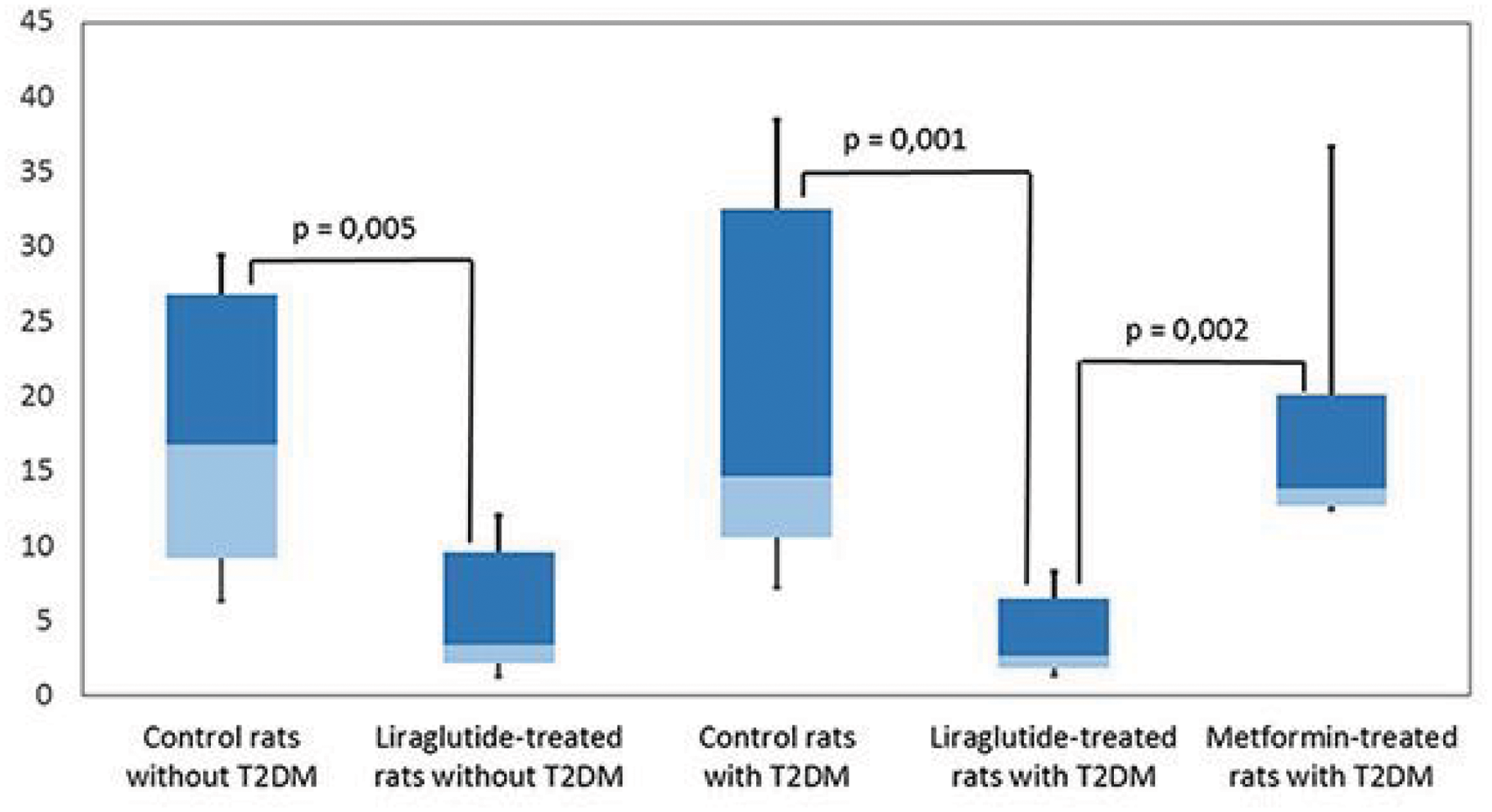
Neuroprotective effect of liraglutide in transient focal brain ischemia in rats with type 2 diabetes mellitus
I. Filchenko1, A. Simanenkova1 and T. Vlasov1
1Pavlov First St. Petersburg State Medical University, St. Petersburg, Russian Federation
Abstract
Objectives: Acute cerebrovascular accident is a common and severe complication of type 2 diabetes mellitus (T2DM). Glucagon-like peptide-1 (GPL-1) receptors are widely present in central nervous system, thus GLP-1 receptor agonists may be used not only to decrease glycaemia but also for neuroprotection. In present study we evaluated neuroprotective effect of GLP-1 receptor agonist liraglutide in transient focal brain ischemia in rats without T2DM and with T2DM.
Methods: The two-phase study was held in male Wistar rats.
In the first phase experimental animals were treated with liraglutide 1 mg/kg subcutaneously for 7 days, while control ones received isotonic NaCl subcutaneously. Then transient focal brain ischemia was modelled in all rats and brain infarct volume was measured. Garcia scale was used to determine neurological deficit.
In the second phase T2DM was induced. In 14 days after T2DM induction the first and the second groups of experimental animals started to receive liraglutide 0,03 mg/kg subcutaneously and metformin 200 mg/kg orally for 7 days, respectively, while control animals were treated with isotonic NaCl subcutaneously. Then transient focal brain ischemia was modelled in all rats, neurological deficit and brain infarct volume were measured.
Results: In the first phase liraglutide-treated rats had significantly smaller brain infarct volume and neurological deficit in comparison with control animals.
In the second phase liraglutide-treated rats had significantly smaller brain infarct volume and neurological deficit in comparison both with animals that received isotonic NaCl and with metformin-treated animals. There were no significant differences in brain infarct volume and neurological deficit between the groups of metformin-treated and control rats.
[Brain infarct volume (%)]
Conclusions: Liraglutide has an independent neuroprotective effect in transient focal brain ischemia, not related to glycemic control improvement. Thus, GLP-1 receptor agonists may be promising in stroke prevention in T2DM.
PS01-020
Poster Viewing Session I
Boosting O-GlcNAcylation, a pro-survival pathway downstream of the unfolded protein response, improves ischemic stroke outcome in mice
M. Jiang1, S. Yu1, H. Sheng1, Z. Yu1, D. Warner1, W. Paschen1 and W. Yang1
1Duke University Medical Center, Durham, United States
Abstract
Objective: The endoplasmic reticulum (ER) plays a pivotal role in folding and processing newly synthesized proteins. These functions are sensitive to a variety of stress conditions. Impairment of ER function activates the unfolded protein response (UPR), an adaptive response to restore ER function and thus cellular protein homeostasis. One pro-survival branch of UPR is controlled by the ER stress sensor protein inositol-requiring enzyme-1 (IRE1). Activated IRE1 induces splicing of X-box binding protein-1 (Xbp1) mRNA, and spliced Xbp1 mRNA is then translated into a new protein, XBP1s, a transcription factor. XBP1s regulates expression of key genes controlling the hexosamine biosynthetic pathway (HBP), and thereby modulates O-linked β-N-acetylglucosamine modification of proteins (O-GlcNAcylation). It is well known that stroke impairs ER function and actives the IRE1 UPR branch. This study is aimed to investigate the role of the IRE1/XBP1/O-GlcNAc axis in stroke outcome.
Methods: Mice with Xbp1 loss- and gain-of-function in neurons were generated. C57Bl/6 were used for the experiments of pharmacologic intervention. Stroke was induced by permanent or transient occlusion of the middle cerebral artery (MCAO) in mice. Infarct volume and neurologic deficit were evaluated.
Results: Deletion of Xbp1 in forebrain neurons significantly worsened outcome in transient and permanent MCAO. Transgenic mice with neuron-specific expression of XBP1s exhibited increased levels of O-GlcNAcylation in the brain, indicating that the XBP1/HBP/O-GlcNAc axis was functional in neurons. Further, after stroke, O-GlcNAcylation was activated in the penumbra region in mice. Finally, pharmacologic increase of O-GlcNAcylation before or after stroke improved outcome in mice.
Conclusions: Our study shows a critical role of the IRE1/XBP1 UPR branch in ischemic stroke, suggesting that restoration of protein homeostasis defines stroke outcome. Boosting O-GlcNAcylation pharmacologically could be a promising strategy to improve stroke outcome.
PS01-021
Poster Viewing Session I
Hippocampal neuroprotection mediated by tyrosine kinase B receptor (TrkB) phosphorylation is estrogen receptor alpha (ERα) dependent only in female neurons after in-vitro ischemia
D. Zafer1,2, V. Chanana1,2, D. Hanalioglu1,2, A. Canturk1,2, D. Kintner1,2, J. Chandrashekhar1,2, K. Freeman1,2, J. Sanchez1,2, P. Ferrazzano1,2, J. Levine3 and P. Cengiz1,2
1University of Wisconsin-Madison, Pediatrics, Madison, United States
2Waisman Center, Madison, United States
3University of Wisconsin-Madison, Neuroscience, Madison, United States
Abstract
Objective: Expression and actions of neurotrophins may account for male neonatal brain susceptibility to hypoxia-ischemia (HI). Treatment with a selective tyrosine kinase B (TrkB) agonist (7,8-dihydroxyflavone [7,8-DHF]) results in hippocampal neuroprotection only in female neonatal mice in an ERα dependent manner post-HI (in vivo). This neuroprotection seems to be mediated by phosphorylated TrkB (p-TrkB) in hippocampal neurons by immunohistochemistry. We hypothesize that sexually differentiated neuroprotection mediated by TrkB phosphorylation is ERα dependent in hippocampal neurons after in vitro ischemia.
Method: Sexed hippocampal primary neurons cultured from P1 ERα+/+ and ERα-/- C57BL/6 J mice. At DIV 7, cells were treated with either normoxic media or 7,8-DHF following normoxia or OGD. Cells were stained, imaged (confocal microscopy) and analyzed (Image J) for cell survival (Calcein, PI), ERα, p-TrkB and MAP-2 at 24 h REOX. ERα mRNA and aromatase expressions detected using RT-PCR. ANOVA with a Bonferroni post-hoc test used for analysis.
Results: OGD/REOX and OGD/REOX + 7,8-DHF increased ERα mRNA expression by 2 (p = 0.06) and 4 (p = 0.004) folds, respectively at 3 h REOX only in ERα+/+ female neurons. 7,8-DHF treatment significantly increased p-TrkB expression only in ERα+/+ female neurons after OGD/REOX (p = 0.0023). OGD/REOX decreased cell survival by 30% in both ERα+/+ and ERα-/- male and female neurons (p = 0.003) whereas, 7,8-DHF rescued cell survival up to normoxic levels only in ERα+/+ female hippocampal neurons (p = 0.048). ANA-12 (TrkB antagonist) administration following OGD/REOX and 7,8-DHF treatment abolished the increased ERα mRNA expression (p = 0.023). Aromatase mRNA expression was similar among the groups (p = 0.62) suggesting similar estradiol content.
Conclusion: 7,8-DHF enhances neuroprotection only in ERα+/+ female hippocampal neurons following in vitro ischemia in an ERα dependent but estradiol independent way. We will investigate the effects of ERα agonist to determine whether the TrkB phosphorylation or cell survival can be rescued in ERα+/+ male hippocampal neurons following in vitro ischemia.
PS01-023
Poster Viewing Session I
MLC901 issued from traditional Chinese medicine favors angiogenesis and associated recovery after ischemic stroke
C. Gandin1, C. Widmann1, M. Lazdunski1 and C. Heurteaux1
1Institut de Pharmacologie Moléculaire et Cellulaire, Valbonne, France
Abstract
Angiogenesis results in improved collateral circulation and impacts on the long-term recovery of patients. MLC901, a preparation of herbal extractsis known to stimulate neurogenesis, neurite outgrowth, and synaptogenesis. Angiogenesis is directly associated with neurogenesis since blood supply is necessary for newly-formed neurons to survive. We aimed to investigate the effect of MLC901 on angiogenesis in a mouse model of stroke. MLC901 was administered in the drinking water (6 g/L) for 5 weeks before ischemia and then during reperfusion. Stroke was induced by 60 min of middle cerebral artery (MCA) occlusion. The expression of vascular endothelial growth factor (VEGF) and angiopoietin 1 (Ang1) / angiopoietin 2 (Ang2), and the regulation of key proteins known to mediate angiogenesis (hypoxic inducible factor (Hif1α) and erythropoietin (EPO)) were analyzed by ELISA, Western blot, and immunohistochemistry.
MLC901 stimulates angiogenesis and modulates the expression of angiogenesis-regulated molecules in response to stroke. While VEGF levels were transiently increased in vehicle-treated ischemic mice compared to sham at 24 hours and 3 days with a tendency to return to baseline at 14 days, MLC901 treatment led to twice as much VEGF levels at 3 and 14 days compared to vehicle. In addition, MLC901 stimulated the growth of new vessels where VEGF was mainly expressed in neurons. VEGF was also expressed in microglial cells. MCAO triggered a 2-fold increase in Hif1α protein levels at 3 days and remained elevated for at least 14 days, mainly restricted to neurons and astrocytes in the peri-infarct area. MLC901 stimulated the late up-regulation of Hif1α. Furthermore, MLC901 stimulated EPO level in neurons and astrocytes surrounding the infarct, and was often colocalized with VEGF around endothelial cells and microvessels .
MLC901 stimulates revascularization and repair of the neurovascular unit after stroke. These findings complement our findings on the effect on neurogenesis and other therapeutic properties of MLC901.
PS01-024
Poster Viewing Session I
Neuroprotective effects of intranasal Guanosine: Wide theurapeutic window and a possible mechanism
G.C. Müller1, S.O. Loureiro1, R.F. Almeida1, L.F. Pettenuzzo1, M. Ganzella2 and D.O. Souza1
1UFRGS, Institute of Basic Health Sciences (ICBS), Department of Biochemistry, Porto Alegre, Brazil
2Max Planck Institute for Biophysical Chemistry, Department of Neurobiology, Göttingen, Germany
Abstract
Stroke is one of the main causes of death and disability worldwide, therefore is an important focus of research. The nucleoside guanosine (GUO) has gained attention as an anti-ischemic drug since several works have shown neuroprotective effects in different animal models. Most of these studies have administered GUO via systemic although it is an endogenous compound and highly metabolizable.
We investigated a new GUO administration route (intranasal) to assess its neuroprotective potential against permanent focal cerebral ischemia.
Thermocoagulation of pial vessels on Wistar rats was used as our model. Three groups were analyzed: Sham, Ischemia and GUO (ischemic animals submitted to three administrations: 3 h, 5 h and 8 h post-surgery). To assess behavioral impairment, we used cylinder test at 48 h hours after ischemia. We evaluated GFAP, β-tubulin III, COX-2, by flow cytometry in both hemispheres and Caspases, Bax, PI3K and GSK3β on ipsilateral hemisphere of lesion.
We found a partial reversion of impairment on behavior of animals treated with GUO at 48 h after stroke. In cytometry analyses, we observed an increase of GFAP and COX-2 expression on contralateral side of lesion on ischemic animals, being prevented by treatment, data strongly correlated. In ipsilateral side we found decreasing expression on β-tubulin III, Bax, GSK3β; and Caspases 2 and 8, parameters not reverted by GUO. We observed alterations in PI3K and Caspase 3 induced by Ischemia and reverted by GUO, correlated with behavioral results.
Although the exact GUO mechanism of action is not fully clear, it seems to counteract different steps following the ischemic cascade events, with repercussions depending the hemisphere. In this work, we assessed a new possible mechanism of a promising neuroprotective drug in a viable therapeutical window. Moreover, intranasal GUO prevented behavioral injury in a lower dose compared to systemic, being an efficient route to deliver GUO to the CNS.
PS01-026
Poster Viewing Session I
CETP genetic variants that increase HDL raise Intracerebral hemorrhage risk
C. Anderson1, G. Falcone1, G. Peloso1, C. Langefeld2, G. Abecasis3, S. Kathiresan1, D. Woo4 and J. Rosand1
1Massachusetts General Hospital, Neurology, Boston, United States
2Wake Forest University, Winston-Salem, United States
3University of Michigan, Biostatistics, Ann Arbor, United States
4University of Cincinnati, Neurology and Rehabilitation Medicine, Cincinnati, United States
Abstract
In observational studies, higher plasma HDL cholesterol (HDL-C) has been associated with increased risk of intracerebral hemorrhage (ICH). DNA variants that decrease cholesteryl ester transfer protein (CETP) activity increase plasma HDL-C; as such, medicines that inhibit CETP and raise HDL-C are in development. Here, we test the hypothesis that CETP variants associated with higher HDL-C also increase risk for ICH.
We performed two analyses of CETP using genomewide data. First, we tested individual CETP variants in a discovery cohort of 1149 ICH cases and 1238 controls from 3 studies, followed by replication in 1625 cases and 1845 controls from 5 studies. Second, we constructed a genetic risk score comprised of 7 independent variants at CETP and tested this score for association with HDL-C as well as ICH risk.
12 variants within CETP demonstrated nominal association with ICH, with the strongest association at the rs173539 locus (odds ratio (OR) 1.25, standard error (SE) 0.06, p = 6.0 × 10⁻⁴) with no heterogeneity across studies (I2 = 0 %). This variant is adjacent to the strongest known determinant of HDL-C. The rs173539 association was replicated in patients of European ancestry (p = 0.03). A genetic score of CETP variants found to increase HDL-C by ∼2.85 mg/dL in the Global Lipids Genetics Consortium was strongly associated with ICH risk (OR 1.86, SE 0.13, p = 1.39 × 10⁻⁶).
Genetic variants in CETP associated with increased HDL-C raise the risk of ICH. Similar to the finding of increased risk of brain hemorrhage with statin use, ongoing therapeutic development of CETP inhibition and other HDL-raising strategies should explore further the potential of adverse cerebrovascular outcomes. Further translational work on the mechanism of HDL's association with hemorrhagic risk is warranted.
PS01-027
Poster Viewing Session I
Role of the endothelium NO-Synthase in early brain injury after experimental subarachnoid hemorrhage
I. Westermayer1, N. Terpolilli1,2, S. Katzdobler1 and N. Plesnila1
1Institute for Stroke and Dementia Research (ISD), University Hospital of the Ludwig-Maximilians-University Munich, Munich, Germany
2University Hospital of the Ludwig-Maximilians-University Munich, Department of Neurosurgery, Munich, Germany
Abstract
Background: Experimental and clinical studies indicate that pathological changes in the early phase (72 hours) after subarachnoid hemorrhage (SAH), summarized as early brain injury (EBI), determine the extent of brain damage and, thus, neurological outcome. A main characteristic of EBI is microcirculatory dysfunction. Potential mechanisms for this include microvascular constriction (microvasospasms) and impaired CO2-reactivity of cerebral microvessels. Both pathophysiological phenomenona have been linked to nitric oxide (NO) depletion after SAH. Endothelial NO-synthase (eNOS) - the main source of cerebrovascular under physiological conditions - is dysfunctional early after SAH but it is unclear how eNOS function/ dysfunction is linked with posthemorrhagic brain damage. In the current study we evaluated parameters of EBI in mice lacking eNOS and respective wildtype controls.
Methods: SAH was induced in male eNOS-/- mice and wt animals using the MCA perforation model under continuous control of ICP, CBF, and MAP. 3 h after SAH, the cerebral microcirculation was directly visualized using in vivo-2-photon-mocroscopy. CO2-vessel-reactivity was studied via IVM before, during, and after induction of hypercapnia (7.5 %); NO was supplied by inhalation (50 ppm) before CO2 challenge.
Results: eNOS-/- mice had significantly increased pathology after SAH with more severe intracranial hypertension and hypoperfusion, significantly more re-bleedings which translated into massively increased post-SAH mortality. The cerebral microcirculation was significantly impaired after SAH. Exogenous NO increased CBF in wt animals but not in eNOS-/- mice. While CO2-reactivity is abolished after SAH, hypercapnia led to significant vasodilatation and CBF increase in wt and eNOS transgenic mice after NO application via inhalation.
Summary & conclusion: eNOS-/- mice have significantly more severe SAH and, subsequently, posthemorrhagic mortality. Early microcirculatory changes were more pronounced in transgenic animals. These findings indicate a key role for eNOS produced NO in the pathophysiology of early brain injury after SAH. Further evaluation of the mechanisms is in planning.
PS01-028
Poster Viewing Session I
Inhibitory effects of Omega-3 on early brain injury after subarachnoid hemorrhage in rats: possible involvement of GPR120 /β-arrestin2/ TGF-β activated kinase-1 binding protein-1 signaling pathway
J. Yin1, Z. Wang1 and G. Chen1
1The First Affiliated Hospital of Soochow University, Suzhou, China
Abstract
Objectives: Omega-3 fatty acids have been reported to improve neuron functions during aging and in patients affected by mild cognitive impairment, and mediate potent anti-inflammatory via G protein-coupled receptor 120 (GPR120) signal pathway [1]. This study was to examine the effects of omega-3 fatty acids on subarachnoid hemorrhage (SAH)-induced early brain injury (EBI).
Methods: Two weeks before SAH, 30% Omega-3 fatty acids was administered by oral gavage at 1 g/kg body weight once every 24 hours. Specific siRNA for GPR120 was exploited.
Results: Terminal deoxynucleotidyl transferase dUTP nick end labeling, fluoro-Jade B staining, and neurobehavioral scores and brain water content test showed that omega-3 fatty acids effectively suppressed SAH-induced brain cell apoptosis and neuronal degradation, behavioral impairment, and brain edema. Western blot, immunoprecipitation, and electrophoretic mobility shift assays results showed that omega-3 fatty acids effectively suppressed SAH-induced elevation of inflammatory factors, including cyclooxygenase-2, monocyte chemoattractant protein-1, and inducible nitric oxide synthase. In addition, omega-3 fatty acids could inhibit phosphorylation of transforming growth factor β activated kinase-1 (TAK1), MEK4, c-Jun N-terminal kinase, and IkappaB kinase as well as activation of nuclear factor kappa B through regulating GPR120/β-arrestin2/TAK1 binding protein-1 pathway. Furthermore, siRNA-induced GPR120 silencing blocked the protective effects of omega-3 fatty acids.
Conclusions: Here, we show that stimulation of GPR120 with omega-3 fatty acids pretreatment causes anti-apoptosis and anti-inflammatory effects via β-arrestin2/ TAK1 binding protein-1/TAK1 pathway in the brains of SAH rats. Fish omega-3 fatty acids as part of a daily diet may reduce EBI in an experimental rat model of SAH.
[Effect of omega-3 on SAH-induced EBI]
References:
1. Oh DY, Talukdar S, Bae EJ, Imamura T, Morinaga H, Fan W, et al. GPR120 is an omega-3 fatty acid receptor mediating potent anti-inflammatory and insulin-sensitizing effects. Cell. 2010;142:687–98.
PS01-029
Poster Viewing Session I
Cyclophilin A/CD147 interactions participate in early brain injury after subarachnoid hemorrhage in rats
B. Dang1, Z. Wang1 and G. Chen1
1The First Affiliated Hospital of Soochow University, Suzhou, China
Abstract
Objectives: Cyclophilin A (CyPA) has been found to be involved in many inflammatory diseases via its receptor, CD147 [1]. This study aimed to estimate the potential role of CyPA/CD147 in subarachnoid hemorrhage (SAH)-induced early brain injury (EBI).
Methods: A prechiasmatic cistern single-injection SAH model was used. The expressions of CyPA and CD147, the interaction between CyPA and CD147, and the secretion of CyPA were assessed using immunofluorescence staining, western blot analysis, and co-immunoprecipitation analysis. Down-regulation of CyPA expression by siRNA was performed and recombinant human CyPA (rh CyPA) and monoclonal antibody of CD147 [3] was exploited to study the role of CyPA/CD147 in SAH-induced EBI.
Results: The expressions of CyPA and CD147 in neurons were higher than that of the sham group, and peaked at 24 h after SAH. Compared with sham group, SAH was found to increase the secretion of CyPA and the interaction between CyPA and CD147. CyPA siRNA and anti-CD147 treatments were found to ameliorate SAH-induced EBI, including cortical apoptosis and necrosis, brain edema, blood-brain barrier damage and neurobehavioral deficits. CyPA siRNA and anti-CD147 treatments also decreased the phosphorylation of ERK 1/2, the protein levels of p53 and caspase-3, and the level of active NF-κB. Finally, rh CyPA treatment resulted in an opposite effect, which was inhibited by anti-CD147 treatment.
Conclusions: CyPA/CD147 interactions may participate in SAH-induced EBI via increasing neuronal apoptosis pathway, at least partly through the ERK1/2-NF-kB pathway. CyPA/CD147 may be a suitable therapeutic target for SAH.
[CyPA/CD147 in SAH]
Reference:
1. Yuan W, Ge H, He B. Pro-inflammatory activities induced by CyPA-EMMPRIN interaction in monocytes. Atherosclerosis 2010;213(2):415–421.
2. Tu Y, Fu J, Wang J, et al. Extracellular matrix metalloproteinase inducer is associated with severity of brain oedema following experimental subarachnoid haemorrhage in rats. J Int Med Res 2012;40(3):1089–1098.
PS01-030
Poster Viewing Session I
Pramipexole-induced hypothermia reduces early brain injury via PI3K/AKT/GSK3β pathway in subarachnoid hemorrhage rats
J. Ma1, Z. Wang1 and G. Chen1
1The First Affiliated Hospital of Soochow University, Suzhou, China
Abstract
Objectives: Previous studies have shown neuroprotective effects of hypothermia [1,2]. However, its effects on subarachnoid hemorrhage (SAH)-induced early brain injury (EBI) remain unclear.
Methods: A SAH rat model was employed to study the effects and mechanisms of pramipexole-induced hypothermia on EBI after SAH. Dose-response experiments were performed to select the appropriate pramipexole concentration and frequency of administration for induction of mild hypothermia (33–36 °C). Western blot, neurobehavioral evaluation, Terminal deoxynucleotidyl transferase-mediated dUTP nick end labeling (TUNEL) and Fluoro-Jade B (FJB) staining were used to detect the effects of pramipexole-induced hypothermia on SAH-induced EBI, as well as to study whether controlled rewarming could attenuate these effects. Inhibitors targeting the PI3K/AKT/GSK3β pathway were administered to determine whether the neuroprotective effect of pramipexole-induced hypothermia was mediated by PI3K/AKT/GSK3β signaling pathway.
Results: The results showed that intraperitoneal injection of pramipexole at 0.25 mg/kg body weight once per 8 hours was found to successfully and safely maintain rats at mild hypothermia. Pramipexole-induced hypothermia ameliorated SAH-induced brain cell death, blood-brain barrier damage and neurobehavioral deficits in a PI3K/AKT/GSK3β signaling-dependent manner.
Conclusions: Therefore, we may conclude that pramipexole-induced hypothermia could effectively inhibit EBI after SAH in rats via PI3K/AKT/GSK3β signaling pathway.
[Pramipexole-induced hypothermia in SAH]
References:
1 Antonic, A. et al. Hypothermia protects human neurons. Int. J. Stroke.9, 544; DOI:10.1111/ijs.12224 (2014).
2 Johansen, F. F. et al. Drug-induced hypothermia as beneficial treatment before and after cerebral ischemia. Pathobiology.81, 42; DOI:10.1159/000352026 (2014).
PS01-031
Poster Viewing Session I
Role of neurexin-1β and neuroligin-1 in cognitive dysfunction after subarachnoid hemorrhage in rats
H. Shen1, Z. Wang1 and G. Chen1
1The First Affiliated Hospital of Soochow University, Suzhou, China
Abstract
Objectives: Neurexin-1β and neuroligin-1 play an important role in the formation, maintenance and regulation of synaptic structures [1-3]. This study is to estimate the potential role of neurexin-1β and neuroligin-1 in subarachnoid hemorrhage (SAH)-induced cognitive dysfunction.
Methods: Experimental SAH model was induced by single blood injection to prechiasmatic cistern. Primary cultured hippocampal neurons were exposed to oxyhemoglobin to mimic SAH in vitro. Specific siRNAs and expression plasmids for neurexin-1β and neuroligin-1 were exploited both in vivo and in vitro. Western blot, immunofluorescence, immunoprecipitation, neurological scoring and Morris water maze were performed to evaluate the mechanism of neurexin-1β and neuroligin-1, as well as neurological outcome.
Results: Both in vivo and in vitro experiments showed the SAH-induced decrease in the expressions of neurexin-1β and neuroligin-1 and the interaction between them in neurons. In addition, the interaction between neurexin-1β and neuroligin-1 was reduced by their knockdown and increased by their overexpression. The formation of excitatory synapses was inhibited by oxyhemoglobin treatment, which was significantly ameliorated by overexpression of neurexin-1β and neuroligin-1 and aggravated by knockdown of neurexin-1β and neuroligin-1. More importantly, neurexin-1β and neuroligin-1 overexpression ameliorated SAH-induced cognitive dysfunction, while neurexin-1β and neuroligin-1 knockdown induced an opposite effect.
Conclusions: Enhancing the expressions of neurexin-1β and neuroligin-1 could promote the interaction between them and the formation of excitatory synapses, which is helpful to improve cognitive dysfunction after SAH. Neurexin-1β and neuroligin-1 might be good targets for improve cognitive function after SAH.
References:
1. Sugita S, Khvochtev M, Sudhof TC. Neurexins are functional alpha-latrotoxin receptors. Neuron. 1999;22:489–496
2. Nam CI, Chen L. Postsynaptic assembly induced by neurexin-neuroligin interaction and neurotransmitter. PNAS 2005;102:6137–6142
3. Chubykin AA, Liu X, Comoletti D, Tsigelny I, Taylor P, Sudhof TC. Dissection of synapse induction by neuroligins: Effect of a neuroligin mutation associated with autism. The Journal of biological chemistry. 2005;280:22365–22374
PS01-032
Poster Viewing Session I
Race/ethnic variation of Apolipoprotein E alleles for lobar intracerebral hemorrhage
D. Woo1, P. Sekar2, J. Rosand3, J. Osborne2, S. Kittner4, C. Langefeld5, C. Moomaw2, M. Flaherty2 and C. Anderson3
1University of Cincinnati, Neurology, Cincinnati, United States
2University of Cincinnati, Neurology and Rehabilitation Medicine, Cincinnati, United States
3Massachusetts General Hospital, Neurology, Boston, United States
4University of Maryland, Neurology, Baltimore, United States
5Wake Forest University, Winston-Salem, United States
Abstract
Introduction: Apolipoprotein E (ApoE) epsilon 2 and 4 (E2 and E4) alleles have been associated with lobar intracerebral hemorrhage (ICH) in predominately white populations. We sought to evaluate if this was true among black and Hispanic populations.
Methods: The Ethnic/Racial Variations of Intracerebral Hemorrhage (ERICH) study is a prospective, multi-center study of ICH among whites, blacks and Hispanics. Controls were identified through random digit dialing to match to cases by age, ethnicity, sex and location. All controls were used for analysis. ApoE genotyping and ICH location was determined blinded to clinical data. Multivariate modeling was performed with backward elimination and controlled for age, sex, hypertension, anticoagulant use, education level, alcohol use, diabetes and ApoE alleles.
Results: As of 4/1/16, 2,997 cases of ICH and 2,756 controls were enrolled into the study. Of these, 887 cases had lobar ICH and ApoE results. Centers that had less than 20 cases of a race/ethnic group were dropped leaving 330 white, 154 black and 207 Hispanic lobar ICH cases. Both ApoE2 (OR = 1.48; 95 % CI 1.1–2.0; p = 0.01) and ApoE4 was (OR = 1.97; 95 % CI: 1.5–2.6; ,< 0.0001) associated with lobar ICH among whites. Yet among blacks, neither ApoE2 (OR = 0.99; 95 % CI 0.67–1.46; p = 0.97) or ApoE4 (OR = 0.98; 95 % CI 0.71–1.35; p = 0.90) were independent risk factors for lobar ICH. Similarly, among Hispanics, neither ApoE2 (OR = 1.04; 95 % CI 0.60–1.81; p = 0.89) or ApoE4 (OR = 1.19; 95 % CI 0.82–1.71; p = 0.35) were associated with lobar ICH. Hypertension was found to be a risk factor for lobar ICH in all 3 race/ethnic groups.
Conclusion: Once controlling for other factors, ApoE alleles were not significantly associated with lobar ICH among black and Hispanic populations. While a small effect is still possible, this finding suggests that other factors affect how ApoE alleles lead to lobar ICH among whites and challenges the notion that hypertension plays little role in lobar ICH.
PS01-033
Poster Viewing Session I
Inhibition of mammalian target of rapamycin attenuates early brain injury through modulating microglial polarization after experimental subarachnoid hemorrhage in rats
W. You1, Z. Wang1 and G. Chen1
1The First Affiliated Hospital of Soochow University, Suzhou, China
Abstract
Objectives: Here, we aimed to study the role and underlying mechanism of mTOR in early brain injury (EBI) after subarachnoid hemorrhage (SAH).
Methods: Experiment 1, the time course of mTOR activation in the cortex following SAH. Experiment 2, the role of mTOR in SAH-induced EBI. Adult SD rats were divided into four groups: sham group (n = 18), SAH + vehicle group (n = 18), SAH + rapamycin group (n = 18), SAH + AZD8055 group (n = 18). Experiment 3, we incubated enriched microglia with OxyHb. Rapamycin and AZD8055 were also used to demonstrate the mTOR's role on microglial polarization in vitro.
Results: The phosphorylation levels of mTOR and its substrates were significantly increased and peaked at 24 h after SAH. Rapamycin or AZD8055 markedly decreased the phosphorylation levels of mTOR and its substrates and the activation of microglia in vivo, and promoted the microglial polarization from M1 phenotype to M2 phenotype. In addition, administration of rapamycin and AZD8055 following SAH significantly ameliorated EBI, including neuronal apoptosis, neuronal necrosis, brain edema and blood-brain barrier permeability.
Conclusions: Our findings suggested that the rapamycin and AZD8055 could attenuate the development of EBI in this SAH model, possibly through inhibiting the activation of microglia by mTOR pathway.
PS01-034
Poster Viewing Session I
Transient receptor potential channel 1/4 reduces subarachnoid hemorrhage-induced early brain injury in rats via calcineurin-mediated NMDAR and NFAT dephosphorylation
Y. Wang1, Z. Wang1 and G. Chen1
1The First Affiliated Hospital of Soochow University, Suzhou, China
Abstract
Objectives: Transient receptor potential channel 1/4 (TRPC1/4) are considered to be related to subarachnoid hemorrhage (SAH)-induced cerebral vasospasm [1].
Methods: SAH rat model was employed to study the roles of TRPC1/4 in the early brain injury (EBI) after SAH. Primary cultured hippocampal neurons were exposed to oxyhemoglobin to mimic SAH in vitro.
Results: The protein levels of TRPC1/4 increased and peaked at 5 days after SAH in rats. Inhibition of TRPC1/4 by SKF96365 [2] aggravated SAH-induced EBI, such as cortical cell death (by TUNEL staining) and degenerating (by FJB staining). In addition, TRPC1/4 overexpression could increase calcineurin activity, while increased calcineurin activity could promote the dephosphorylation of N-methyl-D-aspartate receptor (NMDAR). Calcineurin antagonist FK506 [3] could weaken the neuroprotection and the dephosphorylation of NMDAR induced by TRPC1/4 overexpression. Contrarily, calcineurin agonist chlorogenic acid [4] inhibited SAH-induced EBI, even when siRNA intervention of TRPC1/4 was performed. Moreover, calcineurin also could lead to the nuclear transfer of nuclear factor of activated T cells (NFAT), which is a transcription factor promoting the expressions of TRPC1/4.
Conclusions: TRPC1/4 could inhibit SAH-induced EBI by supressing the phosphorylation of NMDAR via calcineurin. TRPC1/4-induced calcineurin activation also could promote the nuclear transfer of NFAT, suggesting a positive feedback regulation of TRPC1/4 expressions.
References:
1 Gonzalez-Cobos, J. C. & Trebak, M. TRPC channels in smooth muscle cells. Front Biosci (Landmark Ed)15, 1023–1039 (2010).
2 Zhao, Y. et al. Jujuboside B Reduces Vascular Tension by Increasing Ca2+ Influx and Activating Endothelial Nitric Oxide Synthase. PLoS One11, e0149386, doi:10.1371/journal.pone.0149386 (2016).
3 Li, G. et al. The role of calcineurin signaling in microcystin-LR triggered neuronal toxicity. Sci Rep5, 11271, doi:10.1038/srep11271 (2015).
4 Mikami, Y. & Yamazawa, T. Chlorogenic acid, a polyphenol in coffee, protects neurons against glutamate neurotoxicity. Life Sci139, 69–74, doi:10.1016/j.lfs.2015.08.005 (2015).
PS01-035
Poster Viewing Session I
The sGC-activator Bay 60-2770 has no influence on microvasospasms after experimental subarachnoid hemorrhage
S. Katzdobler1, B. Seker1, N.A. Terpolilli1,2, I. Westermayer1 and N. Plesnila1
1Institute for Stroke and Dementia Research Munich, Munich, Germany
2Neurosurgical Clinic of the University of Munich (LMU), Munich, Germany
Abstract
Background: Subarachnoid hemorrhage (SAH) is a severe subtype of stroke characterized by high mortality and adverse neurological long-term outcome. Within the first few hours after the initial bleeding patients start to develop microvasospasms, resulting in a cerebral perfusion deficit despite recovering cerebral perfusion pressure; this was observed in patients as well as in mice after experimental SAH. In a previous study we were able to show that inhalation of nitric oxide reduces the number and severity of spasms after experimental SAH leading to an improved neurological and histopathological outcome. The NO effect on cerebral vessels is mediated by soluble guanylate cyclase (sGC), the NO-inhalation effect can be blocked by inhibition of sGC. In the present study we aimed for direct and NO-independent manipulation of the enzyme soluble guanylate cyclase (sGC) in order to achieve neuroprotection after SAH.
Methods: We subjected C57BL6 mice to either SAH or Sham surgery. Three hours after SAH animals were either treated with i.v. injection of sGC-activator Bay 60–2770 (10 µg/kg) or vehicle only for 30 minutes. For investigation of cerebral microvasospasms two photon microscopy was performed and vessel diameter and spasm severity of pial and parenchymal vessels were analysed. Cerebral blood flow (CBF) as well as mean arterial blood pressure (MAP) were monitored throughout treatment.
Results: Analysis of two photon microscopy data showed no significant changes in neither mean arterial vessel diameter nor spasm severity nor spasm number in the Bay 60–2770 group. No significant differences in mortality, CBF or MAP were observed.
Conclusion: In conclusion, activation of soluble guanylate cyclase by intravenous application of Bay 60–2770 at the dose used in our experiments did not alter posthemorrhagic microcirculatory changes and does not yield promising results for treatment of microvasospams following experimental SAH.
PS01-036
Poster Viewing Session I
Correlation of glymphatic function to cerebral blood flow following subarachnoid hemorrhage
T. Stani1, J. Cetas1 and J. Illif2
1Oregon Health & Science University, Neurological Surgery, Portland, United States
2Oregon Health & Science University, Anesthesia and Perioperative Medicine, Portland, United States
Abstract
Introduction: Subarachnoid hemorrhage (SAH) produces profound disruptions in cerebral blood flow (CBF), and recent data shows that brain-wide glymphatic CSF flux is disrupted as well (Gaberel et al: Impaired Glymphatic Perfusion After Strokes Revealed by Contrast-Enhanced MRI: a new target for fibrinolysis? Stroke 45:3092–6, 2014). This finding offers the possibility for new therapeutic interventions but further characterization of the potential relationships between glymphatic function and blood flow is needed.
Methods: To model SAH, autologous blood was injected into the prechiasmatic cistern of adult male rats (Figure 1, B). A second treatment group received injection of inert fluorescent beads approximately the size of blood cells (Figure 1, C). Controls received artificial CSF injection (Figure 1, A). Blood flow changes following injection were assessed using laser speckle and optical intrinsic signal imaging through a thin skull preparation. 20 µl of 1% Evans Blue (EB) was injected into the cisterna magna 30 minutes after prechiasmatic cistern injections. Rat brains were perfused and fixed 60 minutes after injections. The brain surfaces were imaged under a fluorescent microscope. EB fluorescence intensity was calculated over multiple brain regions and correlated to CBF changes.
[Figure 1]
Results: Injection of subarachnoid blood caused various changes in CBF. Surface flow of EB CSF tracer was correlated to CBF changes. Total EB signal was decreased across the entire brain surface in all SAH-model animals compared to controls. Regional sub-analyses between EB signal and patterns of blood flow were assessed. Injection of beads caused similar disruption in EB tracer flow as well as CBF changes.
Conclusion: SAH led to decreased perivascular CSF flow in all cases. Regional glymphatic disruption may relate to specific post-hemorrhage CBF patterns. Subarachnoid injection of inert beads produces blockade of glymphatic flow and can also cause decreased CBF.
PS01-037
Poster Viewing Session I
Is perlecan domain V a potential therapeutic in preclinical vascular dementia?
A. Trout1, Z. Zhang1, J. Roberts1, A. Hartz1 and G. Bix1
1University of Kentucky, Sanders-Brown Center on Aging, Lexington, United States
Abstract
Vascular contributions to cognitive impairment and dementia (VCID), the second leading cause of dementia behind Alzheimer's disease (AD), is a broad term that encompasses a spectrum of initial asymptomatic cerebrovascular changes (seen in small vessel disease and cerebral amyloid angiopathy where pathologic Amyloid Beta (Aβ)1-42 protein accumulates around brain blood vessels) to the profound symptomatic damage following acute stroke(s). Cerebrovascular remodeling and angiogenesis may represent early compensatory changes to the reduced blood flow seen in VCID by increasing the proteolytic turnover of the surrounding extracellular matrix. We have demonstrated that one such extracellular matrix protein, perlecan (a heparan sulfate proteoglycan), possesses a C terminal domain V (DV) protein that upon cleavage from perlecan greatly enhances brain angiogenesis. Our objective is to determine if DV increases angiogenesis and promotes Aβ clearance in VCID.
We characterized VCID induced changes in a distinct mouse model (diabetic APP/PS1 knock in (db/AD)) that has gradual cognitive decline by 9 months with microangiopathy, Aβ deposition, aneurysms, and microhemorrhages. We also utilized an in vitro model of the blood-brain barrier (BBB) to assess the transport of human Aβ in the presence of DV.
In db/AD animals (3–6 months), we observed a decrease in BBB proteins (i.e.claudin-5), indicating that altered function correlated with an increase in brain DV expression during the asymptomatic angiogenic stage, which precedes cognitive changes (9–12 months). In vitro, DV doubled the transport of Aβ1-42 into the lumen of cerebral microvessels over 24 hours with increased activity and total protein expression of P-glycoprotein (P-gp), one of Aβ's known transport proteins.
Collectively, these data indicate that early cerebrovascular changes induce angiogenic-remodeling that correlates with increased expression of DV. DV, in turn, may further enhance angiogenesis and increase brain Aβ clearance into the vascular compartment through P-gp, suggesting that DV could represent a novel therapeutic for VCID.
PS01-038
Poster Viewing Session I
Endothelial-immune interaction in vascular cognitive impairment
C. Davis1, G. Benedek2, S. Weber2, N. Roese2, W. Zhang2, J. Raber2, H. Offner3 and N. Alkayed1
1Knight Cardiovascular Institute, Oregon Health & Science University, Portland, United States
2Oregon Health & Science University, Portland, United States
3Oregon Health & Science University, Neuroimmunology Research, VA Portland Health Care System, Portland, United States
Abstract
Objectives: The mechanisms of vascular cognitive impairment (VCI) are poorly understood. We previously demonstrated increased expression and activity of soluble epoxide hydrolase (sEH) in microvascular endothelium of human brain tissue from deceased patients with pre-mortem VCI (1). The current study was designed to determine if sEH upregulation is causally linked to cognitive impairment and the mechanisms involved.
Methods: To determine if sEH upregulation impairs cognition, we used mice that constitutively express human sEH in vascular endothelium (Tie2-hsEH). Cognitive performance was assessed using the Morris water maze (MWM) in Tie2-hsEH and wild-type (WT) mice 4 months after unilateral common carotid artery occlusion (UCCAO) or sham surgery. At the end of the study, mice were subjected to measurements of CBF using perfusion MRI, immune cell profiling in brain, blood and spleen using fluorescence-activated cell sorting (FACS) and immunohistochemistry (IHC). Additionally, immune profiling and MWM were performed in WT mice 28 days following surgery, with and without pretreatment with immune modulator DRa1-hMOG (HLA-DR a1 moiety linked to human MOG-35-55 peptide).
Results: Tie2-hsEH mice showed impaired spatial memory retention in the MWM compared to WT mice. No differences were observed in CBF between genotypes. IHC demonstrated a higher density of CD45-positive cells in Tie2-hsEH brain compared to WT. FACS analysis revealed a higher frequency of CD11b+CD45hi cells in Tie2-hsEH vs. WT mice. Similarly, following UCCAO, the frequency of antigen-presenting dendritic cells were increased in brain, but reduced in spleen and blood compared to naïve mice. These changes were reversed by treatment with DRa1-hMOG, which decreased frequency and activation state of these cells.
Conclusions: Our data suggest that infiltration of monocyte-derived macrophages may contribute to cognitive impairment in VCI. Our findings support the therapeutic potential of sEH inhibition and DR-alpha to mitigate VCI-induced cognitive impairments.
References
1. Prostaglandins Other Lipid Mediat. 2014;113-115:30-7.
PS01-039
Poster Viewing Session I
Lacunar infarction and microbleeds in a mouse model of vascular cognitive impairment produced by hypoperfusion
P. Boehm-Sturm1, J. Kuchling2,3, S. Mueller1, M. Foddis1, C. Finke2,3, C. Sassi1, S.P. Koch1, C. Harms1, U. Dirnagl1,4 and T.D. Farr1,5
1Charité University Medicine, Department of Experimental Neurology, Center for Stroke Research Berlin (CSB), Charité Core Facility 7T Experimental MRIs, Berlin, Germany
2Charité University Medicine, Department of Neurology, Berlin Center for Advanced Neuroimaging, Berlin, Germany
3Humboldt University Berlin, Berlin School of Mind and Brain, Berlin, Germany
4German Center for Neurodegenerative Diseases (DZNE), Berlin site, NeuroCure, Berlin, Germany
5University of Nottingham, School of Life Sciences, Nottingham, United Kingdom
Abstract
Objectives: Chronic mouse brain hypoperfusion produces white matter damage; a feature of vascular cognitive impairment (VCI)1,2,3. Despite growing interest in this model, we have struggled to observe a strong phenotype. We increased the hypoperfusion severity in aged mice and observed structural connectome changes with network analysis of diffusion tensor imaging (DTI) data. The present study aimed to examine the effect of extended hypoperfusion (6 m) on structural and functional connectivity.
Methods: Naïve male C57/BL6 mice (6wks) randomly received 160 µm (n = 10) or 500 µm (n = 10, sham) diameter microcoils around both carotid arteries (implantation 24 hrs apart); experimenters were blind. MRI (cerebral blood flow (CBF), T2, spectroscopy, DTI, susceptibility weighted imaging (SWI), and resting state fMRI) were performed out to 6 m. Spatial learning and working memory were examined with the water maze and novel object recognition and tissue was processed for histology and RNA sequencing (6 m).
Results: CBF in the hypoperfused group was 2/3 lower than shams and nearly recovered by 6 m. Severe hypoperfusion had high mortality within 3 m (4/10); 4 animals displayed small subcortical lesions (T2 at 24 hrs) and 2 mice had thalamic microbleeds (SWI at 6 m) (Fig1A). No changes in metabolite concentrations were found in spectroscopy, but structural and functional connectivity were altered in the hypoperfused group (analysis underway). Hypoperfused mice exhibited learning impairments in the water maze (Fig1C).
Fig1. (A) T2, CBF maps, and SWI in a sham and hypoperfused mouse. (B) CBF over time, and (C) escape latencies over 7 days in the water maze.
Conclusions: Increasing hypoperfusion resulted in additional VCI pathological features (lacunar infarctions and microbleeds) as well as alterations in brain organization and function.
References:
(1) Shibata, et al, 2004. Stroke. 35:2598
(2) Holland, et al., 2015. JCBFM. 35:1005
(3) Saggu, et al., 2016. ActaNeuropathComm. 4:76
PS01-040
Poster Viewing Session I
Metabolic regional and network changes in Alzheimer's disease subtypes
K. Herholz1,2, C. Haense1, A. Gerhard1,2,3, M. Jones2,3, J. Snowden2,3, J. Thompson2,3 and C. Kobylecki2,3
1University of Manchester, Wolfson Molecular Imaging Centre, Manchester, United Kingdom
2University of Manchester, Division of Neuroscience and Experimental Psychology, Manchester, United Kingdom
3Salford Royal NHS Foundation Trust, Manchester, United Kingdom
Abstract
Subtypes of Alzheimer's disease (AD) include the amnestic subtype (frequently associated with the ApoE4 genotype), and subtypes characterised by leading visuo-spatial impairment (posterior cortical atrophy) or leading language and working memory problems (multimodal). Their clinical manifestation and course differ especially during the early stages of the disease and the corresponding metabolic patterns on FDG PET scans are also different. We now investigated whether there are also regional metabolic commonalities, and whether metabolic correlations between regions differ between AD subtypes.
The study comprised 37 subjects (9 age-matched controls and 28 patients with mild-to-moderate AD of amnestic [8], visual [8] and multidomain [9] subtypes, mean age 63 years) who underwent extensive clinical and neuropsychological assessment, high-resolution FDG PET and T1-weighted 3D MR imaging. Image processing involved PET-MR coregistration, grey matter segmentation, and Hammers atlas ROI placement adjusted to each individual subject's anatomical space. Regional FDG data were analysed using a linear mixed effects model with significance level p < 0.05 and FDR (Benjamini-Yekutieli) corrections for multiple comparisons where appropriate. Subsequently correlation analysis of the residuals was performed.
Regional metabolic patterns differed significantly between groups, but hypometabolism in posterior cingulate cortex (PCC), angular gyrus and thalamus was common to all subtypes. In controls high positive intercorrelations were seen within a large cluster of fronto-temporo-parietal association cortices, which was negatively correlated with another cluster including somatomotor, primary visual and orbitofrontal regions. Correlation patterns were strikingly different in patients and the normal positive correlation between PCC and hippocampus was lost in all subtypes. Otherwise patterns differed between subtypes, with the most pronounced breakdown of the normal correlation pattern in amnestic patients.
In conclusion, the metabolic correlation breakdown between PCC and hippocampus is a common functional core of all AD subtypes, while most other regional and network impairments differ between subtypes.
Supported by Alzheimer Research UK
PS01-041
Poster Viewing Session I
The importance of cerebral metabolic rate of lactate
Starting with lack of O2 studies on the transport of glucose into the brain in rats in the 70 s we underestimated brain lactate as less efficient in contrast to the oxidation of glucose. But there remains a not […]really been resolved question about the role of lactate: Can lactate overcome the lack of energy caused by a reduced oxidative metabolism only by increasing the rate of glycolysis? Now, great evidence from in vitro and in vivo experiments lactate is a fuel source on enhanced transport from the blood into the brain and may help energetically the neurons by oxidation. In the 80 s we predated the importance of cerebral metabolic rate (CMR) of lactate (lac) as significant indicator for impaired cerebral metabolism.
155 patients with organic brain syndromes of degenerative, vascular, (alcohol)toxic and other etiologies were classified by cluster analysis according to rating variables of the AMDP system. Blood-flow was determined by a modification of the method of Kety and Schmidt and enabled to measure CMRs lactate, glucose and oxygen.
Brain oxidative metabolism differed significantly within the classified syndromes (p (u) = 0.001): CMR lactate was half of normal in the depressive patients and much higher in the patients with organic core symptoms, highest in (alcohol)toxic; delusional patients showed similar changes as the depressives. Multi-infarct- patients were predominating these syndromes.
Deleteriously impaired lactate metabolism, increased or decreased, are not able to account for around 10–12 % in the adult human brain. Therefore, mental fitness has not been saved. There are obvious (pre)morbid clinically correlated biochemical warnings comparing statistically derived clinical data of so-called “organic brain syndromes”, particularly, by CMR of lactate.
PS01-042
Poster Viewing Session I
Transport of solutes along paravascular channels is facilitated by arterial pulsations: a modeling study
D. Österle1, P. Buijsman1, B. Bedussi1, E.N.T.P. Bakker1 and E. van Bavel1
1Academic Medical Center (AMC), Biomedical Engineering and Physics, Amsterdam, Netherlands
Abstract
Introduction: The paravascular space (PVS) around penetrating arteries may be involved in clearance of waste products from the brain. However, the presence and antegrade versus retrograde direction of fluid flow in these channels is strongly debated. Using a modeling approach, we tested the hypothesis that in the absence of net flow in either direction, arterial pulsation facilitates waste removal from the brain by increasing effective diffusion.
Model: The model included a pulsatile intravascular pressure that caused rhythmic distension of the vessel and oscillatory flow in the PVS. There was no net fluid transport between brain interstitium and PVS. Waste material was assumed to diffuse from the brain tissue into the PVS. Transport of waste along the length of the PVS was simulated based on a 1D convection-diffusion process with an effective diffusion coefficient determined by the dimensions and flow oscillation amplitude and frequency. Waste appearing at the entrance of the PVS was taken to be washed away immediately by bulk flow in the sub-arachnoid space. Simulations were run for broad ranges of critical parameters and the rate of waste removal was compared between presence and absence of a pulsatile arterial pressure.
Results: 40 mmHg pulsation increased waste removal from 1.9 to 5.2 %/min. Pulsation caused a 1.7-fold to 4.5-fold higher rate of waste removal, with higher increase in waste removal for higher pulse amplitude, deeper PVS, and larger vessel and PVS diameters.
Conclusions: These simulations predict that an oscillatory flow regime in the PVS substantially enhances the transport of waste products from the interstitium towards the sub-arachnoid space.
Supported by the ISAO and Marie Curie action 606998
PS01-044
Poster Viewing Session I
Effects of omega-3 fatty acids on resting cerebral perfusion in patients with mild cognitive impairment
C. Schwarz1,2, M. Wirth1,2,3, L. Gerischer1,2, T. Köbe1,2 and A. Flöel1,2,3
1Charité - University Medicine Berlin, Department of Neurology, Berlin, Germany
2Charité - University Medicine Berlin, NeuroCure Cluster of Excellence, Berlin, Germany
3Charité - University Medicine Berlin, Center for Stroke Research Berlin, Berlin, Germany
Abstract
Objectives: Alteration of cerebral perfusion is a pathological feature of mild cognitive impairment (MCI) (1) that can potentially be targeted by long-chain omega-3 fatty acids (FA) supplementation. Previous studies showed that omega-3 FA supplementation has the potential to increase cerebral perfusion in animals as well as in healthy humans (2). Therefore, this randomized, placebo-controlled, double-blinded proof-of-concept study assessed effects of omega-3 FA on resting cerebral perfusion in MCI patients.
Methods: In thirteen patients with MCI (omega group: n = 5; placebo group: n = 8), who completed a 26-weeks intervention, resting cerebral perfusion (cerebral blood flow and cerebral blood volume) was measured before and after intervention, using dynamic susceptibility contrast magnetic resonance imaging of the brain at 3T.
Results: The omega group showed on average a sizable increase of cerebral blood flow (26.0 % ± 22.4) and cerebral blood volume (17.8 % ± 13.3) within combined regions-of-interest typically affected in Alzheimer's disease, which was not observed in the placebo group, neither for cerebral blood flow (1.9 % ± 23.8) nor for cerebral blood volume (5.4 % ± 21.0).
Conclusions: These preliminary findings suggest that omega-3 FA supplementation may potentially improve cerebral perfusion in patients that suffer from MCI, often a precursor of Alzheimer's disease, and thus have the potential to delay or even prevent further cognitive decline and the conversion to Alzheimer's disease. Future intervention studies with larger sample size are necessary to further investigate this promising therapeutic effect.
References:
1. Wierenga CE, Hays CC, Zlatar ZZ. Cerebral blood flow measured by arterial spin labeling MRI as a preclinical marker of alzheimer's disease. Journal of Alzheimer's Disease. 2014. p. S411-9.
2. Haast RAM, Kiliaan AJ. Impact of fatty acids on brain circulation, structure and function. Prostaglandins Leukotrienes and Essential Fatty Acids. 2015. p. 3–14.
PS01-045
Poster Viewing Session I
Preserved cerebral autoregulation in clinical Alzheimer's disease during large changes in blood pressure induced by repeated orthostatic maneuvers
B. Witjes1, R. de Heus1, M. Sanders1, M. Oudegeest-Sander1, D. de Jong1 and J.A.H.R. Claassen1
1Radboud University Medical Center, Donders Institute, Dept of Geriatrics, Nijmegen, Netherlands
Abstract
Background: Preclinical studies suggest early amyloid-related cerebrovascular abnormalities in Alzheimer's disease (AD) that could impair neurovascular coupling and contribute to neurodegeneration. Hypertension could aggravate this process and is an important target for AD prevention. However, in animal studies, vascular dysfunction in AD led to severely impaired cerebral autoregulation (CA), which could render antihypertensive treatment unsafe.
Objective: To investigate CA in clinical AD during blood pressure challenges.
Methods: We investigated 35 patients with mild-to-moderate probable AD,17 patients with Mild Cognitive Impairment (MCI) and 35 healthy elderly controls without cognitive problems (HC). Blood pressure (BP, Finapres), cerebral blood flow-velocity (CBFV) in the middle cerebral artery (Multi-Dop X4) were recorded at baseline (5 min sitting) and during repeated sit-to-stand maneuvers at a frequency of 0.05 Hz.
From BP and CBFV beat-to-beat mean arterial pressure (MAP) and mean CBFV (MCBFV) were calculated. CA was quantified by calculating the average maximal changes in MAP and MCBFV and by performing transfer function analysis (TFA) of the induced oscillations (CARNet Matlab script v1, 2016;car-net.org). Groups were compared using ANOVA and post-hoc testing (p < 0.05).
Results: See table 1. At baseline MAP was higher in MCI compared to AD (p = 0.006) and MCBFV was lower in AD (p = 0.002). The induced changes in absolute MAP (≈20%) did not differ between groups; the induced absolute changes in MCBFV were largest in HC (p = 0.001). TFA parameters of CA were similar in all groups.
Conclusion: Sit-to-stand BP changes of 20–25% led to equally large changes in CBFV; these were not higher in MCI or AD.TFA parameters also indicated normal CA in AD and MCI. In contrast with animal studies, we found no evidence of impaired CA in AD.
PS01-046
Poster Viewing Session I
Evaluation of cognitive impairment in senile dementia of the Alzheimer type
and M. Salohiddinov1
1Tashkent Medical Academy, Tashkent, Uzbekistan
Abstract
Objective: To study the diagnostic effectiveness of breach of higher cortical functions in patients with senile dementia of the Alzheimer type by the means of GDR scale. (Global Deterioration Rating) and MMSE (Mini Mental State Examination).
Material and Methods: The study involved 48 patients (23 men and 25 women) aged 65 to 77 years (mean age 70,8 ± 3,3 years) divided into 2 groups: Group 1 (study group) - senile dementia of the Alzheimer's type - 22 patients, group 2 (comparison group) −26 patients with HIM (II- III degree) with vascular dementia.
Results: These neuropsychological studies indicate the state of cognitive functions: in the I group According to GDR, 12 patients were 6,0 ± 0,5 points, amnesia for most of the events of life. In the remaining of 10 patients with an average score of 7,0 ± 0,2 - very serious violations: lack of speech, incontinence, need for enuresis when eating. According to scale of MMSE, 9 patients had mild dementia severity 19,8 ± 2,05 points, In Group II. GDR, 12 patients had 5,0 ± 0,5 score, usually disorientation in time or place. On a scale MMSE, 15 patients had dementia, mild degree 21,0 ± 0,8, the rest of 11 patients had average score of 24 ± 1,0 ball - cognitive impairment.
Conclusions: The total score on a scale of GDR and MMSE is a sensitive indicator of cognitive and higher cortical functions of mild to moderate dementia of Alzheimer's type, effective in determining the tactics of treatment approaches and early prevention in patients with dementia of the Alzheimer type.
References:
1.Teng EL; Hasegawa K; Homma A; "The Cognitive Abilities Screening Instrument (CASI): a practical test for cross-cultural epidemiological studies of dementia"
2. Royall, D; Cordes, J.; Polk, M. "CLOX: an executive clock drawing task".
PS01-047
Poster Viewing Session I
Histone deacetylase inhibition promotes remyelination by the epigenetic control of M2 microglia polarization after a focal demyelinating lesion
G. Wang1,2, L. Ding1, Z. Jiang1, X. Hu2, Y. Gao3 and J. Chen2
1Institute of Nautical Medicine, Nantong University, Neuropharmacology, Nantong, China
2University of Pittsburgh, Center of Cerebrovascular Disease Research, Pittsburgh, United States
3Fudan University, State Key Laboratory of Medical Neurobiology and Institute of Brain Science, Shanghai, China
Abstract
Objectives: Demyelinating disorders are a group of diseases characterized by myelin loss, and frequent failure of remyelination contributes to neurodegeneration that correlates with chronic disability in patients. Although many attempts have focused on promoting remyelination and the control of oligodendrocyte progenitor cells (OPCs) differentiation, the underlying mechanisms remain unclear. One promising candidate that may fulfill this role is inhibition of class I/II histone deacetylases (HDACs). In the present study we investigated the effects and potential mechanisms of HDAC inhibitor, Sodium valproate (VPA) on demyelination and remyelination
Methods: Focal demyelinating lesion (FDL) was induced by a stereotaxic injection of 1.5 µl lysolecithin (LPC, 1% LPC in 0.9% NaCl) in the corpus callosum in 8-week C57/BL6 mice. Class I/IIHDAC inhibitor VPA was injected at 100 mg/kg 3 days after injury, and repeated daily for the following 2 days. Behavior tests, immunological analyses, and microglial phenotypic changes were assessed for up to 21 days post-FDL. Functional changes in nerve conduction were examined by measuring compound action potential in the corpus callosum. In vitro, primary co-cultures of microglia and oligodendrocytes were used.
Results: We demonstrated that VPA inhibited the release of nitric oxide and tumor necrosis factor-a from primary microglia upon IFN-γ and myelin stimulation. VPA can inhibit inflammation while at the same time enhancing beneficial immune responses such as microglial phagocytosis. In vivo studies demonstrated that VPA reduced LPC-induced demyelination and improved motor and cognitive function. Moreover, VPA promoted OPCs recruitment, proliferation and the efficiency of subsequent remyelination on day 21 post-FDL.
Conclusions: The positive effects of VPA were accompanied by a shift in microglial polarization toward the beneficial M2 phenotype both in vitro and in vivo. These results suggest that VPA may be clinically useful as immunomodulatory agents for remyelination through a novel mechanism involving microglial phenotype switching.
PS01-048
Poster Viewing Session I
Therapy with endothelial progenitor cells secretome induces white matter repair in a mouse model of prolonged cerebral hypoperfusion
T. Maki1, A. Morancho2, P. Martinez-San Segundo2, K. Hayakawa1, H. Takase1, A.C. Liang1, E. Medina2, K. Washida1, J. Montaner2, E.H. Lo1, K. Arai1 and A. Rosell2
1Massachusetts General Hospital and Harvard Medical School, Departments of Radiology and Neurology, Neuroprotection Research Laboratory, Charlestown, United States
2Hospital Vall d'Hebron Research Institute, Neurovascular Research Laboratory, Barcelona, Spain
Abstract
Objectives: White matter damage associated to chronic hypoperfusion might cause vascular dementia and stroke. To overcome these serious complications new therapeutic approaches are needed to potentiate repair processes of the white matter. Our aim was to demonstrate the therapeutic actions of endothelial progenitor cells (EPCs) secretome on oligovascular remodeling in a mouse model of prolonged cerebral hypoperfusion.
Methods: Murine EPCs' secretome was administered in mice subjected to bilateral common carotid artery stenosis at 24 hours and at 1 week. At day 28 cognitive function was evaluated with the Y maze test together with OPC proliferation/differentiation and vessel density in the injured corpus callosum. In parallel the proteome profile of EPCs secretome was analyzed and proliferation/differentiation of Oligodendrocyte Progenitors (OPCs) and mature endothelial cells (hCMED/D3) evaluated in vitro in the presence of EPCs secretome.
Results: EPCs secretome preserved mice cognitive function accompanied by an increase of vessel density, myelin and mature oligodendrocytes in areas with white matter injury. Several growth factors, cytokines and proteases were identified in the therapeutic secretome such as Angiogenin, SDF1, VEGF or MMPs. In vitro, EPCs secretome significantly enhanced EPCs and OPCs proliferation and stimulated EPCs maturation by increasing myelin basic protein. Finally, Angiogenin was identified as an important factor for the secretome therapeutic actions on endothelial cells, since its pharmacological blockage with neomicyn inhibited cell proliferation in vitro.
Conclusions: Factors secreted by EPCs can reverse the white matter damage caused by prolonged cerebral hypoperfusion by boosting the oligovascular remodeling.
References
Rosell A et al. Factors secreted by endothelial progenitor cells enhance neurorepair responses after cerebral ischemia in mice. PLoS One. 2013;8:e73244.
Hayakawa K et al. Vascular endotelial growth factor regulates the migration of oligodendrocyte precursor cells. J Neurosci. 2011; 31:10666.
Miyamoto N et al. Crosstalk between cerebral endothelium and oligodendrocyte. Cell Mol Life Sci. 2014; 71:1055.
PS01-049
Poster Viewing Session I
Cerebral blood flow in patients with white matter hyperintensities and effect of co-variates
Y. Shi1, M. Thrippleton1, G. Blair1, I. Marshall1, I. Hamilton1, D. Dickie1, F. Doubal1 and J. Wardlaw1
1University of Edinburgh, Centre for Clinical Brain Sciences, Edinburgh, United Kingdom
Abstract
Objectives: White matter hyperintensities (WMH) are highly prevalent in older people and are associated with increased risk of stroke and dementia. It has been hypothesized that reduced cerebral blood flow (CBF) causes WMH, however previous studies did not account for age and brain atrophy [1]. In this cross-sectional study, we investigated the relationship between CBF and WMH after adjustment for confounders.
Methods: We scanned 60 patients with mild ischemic stroke and WMH in a 1.5T GE scanner. CBF was measured using phase-contrast MRI, calculating total CBF from blood flow in internal carotid and vertebral arteries. WMH, brain volume, and intra-cranial volume (ICV) were computed from validated tissue processing methods from structural MRI. We performed linear regression to investigate the relationships between variables. WMH/ICV ratio was log-transformed in all regression models.
Results: Complete CBF and WMH data were obtained from 56/60 patients (mean age 67.95 ± 8.69 yrs; 40 male). Mean total CBF was 654.32 ± 113.00 ml/min or 60.17 ± 9.52 ml/min per 100 ml of brain volume. The median WMH volume was 10.74 ml (range 1.40–74.97 ml), representing median WMH/ICV 0.74 % (range 0.11–5.17 %). In univariate analysis, higher WMH/ICV ratio was significantly related to lower total CBF (ml/min) (β = −0.268, P = 0.046), but the relationship weakened using normalised CBF to brain volume data (β = −0.248, P = 0.065). After adjustment for age, gender and brain volume/ICV ratio, the relationship between WMH/ICV and CBF further decreased (β = −0.127, P = 0.306), whereas more WMH remained associated with older age (β = 0.540, P < 0.001).
Conclusions: WMH seemed to be more related to older age in cross-sectional analysis, rather than lower CBF per 100 ml of brain volume. Future studies should seek alternative mechanisms for WMH, such as altered dynamic function of cerebral small vessels.
References
[1] Shi Y, et al. Cerebral blood flow in small vessel disease: a systematic review and meta-analysis. JCBFM 2016;38:1653–1667.
PS01-050
Poster Viewing Session I
Does location of lesion of White Matter? Effects on cognition and functional connectivity in mild cognitive impairment (MCI)
G. Benson1,2, M. Wirth1,2, C. Lange3, K. Prehn1,2, T. Köbe1,2 and A. Flöel1,2,4
1Charité - Universitätsmedizin, Neurocure Clinical Research Center, Berlin, Germany
2Charité - Universitätsmedizin, Department of Neurology, Berlin, Germany
4Charité - Universitätsmedizin, Center for Stroke Research, Berlin, Germany
Abstract
Objective: Besides global White Matter Hyperintensities (WMHs), regional (particularly posterior) WMHs have been associated with increased risk of Alzheimer's Disease1. The present study aimed to explore differential effects of global and regional (posterior and anterior) WMHs on cognitive domains and functional connectivity in the default mode network (DMN) in patients with MCI.
Methods: 86 patients with MCI (mean age 68.6 ± 7.5 years) underwent cognitive evaluation, FLAIR-MRI, and resting-state fMRI. Whole brain (global) and regional (anterior and posterior) WMHs were calculated using an automatic lesion segmentation algorithm. Cognitive composite scores were created for memory and executive function. Functional connectivity (FC) was explored in 59 patients with local seeds in the anterior and posterior DMN. Analyses were run using non-parametric tests.
Results: Higher whole brain and regional posterior and anterior WMHs volumes were significantly correlated to lower scores on memory (r's = −.303, −.248, −.271 p′s < 0.05, respectively) and executive function (r's = −.242, −.226, −.234, p′s < 0.05). In the anterior DMN, global WMHs were associated with higher FC in frontal and precuneus regions, while posterior and anterior WMHs were associated with higher FC in the precuneus and parahippocampal gyrus. Moreover, higher load of global and regional WMHs were correlated with higher FC in the posterior DMN, namely in the precuneus, cingulate, and paracingulate gyrus (p′s < 0.005, uncorrected).
Conclusion: Our preliminary findings suggest that higher WMH load was associated with lower cognitive scores but higher functional connectivity within the anterior and posterior DMN, the latter possibly indicating compensatory mechanisms to counteract adverse effects of WMHs in MCI. Regional versus global WMHs was not relevant for these associations. Further analyses will be conducted to better understand detrimental contributions of regional WMHs.
References
Brickman, A. M. et al. Reconsidering harbingers of dementia: Progression of parietal lobe white matter hyperintensities predicts Alzheimer's disease incidence. Neurobiol. Aging 36, 27–32 (2015)
PS01-051
Poster Viewing Session I
Computational modeling of intrathecal magnetic drug targeting for treatment of central nervous system diseases
K. Tangen1, I. Venugopal1 and A. Linninger1
1University of Illinois at Chicago, Chicago, United States
Abstract
Objectives: Intrathecal drug delivery offers a unique treatment option for diseases of the central nervous system (CNS) and bypasses the blood brain barrier which restricts therapeutic compounds from reaching their target destination. Magnetic nanoparticle(MNP) drug targeting utilizes therapeutics conjugated to magnetic carriers and localized with magnetic fields. Both methods require smaller doses than systemic delivery which can minimize side effects. Coupling these two methods, intrathecal magnetic drug targeting (ITMDT) could provide a steerable treatment modality for the CNS to improve cancer treatment, chronic pain, or gene delivery. Using computational fluid dynamics and magnetohydrodynamics we aim to develop a virtual platform to predict intrathecal magnetic drug targeting in the CNS.
Methods: We acquire human MRI data and segment a computational mesh of the cerebrospinal fluid spaces. We conduct simulations which predict pulsatile CSF fluid flow. Additionally, we then predict drug distribution in the CSF-filled spaces following IT injection using ANSYS Fluent. We solve a magnetic field for an externally placed magnet adjacent to the spinal cord. The magnetic field and species transport in the CSF are coupled to predict localization efficiency of MNP in the CNS. Separately we have experimentally tested MNP localization efficiency in a model of the human spine for validation.
Results: The computational model will predicts MNP capture efficiency at the site of the magnet. Experimental models have already shown up to 98% capture efficiency at the target region in the CNS. The computational results will be compared to the experiments for validation of the virtual platform.
[IT Magnetic Drug Targeting - CFD and Experimental]
Conclusions: IT drug targeting with MNPs has been experimentally demonstrated to achieve high localization in a bench-top model of the human CNS. A virtual platform to predict ITMDT could improve treatment options for CNS tumor patients, gene therapy, and chronic pain.
PS01-052
Poster Viewing Session I
Blood-brain barrier permeability in Parkinson's disease with or without levodopa-induced dyskinesia: A rubidium-82 PET study during saline or levodopa infusion
K. Fujita1, V. Dhawan1, S. Peng1, Y. Ma1 and D. Eidelberg1
1The Feinstein Institute for Medical Research, Center for Neurosciences, Manhasset, United States
Abstract
Objectives: It has been proposed that levodopa-induced dyskinesia (LID) in Parkinson's disease (PD) is associated with localized alterations in blood-brain barrier (BBB) permeability. Evidence is scant, however, for significant BBB changes in living PD patients. We investigated the changes of BBB influx during levodopa infusion in PD patients with or without LID using the potassium analogue rubidium-82 (82Rb, half-life 76 sec) and PET imaging.
Methods: We studied 10 PD patients (age, 59.6 ± 6.8 years; M/F, 8/2; 8 with chronic levodopa treatment and 2 without; 5 with LID and 5 without) and 10 healthy controls (age, 61.1 ± 6.5 years; M/F, 4/6). All the subjects underwent 82Rb scan during saline infusion (OFF). The 8 patients with chronic treatment further underwent 82Rb scan during levodopa infusion (ON). 82Rb was injected as an intravenous bolus, and serial PET images were acquired for 6 min. The images were realigned and normalized using a transmission scan in SPM5. Concentration in tissue (Ct) was obtained in a set of atlas-based brain regions, with concentration in blood (Cb) measured in the superior sagittal sinus. Volume of distribution (Vd) was calculated in each region.
Results: In the OFF condition, there were no significant Vd differences between the 10 PD and 10 control subjects in DOPA decarboxylase-rich (thalamus, putamen, medial frontal gyrus) areas or a reference (superior temporal gyrus) region (p = 0.268–0.981, Student's t-test). Likewise, no Vd difference was present in these regions between the 5 patients with and the 5 without LID (p = 0.100–0.979). Levodopa-mediated changes in Vd were not significant in the 8 patients who were scanned OFF and ON medications (p = 0.162–0.457).
Conclusions: The findings do not support the presence of significant alterations in BBB permeability in PD patients with or without LID.
[Figure: Representative images and Vd comparisons]
PS01-053
Poster Viewing Session I
Neurovascular protection by Adropin in experimental ischemic stroke
C. Yang1, K. Demars1 and E. Candelario-Jalil2
1University of Florida, College of Medicine, Gainesville, United States
2University of Florida, College of Medicine, Neuroscience, Gainesville, United States
Abstract
Adropin has been shown to improve peripheral endothelial dysfunction, reduce insulin resistance, and increase glucose utilization in obesity and diabetes. We recently showed that adropin reduces paracellular permeability of brain endothelial cells exposed to ischemia-like conditions in vitro. Here we hypothesized that adropin exerts neuroprotection through activation of endothelial nitric oxide synthase (eNOS)/NO signaling pathway and reduction of blood-brain barrier (BBB) damage. Male mice were subjected to permanent middle cerebral artery occlusion (MCAO) and treated intravenously with either vehicle or synthetic adropin (90, 900 and 2700 nmol/kg) at the onset of ischemia and sacrificed to determine infarct size at 48 h. Brain tissue was collected at 24 h after stroke to determine BBB damage, matrix metalloproteinase (MMP)-9 activity, and levels of tight junction proteins, eNOS, and gp91phox-containing NADPH oxidase. We determined the effects of a 3-h delayed administration on stroke outcomes to evaluate the translational value of adropin therapy. Adropin given at the onset of MCAO dose-dependently reduced infarct size. More importantly, delaying adropin administration to 3 h post-MCAO profoundly reduced infarct size compared to the vehicle group. Ischemia slightly increased eNOS phosphorylation at Ser1176, significantly increased gp91phox (a major source of reactive oxygen species), and reduced endogenous brain adropin levels. Adropin treatment induced a dramatic increase in eNOS phosphorylation, significantly reduced gp91phox levels, and restored endogenous adropin levels compared to the vehicle group. Additionally, adropin protected against a stroke-induced increase in MMP-9 activity, loss of zona occludens-1 and occludin, and BBB disruption as assessed by extravasated plasma proteins (IgG, albumin, and hemoglobin). Adropin protection was completely abolished in eNOS deficient mice suggesting an eNOS-dependent mechanism underlying the protective effects of adropin in stroke. Collectively, these findings indicate that adropin is a novel mediator of neurovascular protection following ischemic stroke.
PS01-054
Poster Viewing Session I
Neuroprotective effects of nanowired cerebrolysin in regional cerebral blood flow disturbances, blood-brain barrier breakdown, edema formation and brain pathology following a focal blast brain injury
H.S. Sharma1, A. Sharma1 and D.F. Muresanu2,3
1Uppsala University Hospital, Surgical Sciences, Anesthesiology & Intensive Care Medicine, Uppsala, Sweden
2University of Medicine and Pharmacy, Clinical Neurosciences, Cluj-Napoca, Romania
3RoNeuro Institute for Neurological Research and Diagnostic, Neurology, Cluj-Napoca, Romania
Abstract
Military personnel are prone to blast traumatic brain injury (bTBI). The pathophysiology of bTBI requires further exploration for effective therapeutic measures. bTBI caused by various factors including pressure waves, penetrating injuries and chemical explosives. Thus, each case of bTBI is different and requires suitable treatment for life saving of soldiers. We used a shock tube blast devise using compressed air-and compressed helium-driven membrane rupture induced pressure waves to simulate some aspects of bTBI in a rat model. Equithesin anesthetized rats head was exposed to overpressure blast (100, 150 or 200 kPa) in the shock-tube (shockwave velocity 400 to 450 m/sec). After 4 and 8 h bTBI, breakdown of the blood-brain barrier (BBB) permeability to Evans Blue albumin (EBA) and Radioiodine ([131-I]) was measured in 8 brain regions Regional cerebral blood flow (rCBF) was also measured using radiolabelled microspheres. Morphological examination was done for neuronal, glial, myelin changes and albumin leakage. Profound progressive increase in the BBB permeability to EBA and radioiodine in the cerebral cortex, hippocampus, cerebellum, thalamus, hypothalamus and brain stem was seen with time that was related with the blast overpressure strength. In these brain areas severe reduction in the rCBF occurred associated with brain edema, sponginess and neuronal, glial and myelin damages. Activation of astrocytes and albumin leakage were most pronounced in the areas associated with cellular injuries. Cerebrolysin (a multimodal drug comprising neurotrophic factors and active peptide fragments) given 30 min to 1 h after bTBI (5 to 10 ml/kg, i.v.) significantly reduced brain pathology following 4 h trauma. However, TiO2 nanodelivery of cerebrolysin (5 ml/kg, i.v.) is needed to induce significant neuroprotection in bTBI after 8 h. These results are the first to show that cerebrolysin has the potential to reduce brain pathology in bTBI, not reported earlier.
PS01-055
Poster Viewing Session I
DCE-MRI blood-brain barrier assessment in acute ischemic stroke
K. Villringer1, B. Sanz Cuesta2, A.-C. Ostwaldt1, U. Grittner1,3, P. Brunecker1, A.A. Khalil1, K. Schindler1, O. Eisenblätter1, H. Audebert1,4 and J.B. Fiebach1
1Charité-Universitätsmedizin, Center for Stroke Research, Berlin, Germany
2Autonomous University of Madrid, Neuroscience Area of IdiPAZ Health Research Institute, Department of Neurology and Stroke Center, Madrid, Spain
3Charité-Universitätsmedizin, Department of Biostatistics and Clinical Epidemiology, Berlin, Germany
4Charité-Universitätsmedizin, Department of Neurology, Berlin, Germany
Abstract
Objective: To quantitatively evaluate blood-brain barrier changes in ischemic stroke patients using dynamic contrast enhanced (DCE)-MR.
Methods: We examined 54 stroke patients (clinicaltrials.gov NCT00715533, NCT02077582) in a 3 Tesla MR scanner within 48 hours after symptom onset. Twenty-eight patients had a follow-up examination on day 5–7. DCE T1 mapping and Patlak analysis were employed to assess BBB permeability changes.
Results: Median stroke Ktrans values (0.7*10−3 min−1, (IQR: 0.4–1.8)*10−3 min−1) were more than 3 fold higher compared to median mirror Ktrans values (0.2*10−3 min−1, IQR: 0.1–0.7*10−3 min−1, p < 0.001) and further increased at follow-up (n = 28, 2.3*10−3 min−1, IQR: 0.8–4.6*10−3 min−1, p < 0.001). By contrast, mirror Ktrans values decreased over time with a clear interaction of time point and stroke/mirror side (p < 0.001). Median stroke Ktrans values were 2.5 times lower than in haemorrhagic transformed regions (0.7 versus 1.8 10−3 min−1; p = 0.055). There was no association between stroke Ktrans values and the delay from symptom onset to baseline examination, age and presence of hyperintense acute reperfusion marker.
Conclusion: BBB in acute stroke patients can be successfully assessed quantitatively.
The decrease of BBB permeability in unaffected regions at follow-up may be an indicator of global BBB leakage even in vessel territories remote from the index infarct.
PS01-056
Poster Viewing Session I
Impact of head-down suspension on prefrontal cortex neurons and blood-brain barrier
and J. Ashaolu1
1University of Medical Sciences, Anatomy, Ondo, Nigeria
Abstract
The need for human exploration of the space is becoming increasingly important and it is necessary to determine the influence of such extreme environment on the biological system. However, cranial fluid shift and increased intracranial pressure has been associated with microgravity.
This work is targeted towards examining the effect of simulated microgravity on the histo-architectural properties of neurons of the prefrontal cortical region of the brain and blood-brain barrier (BBB) morphology.
Twelve wistar rats divided into two groups, control (n = 6) and simulated microgravity group (n = 6) were used for this experiment. The simulated microgravity group underwent two weeks of 30o head-down tilting (HDT) while the control rats were maintained in their normal cages. The prefrontal cortex of the brains were excised and processed for Haematoxylin and Eosin, Cresyl Violet and Sudan Black B staining while SMI 71 immunohistochemical staining was used to determine the BBB structural integrity at hippocampal lacunosum maculare layer. Pyramidal neuronal density, length and width were compared using standard Image J software and statistical significance were determined at p < 0.05.
The result of the study showed most of neurons in the HLS group external pyramidal layer were no longer pyramidal but neuronal clustering was observed. Degeneration neurons were also observed. Reduced cellular density, lipidosis and cerebral haemorrhage were also observed. Structural impact of simulated microgravity on the BBB will be discussed. The neuronal deformity observed shows that extreme exposure to simulated microgravity might be detrimental and can compromise neuronal functionality.
This study recommends that extreme exposure to simulated microgravity should be prevented and if need be appropriate counter-measures should be employed.
PS01-057
Poster Viewing Session I
Perivascular HIF-1α deletion at the BBB improves hypoxic vascular stability in vivo
S. Patkar1, S.-F. Huang1, C.-C. Tsao1, S. Francia1, J. Baumann1, N. Kachappilly1 and O.O. Ogunshola1
1University of Zurich, Institute of Veterinary Physiology, Zurich, Switzerland
Abstract
The importance of the blood-brain barrier (BBB) in maintaining brain homeostasis cannot be better appreciated than during disease states, where disruption of its function is associated with dramatic detrimental clinical outcome. Hypoxia and hypoxia/reoxygenation are key components of many neurological diseases shown to significantly induce barrier disturbance and dysfunction. Since hypoxia inducible factors (HIFs) and their target genes are major regulators of cellular alterations and adaptations in response to oxygen deprivation, temporal-spatial modulation of HIF signaling likely impacts BBB integrity.
Our previous in vitro studies suggested that HIF-1 stabilisation in both vascular and perivascular compartments has detrimental outcome on barrier stability. To better understand the functional relevance of these findings we have taken advantage of Cre/Lox technology to generate conditional mutant mice with BBB associated cell-specific deletion of HIF-1α. We exposed 8–12 week old adult mice to hypoxia (10% O2) for 24–96 h and assessed the consequence of HIF-1α deficiency on vascular characteristics, barrier integrity and selected downstream signaling pathways. HIF-1 knockdown in the pericyte-targeted (SMMHC-CreERT2:HIF1αfl/fl) mouse line prevented hypoxia-mediated vessel diameter dilation, delocalization of tight junction proteins, as well as reduced brain water and tracer flux. In correlation a significant improvement in tight junction frequency and structure was observed by electron microscopy. We are currently investigating the mechanisms driving these effects and additionally the impact of astrocyte HIF-1 deletion (GLAST-CreERT2:HIF1αfl/fl) on barrier characteristics. Overall our data supports the notion that perivascular HIF-1 stabilisation significantly impairs barrier function.
Supported by an SNF grant (31003A_150062) to O.O.
PS01-058
Poster Viewing Session I
Ultra high-field (11.7T) cryocoil non-contrast-enhanced brain angiography MRI of APPPS1 and C57BL/6 J mice, and a novel method for mapping arterial diameter, signal intensity and distribution
R. Saggu1 and G. Petzold1
1German Center for Neurodegenerative Disease (DZNE), Bonn, Germany
Abstract
Objectives: To develop and optimise a short-scan-time in vivo non-contrast-enhanced mouse brain angiography MRI sequence using a cryogenic coil at ultra high-field 11.7T. To examine the evolution of cerebral arterial vasculature in WT mice aged 10 week-old to 14 month-old and compare with age-matched 14 month-old APPPS1 mice. To develop a novel objective analysis method for quantifying vessel size, signal and distribution in contrast with current subjective methods in the field.
Methods: A high-resolution (56 µm3) FlashTof2D MRI sequence was developed for use with a transmit/receive 1H surface cryocoil on an 11.7T horizontal small-bore magnet (Biospec 117/16, Bruker, Germany). 10 week-old, 6- and 14-month-old WT male C57BL/6 J and 14 month-old APPPS1 mice were imaged (n = 10 each group). A novel method of mapping arterial diameter and signal intensity was developed using a combination of registration, segmentation and distance transformation. Data sets were aligned and averaged over the whole population generating spatially-resolved 3D Average Maps and Difference Maps.
Results: APPPS1 versus WT mice Average Maps showed blood vessel diameter and signal intensity were reduced centrally but increased laterally within the brain. Significance Maps demonstrated that the two distributions were well separated, confirming the observations.
[WT Angiography MRI]
Conclusions: The high-resolution non-contrast-enhanced MRI sequence enabled imaging of the cerebral vasculature such that a novel method of blood vessel quantification could be developed. Together, these methods have demonstrated differences in brain arterial diameter, signal intensity and distribution in WT ageing and age-matched WT versus APPPS1 mice, opening the way for the study and quantification of other mouse brain disease models.
PS01-059
Poster Viewing Session I
Injectable hydrogel for promoting angiogenesis, tissue repair and functional recovery in an animal stroke model
P. Yanev1,2, G.A.F. van Tilborg2, A. van der Toorn2, X. Kong1, A.M. Stowe1 and R.M. Dijkhuizen2
1UT Southwestern Medical Center, Department of Neurology and Neurotherapeutics, Dallas, United States
2University Medical Center Utrecht, Center for Image Sciences, Biomedical MR Imaging and Spectroscopy Group, Utrecht, Netherlands
Abstract
Objectives: Long-term neural repair and functional recovery after stroke involve cerebrovascular reorganization, including angiogenesis1. Novel hydrogel carrier systems, which enable prolonged exposure to therapeutic doses of drugs, including pro-angiogenic factors such as vascular endothelial growth factor (VEGF) and Angiopoietin-1 (Ang-1), may stimulate post-stroke recovery. Our goal was to demonstrate the efficacy and mechanism of this treatment strategy by measurement of sensorimotor performance and microvessel density (MVD) in a rat stroke model.
Methods: Fourteen days after photothrombotic stroke to the sensorimotor cortex, a randomized cohort of male Wistar rats received a stereotaxic injection of self-assembling peptide-based hydrogel (PuraMatrix®) mixed with VEGF and Ang-1 (n = 10) or saline (n = 10) into the lesion. The injection location and volume were determined from MRI. In a blinded manner, sensorimotor performance score (SPS) was serially measured from one day before up to 70 days post-stroke. Tandem alkaline-phosphatase (vessel)- and NeuN (neuron)-stained 50 µm brain sections were used for unbiased quantification of MVD and neuronal counts (Stereo Investigator), taking into account vessel branching valence and nodes.
Results and conclusion: SPS was significantly improved in hydrogel- versus saline-treated rats from day 49 onwards (p < 0.05). This was concomitant with increased MVD and NeuN+ cells in the peri-infarct sensorimotor cortex compared to contralateral homotopic areas in the hydrogel-treated animals (p < 0.01), which was not observed in saline-treated controls. Our results indicate that an intra-lesional injection of angiogenesis-promoting hydrogel carrier system, which releases VEGF and Ang-1, is an effective approach to stimulate long-term blood vessel formation around the infarct, thereby creating a favorable niche for chronic neural repair after stroke.
References
1. Yanev and Dijkhuizen, Stroke 2012:43;3436–41.
PS01-060
Poster Viewing Session I
Genome-wide sequencing reveals microRNAs differentially expressed in cerebral cavernous malformations
and S. Kar1
1International Neuroscience Institute, Neurosurgery, Hannover, Germany
Abstract
Cerebral cavernous malformations (CCM) are common vascular lesions of the central nervous system associated with leaky blood vessels. Loss of function mutations in the genes encoding KRIT1 (CCM1), CCM2 and PDCD10 results in severe headaches, focal neurological deficits and seizures. Currently, the only available method of treatment for accessible lesions is surgical intervention. Therefore, identification of new molecular targets is crucial for developing better treatment strategies. MicroRNAs (miRNAs) are recently identified small noncoding RNA molecules of about 20 ∼ 22 nucleotides in length and are involved in the regulation of gene expression at post-transcriptional level. Recent studies have demonstrated the role of miRNAs in the regulation of angiogenesis and in stroke pathogenesis. Since the relevance of miRNAs in CCM vascular pathophysiology has not been well elucidated, our goal was to explore the miRNAs expression profiles in CCM patients with respect to normal brain tissues. We carried out a genome-wide study, using Illumina HiSeq2500 sequencer and identified a total of 764 matured miRNAs differentially expressed in CCM patients. From 764, 327 miRNAs with high abundance (RPM values ≥ 30 in at least 2 of the 6 libraries studied) were selected and were subsequently filtered to a subset of 52 after FDR correction. Application of additional stringency (Bonferroni correction) yielded five miRNAs (miR-95-3p, miR-370-3p, let-7b-5p, miR-181a-2-3p and miR-361-5p) which were significantly downregulated in CCM patients. The expression of the selected miRNAs was validated by qRT-PCR and the results were found to be consistent with the sequencing data. Additionally, in silico analysis revealed that the downregulated miRNAs have direct functional relation to genes previously characterized as key regulators of CCM pathology. To date, this is the first study reporting global miRNAs expression profiling in CCM patients and provides a rich source of data set to understand detailed molecular machinery involved in CCM pathophysiology.
PS01-061
Poster Viewing Session I
Proangiogenic functions of osteopontin icosamer peptide via involving with αvβ3 integrin
H. Lee1, S.-W. Kim1, I.-D. Kim1, H.-K. Lee1, L. Luo1 and J.-K. Lee1
1Inha University School of Medicine, Incheon, Korea, Republic of
Abstract
Osteopontin (OPN) is a phosphorylated glycoprotein that is secreted into body fluid after being synthesized in various cells and tissues. OPN contains arginine, glycine, aspartate (RGD) and serine, leucine, alanine, tyrosine (SLAY) motifs. They bind to several cell surface integrins which mediate a wide range of cellular processes such as adhesion, migration, and proliferation of a variety of cell types. In the present study, authors examined the pro-angiogenic effects of a RGD and SLAY-containing 20 amino acids OPN peptide (OPNpt20) in human umbilical vein endothelial cells (HUVECs) and in a rat model of focal cerebral ischemia and reperfusion injury. We found that OPNpt20 exerted a robust pro-angiogenic effect in HUVEC cultures, including proliferation, migration, and tube formation. However, a mutant peptide (OPNpt20-RAA) replaced RGD to RAA had less effect on proliferation and migration assays. Moreover, a mutant peptide (OPNpt20-Db) replaced RGD and SLAY by RAA and SLAA failed to activate all of pro-angiogenic processes. It shows that both of motifs play a role in the angiogenic functions of OPNpt20 in HUVEC cultures. In OPNpt20-treated HUVEC cultures, PI3K/AKT and ERK signaling pathways were activated. Futhermore, these signaling pathways and tube formation were suppressed by blocking αvβ3 integrin with anti-αvβ3 antibody. It indicates that OPNpt20 stimulates angiogenesis via αvβ3/PI3K/AKT and ERK signaling pathways in HUVEC cultures. Pro-angiogenic function of OPNpt20 was further confirmed in the postischemic brain. OPNpt20 induced an increase of the rat endothelial cell antigen (RECA-1) immunoreactivity as well as the expression of angiogenesis related proteins such as vascular endothelial growth factor (VEGF) and alpha smooth muscle actin (α-SMA) in remote cortical areas of OPNpt20 administered animals. Together these results demonstrate that the RGD and SLAY motifs containing OPN icosamer peptide has a robust pro-angiogenic effects and it might contribute to a robust neuroprotective effects in the postischemic brain.
PS01-062
Poster Viewing Session I
MiR-126 regulates the angiogenic function of endothelial cells after hypoxia via Hif-1a
M. Qu1, F. Yuan1, M. Mamtilahun1, S. Wang1, Y. Wang1, Z. Zhang1 and G. Yang1
1Shanghai Jiao Tong University, Shanghai, China
Abstract
Objectives: Angiogenesis is beneficial for the recovery during chronic phase of ischemic stroke. MiR-126 is the most abundant microRNA in endothelial cells and play a critical role during angiogenesis. Therefore, to clarify the regulative mechanisms and function of miR-126 is necessary. Here we investigate whether hif-1a regulates miR-126 expression, and whether miR-126 two strains have different function after hypoxia.
Methods: Human umbilical vein endothelial cells (HUVECs) were cultured with 1% oxygen and serum-free medium. HUVEC and Endothelial progenitor cell (EPC) proliferation, migration, and tube formation were detected after overexpression of miR-126-3p/miR-126-5p. MiR-126-3p/miR-126-5p were inhibited by lenti-viral sponge. Luciferase reporter was performed to determine the downstream microRNA of hif-1a.
Results: We demonstrated that hypoxia induced the upregulation of hif-1a and miR-126-3p/miR-126-5p in a time-dependent manner. Overexpression of miR-126-3p/miR-126-5p promoted HUVEC proliferation, migration, and tube formation compared to the control (p < 0.05). Inhibiting miR-126-3p/miR-126-5p reversed these cell functions (p < 0.05). It was noted that miR-126-3p mainly promote tube formation and miR-126-5p mainly promote proliferation. Luciferase reporter gene assay demonstrated miR-126-3p/miR-126-5p was the downstream target of hif-1a.
Conclusions: Our results suggested miR-126-3p/miR-126-5p was potential therapeutic target for hypoxia. Two strains performed different function. Utilizing the binding site of hif-1a, we can construct a lenti-miR-126 overexpression virus with hif-1a binding site to modulate mir-126 expression in ischemic area.
References:
1. Brain angiogenesis in developmental and pathological processes: neurovascular injury and angiogenic recovery after stroke.
2. MicroRNA-126-5p promotes endothelial proliferation and limits atherosclerosis by suppressing Dlk1.
PS01-063
Poster Viewing Session I
Subnormal neurogenesis and cortical growth in a piglet model of congenital heart disease
P. Morton1, L. Korotcova1, B. Lewis2, S. Bhuvanendran3, S. Ramachandra1, V. Kumar1, F. Shaikh1, E. Short1, D. Zurakowski4, J. Zhang5, S. Mori5, J. Frank2, R. Jonas1, V. Gallo6 and N. Ishibashi1
1Children's National Health System, Neuroscience and Heart Institute, Washington, United States
2National Institutes of Health, Diagnostic Radiology Research, Bethesda, United States
3Children's National Health System, Center for Genetic Medicine Research, Washington, United States
4Children's Hospital Boston, Anesthesia and Surgery, Boston, United States
5Johns Hopkins University School of Medicine, Radiology and Radiological Science, Baltimore, United States
6Children's National Health System, Neuroscience, Washington, United States
Abstract
Many patients suffering from severe/complex congenital heart disease (CHD) display significant neurodevelopmental deficits and subnormal cortical development associated with reduced cerebral oxygenation during fetal life and early infancy. Due to technical/ethical barriers, the cellular mechanisms underlying cortical dysmaturation and function in CHD remain elusive and hinder therapeutic advances. Therefore, novel models utilizing gyrencephalic species to recapitulate subnormal cortical brain development in CHD will be essential in determining the cellular mechanisms underlying brain dysmaturation. We have developed a neonatal porcine hypoxia model to address these key issues. Magnetic resonance imaging (MRI) revealed that chronic hypoxia reduced cortical volume and folding of the frontal cortex. Under normal physiological conditions, we found that the porcine subventricular zone (SVZ) shares unique anatomical/structural similarities with the human SVZ; including nearly identical laminar organization with an astrocyte ribbon. In addition, we identified a subregion of the SVZ with an abundance of multipotent neural stem progenitor cells (NSPCs) that supply the frontal cortex with newborn neurons during early postnatal development. In vivo cell labeling demonstrated that chronic hypoxia limits the contribution of SVZ-derived neurons to postnatal cortical development. Finally, a decrease in the number of immature neurons was displayed within the frontal cortices with no changes in apoptosis. These findings suggest that chronic hypoxia reduces the generation of neuronal producing NSPCs in the SVZ which delays/impairs corticogenesis. Since the SVZ generates neural NSPCs capable of replenishing damaged neurons and glia in the brain throughout the human lifespan, novel therapies designed to protect or replenish SVZ-derived NSPCs may restore cortical growth and improve neurological function in patients born with CHD.
PS01-064
Poster Viewing Session I
MiR-451 accelerates neuronal differentiation in vitro and in vivo
S. Patz1, C. Trattnig1, M. Ücal1, G. Grünbacher1, U. Zefferer1, M. Absenger-Novak2, C. Tams-Amersdorfer3 and U. Schäfer1
1Medical University Graz, Neurosurgery/Experimental Neurotraumatology, Graz, Austria
2Medical University Graz, Centre of Medical Research, Graz, Austria
3Medical University Graz, Institute of Pathophysiology und Immunology, Graz, Austria
Abstract
Introduction: MiR-451 is present exclusively in microparticles isolated from CSF of traumatic brain injured patients. Uptake of these MPs by human NT2 cells in vitro results in miR-451 specific down-regulation of FGFR1 and CD133 expression. We therefore hypothesize that high cerebral miR-451 levels influence adult neurogenesis. Thus we analyzed the role of miR-451 during neuronal differentiation in vitro and in vivo.
Methods: NT2 cells were transduced with AB.G.miR-451 and AB.G.ct vectors. Neuronal differentiation was induced and cellular development observed; miRNA and target gene expression were monitored by qRT-PCR; immunofluorescence was performed to visualize neurites. Brains of miR-451-/- and wildtype mice were analyzed by ISH, immunofluorescence and qRT-PCR.
Results: MiR-451 overexpression led to a significantly shifted mRNA expression of developmental makers (e.g. Nestin, Tuj1, MAP2) to earlier developmental time points. In line with this we observe an accelerated neuronal network building by more/longer neurites as well as higher interconnectedness. MiR-451 targets are either constantly downregulated (e.g. AKT1, CAB39, MIF) or show shifted expression profiles (e.g. CDKN2D, CXCL16, IL6R). Further miR-9 and miR-124, which play a pivotal role in neurogenesis, are significantly upregulated upon miR-451 overexpression. In vivo, endogenous miR-451 expression is upregulated during development in the hippocampus of wildtype mice. ISH reveals single intensively stained cells in the SGZ and hilus of the dentate gyrus. Moreover genetic ablation of miR-451 promotes proliferation and impairs neuronal differentiation, as shown by Ki67 and DCX staining.
Conclusion: These results clearly support a role of miR-451 in neuronal maturation processes in vitro and in vivo.
PS01-065
Poster Viewing Session I
Juvenile neurogenesis: a new target for endogenous regeneration, repair, and improved functional outcome following ischemic stroke
K. Rodgers1, O. Patsos2, J. Ahrendsen3, F. Strnad2, J. Yonchek2, R. Traystman1, W. Macklin3 and P. Herson1
1University of Colorado Denver | Anschutz Medical Campus, Departments of Anesthesiology, Pharmacology, and Neuronal Injury Program, Aurora, United States
2University of Colorado Denver | Anschutz Medical Campus, Department of Anesthesiology and Neuronal Injury Program, Aurora, United States
3University of Colorado School of Medicine, Department of Cell and Developmental Biology, Aurora, United States
Abstract
Neurons are severely damaged or die following stroke, impairing local brain function and contributing to long-term disability in adults. Stroke is commonly associated with sensory and motor deficits, and a major goal of stroke treatment in patients is the restoration of behavioral function. Neurogenesis is one emerging approach, which involves the generation of functionally integrated neurons from progenitor cells.
Stroke-induced neurogenesis in adult mice involves vigorous proliferation and migration of neural progenitor cells, but most die within 4 weeks. To test the hypothesis that neurogenesis is enhanced in the immature brain, we examined bromodeoxyuridine (BrdU) labeling following 45-min transient middle cerebral artery occlusion (MCAo) in juvenile (P21–25) and adult (P60) mice. Despite extensive neuronal cell death in the injured striatum of both age groups at acute time points (24 hr and 7 d), we demonstrated a robust regenerative response at 30 d, with more mature newborn neurons in juveniles (49.3 %) than in adults (7.9 %). To determine if newborn neurons were region-specific medium spiny neurons (MSN), co-labeling of COUPTF1-interacting protein 2 (Ctip2, marker of MSNs) and BrdU was assessed, revealing more newly generated MSNs in juveniles (39.4 %) compared to adults (7.6 %).
We also tested the hypothesis that ischemia-induced neurogenesis would diminish stroke-induced deficits and improve functional recovery in juvenile mice compared to sham-operated and adult mice. We performed an array of sensorimotor, locomotive, and limb use tests at baseline, 7 d and 30 d following MCAo. Our behavioral data mirrored the molecular results, showing impairments in both juveniles and adults at acute time points (7 d) after stoke. However, by 30 d, MCAo-injured juvenile mice were not different from sham-operated controls, while adult MCAo-injured mice showed continued impairment in all tasks. This ability for intrinsic repair occurred only in juveniles, demonstrating an incredible potential for improved functional recovery that may have major biological significance following CNS damage.
PS01-066
Poster Viewing Session I
Astrogenesis contributes to new astrocyte-blood vessel contacts in the barrel cortex of neonate and juvenile mice
A. Rodriguez-Contreras1 and L. Shi2
1City University of New York, City College, Biology, New York, United States
2Columbia University, Chemistry, New York, United States
Abstract
Objectives: The main goal of this study is to test the hypothesis that new astrocytes will form more contacts with newly developed blood vessels during the critical period of angiogenesis between P6-P12 than between P16-P21, when vessel growth slows down1, 2.
Methods: 5-ethynyl-2′-deoxyuridine (EdU) labeling and immunolabeling experiments were performed in the barrel cortex of transgenic mice expressing Cre recombinase and the calcium indicator GCaMP6 under the control of the GFAP promoter. EdU was administered by daily intraperitoneal injections between P6-P10 or between P16-P21 (40 µg EdU per gram of body weight; n = 4 pups per group). At P21, all mice were perfused with 4% formaldehyde and their brains processed for multiple fluorescence labeling using click chemistry to label EdU, IB4 histochemistry to label blood vessels and anti-GFAP/anti-ALdh1L1 mix or anti-GCaMP6 immunohistochemistry to label astrocytes. Confocal imaging and 3D segmentation analyses were used to visualize and quantify astrocyte-blood vessel contacts in the barrel cortex, and to quantify the proportion of EdU labeled cells expressing an astrocyte phenotype.
Results: The results show no significant differences in vessel or astrocyte volume between different EdU injection groups. However, we found a noticeable difference in EdU cell density between animals injected with EdU at P6-P10 (22,678 ± 3,402 EdU labeled cells per mm3) and animals injected with EdU at P16-P21 (4536 ± 10,205 EdU labeled cells per mm3). Furthermore, we estimated that 3,402 ± 2,268 cells per mm3 formed contacts with blood vessels in animals injected with EdU at P6-P10 compared to 454 ± 454 cells per mm3 in animals injected at P16-P21.
Conclusions: These results support the hypothesis and motivate future studies to characterize calcium signaling at new sites of astrocyte-blood vessel contacts during a critical period of development.
References:
1. Lacoste et al. 2014. Neuron 83:1117–30.
2. Whitheus et al. 2014. Nature 505:407–11.
PS01-067
Poster Viewing Session I
Manipulating serotonin signaling in depression - The key to happiness?
M. Petermann1, G. Kronenberg2, N. Alenina1 and F. Klempin1,3
1Max Delbrück Center for Molecular Medicine, Berlin, Germany
2Campus Charité Mitte, Department of Psychiatry and Psychotherapy, Berlin, Germany
3Campus Charité Mitte, Berlin, Germany
Abstract
Objective: Serotonin, well known as the "molecule of happiness" is target for antidepressants. However, research into new mechanisms of antidepressant action revived. Here, we aim to determine the underlying mechanisms in mice altered in serotonin concentrations. Nowadays, manipulating serotonin levels is the favored medical treatment by applying selective serotonin re-uptake inhibitor (SSRI). More recently, SSR-enhancer (SSRE), the “hypothesis killer”, attract clinical attention since it also improves the patient's mood. Accompanied with the dysregulation of central serotonin, a decline in hippocampal neurogenesis has been observed leading to depressive-like behavior in rodents. In turn, clinical improvement goes along with increased adult neurogenesis. Whether SSRI/E act solely via manipulating serotonin levels or also target alternative pathways such as neurotrophic signaling (e.g. BDNF) needs to be determined.
Methods: We used a mouse model deficient in the central serotonin synthesizing enzyme tryptophan hydroxylase (Tph2-/- mice) to investigate changes in neurogenesis following antidepressant treatment. This exciting genetic loss-of-function model was compared with a second mouse model deficient in the serotonin transporter (SERT mice); and BDNF and behavioral changes were measured.
Results: We discovered that no alterations in baseline proliferation occurs in the dentate gyrus of the hippocampus in Tph2-/- mice. However, both transgenic models revealed increased BrdU numbers following SSRI treatment compared to wild type control. Excitingly, our data showed the impact of stress on serotonin-induced survival of precursor cells.
Conclusion: Our study sets an interesting point towards the mechanism of antidepressant action and supports the idea of alternative pathways that could be used as therapy.
PS01-068
Poster Viewing Session I
Neural activity and behavioural experience adjust steady-state intracellular astrocyte Ca2+ which affects arteriole tone
E.M. Mehina1,2 and G.R. Gordon1
1University of Calgary, Calgary, Canada
2University of Victoria, Victoria, Canada
Abstract
Astrocyte resting Ca2+ activity can influence arteriole tone and affect basal synaptic strength. However, whether resting astrocyte Ca2+ concentrations can be altered to achieve a new steady-state level with an impact on surrounding brain cells and vasculature remains unknown. Using two-photon Ca2+ imaging in acute rat brain slices from the somatosensory neocortex, we show that theta burst neural activity produces an unexpected long-lasting reduction in astrocyte free Ca2+ that is compounded by successive stimulations (Figure 1A).
[Astrocyte Ca2+ Decrease and Vessel Tone Change]
This drop in Ca2+ was completely abolished by antagonists (individually applied) targeting multiple ionotropic and metabotropic glutamate receptors, and intracellular cascades culminated in effects partially involving internal Ca2+ stores and nitric oxide. We were unable to trigger this drop in astrocyte free Ca2+ in animals from an enriched environment, suggesting previous in vivo plasticity occluded our ability to induce the effect ex vivo. The decrease in steady-state astrocyte Ca2+ reliably corresponded with an increase in arteriole tone, both of which could be prevented by an NMDA receptor antagonist (Figure 1B). These data suggest that patterns of neural activity and experience can adjust steady-state astrocyte Ca2+ which affects basal arteriole diameter.
PS01-069
Poster Viewing Session I
Astrocyte calcium mediates peri-infarct depolarizations in a rodent stroke model
C. Rakers1 and G.C. Petzold1,2
1German Center for Neurodegenerative Diseases (DZNE), Bonn, Germany
2Institute of Neurology, University Hospital Bonn, Bonn, Germany
Abstract
Stroke is one of the most common diseases, and a leading cause of death and disability. In the stroke penumbra, i.e. the potentially salvageable tissue surrounding the infarct core, detrimental peri-infarct depolarizations (PIDs) contribute to secondary infarct growth and negatively affect stroke outcome, but the cellular pathways underlying these PIDs have remained unclear. In this study, we investigated the role of the astroglial inositol triphosphate (IP3) pathway in PIDs during focal ischemia.
We performed in vivo multiphoton microscopy, laser speckle contrast imaging of cerebral blood flow, and electrophysiological recordings in a permanent middle cerebral artery occlusion (pMCAO) model. Employing Ip3 receptor type 2 (Ip3r2)-deficient mice and pharmacological metabotropic glutamate receptor (mGluR) inhibitors in wildtype mice, we studied calcium changes, cerebrovascular changes, and total PID burden. Moreover, we measured glutamate release near astrocytes during PIDs using virus-mediated expression of the glutamate sensor iGluSnFR. Infarct volumetry and cell death were estimated on brain sections 72 hrs after transient MCAO.
We showed that PIDs are associated with a strong increase of intracellular calcium in astrocytes and neurons. We found that astroglial calcium elevations during PIDs are mediated by IP3R2-dependent release from internal stores. Ip3r2-deficient mice showed a reduction of PID frequency, overall PID burden and neurodegeneration after stroke. These effects were not related to local blood flow changes in response to PIDs. However, release and extracellular accumulation of glutamate during PIDs were strongly curtailed in Ip3r2-deficient mice and after mGluR inhibition in wildtype mice, resulting in ameliorated calcium overload of neurons and astrocytes.
Together, these data show that the metabotropic IP3 pathway mediates PID-related calcium transients in neurons and astrocytes as well as glutamate release and, thus, implicate astroglial calcium pathways as potential new targets for stroke therapy.
PS01-070
Poster Viewing Session I
Microglia are key contributors to induction and propagation of spreading depolarization in the intact mouse brain
D.P. Varga1, Á. Menyhárt1, E. Császár2, B. Martinecz2, N. Lénárt2, E. Farkas1 and Á. Dénes2
1University of Szeged, Department of Medical Physics and Informatics, Szeged, Hungary
2Institute of Experimental Medicine, Hungarian Academy of Sciences, Laboratory of Neuroimmunology, Budapest, Hungary
Abstract
Objectives: The selective elimination of microglia has been recently shown to increase infarct size in a mouse model of focal cerebral ischemia by the dysregulation of Ca2+ signaling, and to reduce concurrently the incidence of spreading depolarization (SD). Here we set out to resolve whether the presence of microglia is critical for the elicitation and propagation of SD.
Methods: Microglia depletion in adult male C57Bl/6 J mice (n = 6) was achieved by feeding a chow diet containing the CSF1R antagonist PLX5622 for 3 weeks. Mice on control diet served as reference (n = 7). On the day of experiments, two craniotomies were created on the right parietal bone of the animals under isoflurane-anesthesia. Four SDs were triggered by cathodal constant current stimulation in the rostral craniotomy. SD occurrence was confirmed in the caudal window by the acquisition of direct current (DC) potential.
Results: The electrical threshold of SD elicitation elevated significantly for recurrent SDs in the microglia-depleted mice with respect to controls (182 ± 119 vs. 85 ± 30 µC) (Fig. 1A). Microglia depletion also altered the direct current (DC) potential signature of SD (Fig. 1B), reflected by its attenuated amplitude (15.2 ± 2.3 vs 18 ± 2 mV), shorter duration (23.7 ± 8.3 vs. 36 ± 7.6 s) and smaller area under the curve (387 ± 137 vs. 645 ± 157 mV x s) with respect to control. In addition, recurrent SDs were associated with alterations in neuronal activation, ion channels and induced neuronal injury in microglia depleted animals.
Conclusions: The data suggest that the presence of activated microglia lowers the threshold of SD elicitation and makes SD occurrence more likely. This supports the previous observation that microglia depletion lowers the incidence of spontaneous SDs in focal cerebral ischemia. Mechanisms through which microglia contribute to SD are currently being investigated.
PS01-071
Poster Viewing Session I
Relationship between efficient brain networks and glucose consumption in simultaneously recorded resting state fMRI - FDG PET
I. Neuner1,2,3, S. Ramkiran1, R. Rajkumar1,2,3, J. Mauler1, E. Rota Kops1, J. Arrubla1, L. Tellmann1, C. Lerche1, K.J. Langen1,4 and H. Herzog1
1Forschungszentrum Juelich GmbH, Institute of Neuroscience and Medicine 4 (INM4), Juelich, Germany
2RWTH Aachen University, Department of Psychiatry, Psychotherapy and Psychosomatics, Aachen, Germany
4RWTH Aachen University, Department of Nuclear Medicine, Aachen, Germany
Abstract
The brain is said to be intrinsically organized as efficient small world networks. Functional activity of the brain can be imaged by fMRI while [18F]FDG-PET can be applied to map the glucose consumption. By combining the two imaging modalities, we aim to test the hypotheses whether a highly efficient network organization would require a higher amount of glucose or a lower amount of glucose being highly energy efficient. Using a 3T MR-BrainPET scanner, resting state fMRI (rs-fMRI) and [18F]FDG-PET data were recorded simultaneously in 11 healthy males (28.6 ± 3.4 years). rs-fMRI: T2*-weighted EPI sequence (TR2.2 s/TE30 ms/FOV200mm/6 minutes/eyes closed). PET data: list mode, iteratively reconstructed from 30 to 60 minutes after injection of app. 200 MBq [18F]FDG. The average activity (in kBq/cc) was extracted in ROIs defined in Harvard-Oxford Cortical atlas and AAL atlas). rs-fMRI data were pre-processed and functional and effective connectivity maps were obtained for each subject using ROI based correlation and regression analysis respectively. Thresholded (values > 0.4 or < −0.4) functional and effective connectivity maps were used to compute graph theory metrics such as degree, strength, efficiency, clustering coefficient and path length. Subject-wise correlation of graph theory metrics from rs-fMRI with average activity from PET images showed correlation above 0.3 (p < 0.001) in 8 subjects for functional-degree and -strength as well as effective efficiency (computed on binarized graph). As degree (number of connections) and strength (sum of weights of connections) indicate the importance of a node in communication, this result suggests that a functionally important or more connected node would consume a higher amount of glucose as compared to others. Similarly we can conclude that a highly efficient node, having well connected neighbours, would play an important role in maintaining clusters and segregating information and thus would require more glucose in order to meet its demands.
PS01-072
Poster Viewing Session I
Monte-Carlo PET simulation with high-resolution anatomic template from BigBrain
T. Funck1, A.C. Evans1 and A. Thiel2
1McGill University, Montreal Neurological Institute, Montreal, Canada
2McGill University, Lady Davis Institute, Jewish General Hospital, Montreal, Canada
Abstract
Objectives: Monte-Carlo simulation of the physics of the PET system allows for the generation of simulated PET data based on a radioactivity distribution defined on an anatomic template. Our aim was to determine if there is a potential advantage to using high-resolution templates for PET simulation.
Method: Two templates were created based on a 0.1 mm classified version of the BigBrain [1] (Fig.1.A) by upsampling the latter to 0.5 mm (Fig.1.C) and 1 mm (Fig.1.D) using nearest-neighbor interpolation. Radioactivity values were assigned to the white matter (300 Bq), cortical gray matter (1000 Bq in layer 1 and 900 Bq elsewhere). PET simulations of the HRRT [2] were performed with Gate (v. simulation7.0) [3]. 690 simulations of 0.06 s were performed for the 1 mm template and 259 for the 0.5 mm template. The simulated data were compared with the Welch two-sample t-test for the total number of detected photon coincidenences (counts), the deposition energy of the photons, and their respective sensitivities.
Results: The mean counts (p < 0.001, t = −7989.1), sensitivity (p < 0.001, t = 9680.6), and energy depositions (p < 0.001, t = 460.98) were all significantly different between PET data generated with the the 0.5 mm and 1 mm templates. Whereas the effect size was large for the counts (1 mm: 228,383 ± 501; 0.5 mm: 65,1079 ± 794; see Fig.1.E-F) and sensitivity (1 mm: 0.23 % ± 0.0005; 0.5 mm: 0.016 % ± 0.00006), it was very small for the photon deposition energy (1 mm: 0.475 ± 0.0001; 0.5 mm: 0.472 ± 0.00007).
[simulation]
Conclusions: The statistical differences in the simulated data suggest that the spatial resolution of the anatomic template may have an important effect on PET simulation. Future work will use higher levels of radioactivity, a wider range of resolutions for the template, and reconstruct the count data into 3D images.
References:
[1] Amunts. (2013). Science. 340(6139), 1472–5.
[2] Bataille. (2004). IEEE. 4, 2570–4.
[3] Jan. (2004) Phy Med Bio. 49(19), 4543–61.
PS01-073
Poster Viewing Session I
[11C]DASB bolus plus constant infusion to rapidly quantify serotonin transporter binding
G. Gryglewski1, L. Rischka1, C. Philippe2, A. Hahn1, G.M. James1, E. Klebermass2, M. Hienert1, L. Silberbauer1, T. Vanicek1, A. Kautzky1, N. Berroterán-Infante2, L. Nics2, T. Traub-Weidinger2, M. Mitterhauser2,3, W. Wadsak2,4, M. Hacker2, S. Kasper1 and R. Lanzenberger1
1Medical University of Vienna, Department of Psychiatry and Psychotherapy, Vienna, Austria
2Medical University of Vienna, Department of Biomedical Imaging and Image-guided Therapy, Division of Nuclear Medicine, Vienna, Austria
3Ludwig Boltzmann Institute for Applied Diagnostics, Vienna, Austria
4Center for Biomarker Research in Medicine, Graz, Austria
Abstract
Objectives: Standard methods for quantification of serotonin transporters (SERT) in-vivo in human brain require analysis of dynamic data acquired for over 90 minutes following tracer bolus [1]. We aimed to establish and validate a protocol using [11C]DASB bolus plus constant infusion in order to increase the efficiency of SERT quantification and reduce necessary PET acquisition times.
Methods: Eleven study participants underwent one PET scan during [11C]DASB bolus plus constant infusion (mean Kbol 170 min) and one scan after the traditional [11C]DASB bolus protocol. In 9 subjects arterial blood samples were drawn. For data acquired after tracer bolus, distribution volumes (VT) were calculated using Logan plots and binding potentials (BPND) were calculated using multilinear reference tissue modeling (MRTM2) with cerebellar gray matter as reference region [1]. For scans acquired during tracer bolus plus infusion, average tissue (CT) and plasma parent (CP) activity measured from 75 to 90 minutes after start of tracer application were used. VT was calculated as VT = CT/CP, BPND was calculated as BPND = CT/CND-1.
Results: VT in thalamus and striatum had intraclass correlation coefficients (ICCs) of 0.78 and 0.73, respectively with bias ≤ 2.1 % between methods. For, BPND ICCs were 0.83 and 0.72, respectively with bias ≤ 3.5 %. Binding in amygdala was lower when bolus plus infusion was used by −5.5 % with ICC 0.70 for VT and −5.1 % with ICC 0.84 for BPND.
Conclusions: A bolus plus constant infusion prptocol using [11C]DASB as radioligand was successfully established. Using 15 minutes of PET data acquisition in this set-up, SERT quantification in principal regions of interest can be performed without arterial blood sampling with comparable results to current standard methods. Thus, up to three measurements can be performed with one [11C]DASB synthesis of 7GBq and vulnerable populations can be rapidly investigated.
References
[1] Ichise, M. et al. JCBFM, 2003. 23: 1096–112.
PS01-074
Poster Viewing Session I
Attenuation correction with a multi-atlas method for brain PET-MR imaging: assessment with realistic simulated [11C]raclopride bolus-infusion PET data
I. Mérida1,2, A. Hammers3,4, J. Redouté1, C. McGinnity5, C. Fonteneau6,7, M.-F. Suaud-Chagny7,8, A. Reilhac1 and N. Costes1
1CERMEP-Imagerie du vivant, Bron, France
2Siemens Healthcare France, Sant-Denis, France
3King's College London & Guy's and St Thomas' PET Centre, Division of Imaging Sciences and Biomedical Engineering, London, United Kingdom
4Neurodis Foundation, Lyon, France
5King's College London & Guy's and St Thomas' PET Centre, London, United Kingdom
6Centre de Recherche en Neurosciences de Lyon, Equipe PSYR2, Lyon, France
7Centre Hospitalier Le Vinatier, Lyon, France
8Centre de Recherche en Neurosciences de Lyon, Lyon, France
Abstract
Objectives: We have recently shown that inaccurate MR-based attenuation maps used in PET-MR systems can induce an error on dynamic PET data that depends on tracer distribution and varies over time [1]. Here we assess the impact of different MR-based attenuation correction (AC) methods on PET quantification and kinetic modelling. We compare our multi-atlas technique (MaxProb) [2] and the segmented-UTE [3] to ground-truth CT.
Methods: Brain PET data was simulated for twelve subjects with PET-SORTEO [4] to reproduce a bolus-infusion [11C]raclopride protocol. For the simulations, emission phantoms were defined with 15 regions of interest on MRI [5], and the input 90-minute time-activity-curves were derived from real PET/CT data. Simulated PET data was reconstructed using MaxProb, segmented-UTE and ground-truth CT AC. Simple tissue-to-reference ratios were used to estimate the BPND in caudate, accumbens and putamen, at equilibrium, with cerebellum as the reference region.
Results: For the cerebellum, mean bias on time-activity-curves varied over time from −7.7 to −13.1% with segmented-UTE and from −2.5 to −5.2% with MaxProb. Mean bias in caudate, accumbens and putamen varied from −2.8 to −7.1% for segmented-UTE and from −2.2 to −4.6% for MaxProb. The bias tended to increase at later time-points. Mean error on tissue-to-reference ratios reached +5.4% for UTE but remained below +1% for MaxProb.
Conclusions: Compared with segmented-UTE, MaxProb produces less bias for time-activity-curves and hardly any bias for tissue-to-reference ratios. Multi-atlas AC may enhance sensitivity to detect physiological variations between groups of subjects or experimental conditions. Further work will focus on the sensitivity of the AC method to detect tracer displacement induced by endogenous dopamine.
References:
[1] Mérida I. et al. PSMR 2016
[2] Mérida I. et al. ISBI 2015
[3] Keerman V. et al. JNM 2010
[4] Reilhac A. et al. PSMR 2016
[5] Heckemann R.A. et al. Neuroimage 2010
PS01-075
Poster Viewing Session I
Improved Major Depressive Disorder (MDD) biomarker performance with a novel image reconstruction approach
N. Joshi1, A. Mikhno2, C. DeLorenzo1 and R.V. Parsey1
1Stony Brook University, Center for Understanding Biology using Imaging Technology (CUBIT), Stony Brook, United States
2Independent Researcher, Princeton, United States
Abstract
Objectives: MDD is a chronic recurrent illness and second leading cause of global disease [1]. Measuring the serotonin 1A receptor density with [11C]WAY-100635 PET within the raphe, the principle site of serotonin release in the brain, may be an objective biomarker for MDD. Obtaining an accurate signal from the raphe is difficult due to its small size and subsequent partial volume error. This study evaluated the utility of a previously developed PET reconstruction approach, Gradual Point Spread Function-MLEM (GPSF-MLEM) [2], shown to improve signal recovery in the raphe [3].
Methods: Imaging data were obtained for 35 male subjects (20 healthy controls, 15 MDD) from a previous study [4]. [11C]WAY-100635 ECAT HR + PET scans (120 minutes) were reconstructed with Filtered Back Projection (FBP) and GPSF-MLEM using Software for Tomographic Image Reconstruction (STIR) [5], motion corrected, and coregistered with the MRI. One control subject deemed an outlier was excluded. Raphe (derived from a PET atlas) [6] time-activity curves were extracted and BPF quantified using a two-tissue compartment constrained model with cerebellar white matter as a reference with one-tissue compartment fit; metabolite corrected arterial input function. The performance of classifying diagnosis was assessed with GPSF-MLEM and FBP.
Results: Receiver operating characteristic area (ROC) area under the curve was 0.98 for GPSF-MLEM and 0.92 for FBP. The sensitivity/specificity/accuracy at a balanced and high specificity BPF cutoff were 93/89/91 and 87/100/94 for GPSF-MLEM, and 87/79/82 and 53/100/79 for FBP.
Conclusions: Measuring raphe signal from GPSF-MLEM reconstructed PET images improves the accuracy of diagnostic classification as compared to FBP reconstruction; aiding the development of a MDD biomarker by potentially allowing for earlier identification of failed treatment and early intervention.
References:
[1]Vos, Lancet, 2015;
[2]Mikhno, Doctoral Dissertation, Columbia University, 2015;
[3]Joshi, NRM, 2016;
[4]Kaufman, Neuropsychopharmacology, 2015;
[5]Thielemans, Physics Med. Bio., 2012;
[6]DeLorenzo, Frontiers in Psychiatry, 2013
PS01-076
Poster Viewing Session I
Planckian information calculated from fMRI signals indicates that psilocybin increases neurodynamic organizations in human brains
and S. Ji1
1Rutgers University, Pharmacology and Toxicology, Kendall Park, United States
Abstract
Planckian information (IP) [1] is defined as the binary logarithm of the ratio between the area under the curve (AUC) of a long-tailed histogram fitting the Planckian distribution equation (PDE) [1, 2] and the AUC of the conjugate Gaussian-like equation. The first term in PDE, y = (A/(x + B)5)/(eC/(x + B) -1), is related to the number of standing waves formed in the system and the second term to the average energy of the standing waves [1, 2]. PDE has been found to fit many long-tailed histograms [1, 2] including the fMRI signals reported by Carhart-Harris et al. [3] whose subjects were 15 healthy volunteers before and after psilocybin infusion. Four brain regions exhibited significant changes in fMRI signals characterized by increases in the deviations of the local signals from their mean. When the distances of the signals of individual voxels from the group-mean fMRI signal are grouped and their frequencies counted, long-tailed histograms were obtained which fitted PDE. The numerical values IP increased from 1.04 bits to 1.31 bits. One interpretation of this observation is that the psilocybin infusion increased the neurodynamic organizations in the human brain. This interpretation differs from the one offered by Carhart-Harris et al., and possible reasons for this discrepancy will be presented.
References:
[1] Ji, S. (2016). Planckian Information (IP): A Measure of the Order in Complex Systems. In: Information and Complexity (M. Burgin, C. S. Calude, eds.) (in press).
[2] Ji, S. (2015). Planckian distributions in molecular machines, living cells, and brains: The wave-particle duality in biomedical sciences. Proc Internat Confer Biology and Biomedical Engine Vienna, March 15–17, pp. 115–137
[3] Carhart-Harris, R.L. et al. (2014) The entropic brain: a theory of consciousness informed by neuroimaging research with psychedelic drugs. Front Human Neurosci8:1–22.
PS01-077
Poster Viewing Session I
Quantification of [18F]FET tracer kinetics in glioblastoma
T. Koopman1, N. Verburg2, O.S. Hoekstra1, P.C. Versélewel de Witt Hamer2, A.A. Lammertsma1, R. Boellaard1,3 and M.M. Yaqub1
1VU University Medical Center, Department of Radiology & Nuclear Medicine, Amsterdam, Netherlands
2VU University Medical Center, Department of Neurosurgery, Amsterdam, Netherlands
3University Medical Center Groningen, Department of Nuclear Medicine and Molecular Imaging, Groningen, Netherlands
Abstract
Aim: O-(2-[18F]Fluoroethyl)-l-tyrosine (FET) may be used to guide delineation of glioblastomas and to assess their grade. Most PET studies have used standardized uptake values (SUV) at 20–40 min p.i., but this approach has not been validated. Therefore, the optimal tracer kinetic model was identified and the performance of various parametric methods for quantification of dynamic FET studies evaluated.
Methods: Ninety minutes dynamic FET scans of six patients were evaluated. Tumour volumes of interest (VOI) were defined using various MR images (flair, T1, T2, T1 + gadolinium). These VOIs were divided into high, medium and low SUV (20–40 min p.i.). A contralateral reference region was defined manually. VOI derived time-activity curves (TAC) were analysed using one-tissue irreversible (1T1k) and reversible (1T2k), and two-tissue irreversible (2T3k) and reversible (2T4k) models with metabolite corrected plasma input curves. Parametric methods evaluated were SUV, SUV ratio (SUVR) to reference region, SUVR to blood, and Logan graphical analysis. For SUV and SUVR various time-intervals were considered.
Results: Reversible models were preferred based on the Akaike information criterion, visual analysis of the fits and uncertainties of the kinetic parameters. The volume of distribution VT showed high correlation between the two reversible models (R2 = 0.997). Logan VT provided the best correlation with 2T4k VT when using the first 40 min of the scan (R2 = 0.940). However, visual inspection of parametric Logan VT maps showed that use of longer scan durations resulted in less noisy images. Among the static scanning procedures, the 60–90 min SUVR to parent plasma yielded the highest correlation with 2T4k VT (R2 = 0.900). The non-invasive parameters, SUV and SUVR, showed best correlations with 2T4k VT for the 60–90 min interval (R2 = 0.789 and R2 = 0.793 respectively).
Conclusion: The preferred tracer kinetic model is reversible. Logan VT has high correlation with 2T4k VT, but the optimal scan interval for delineation needs further exploration.
PS01-078
Poster Viewing Session I
Statistical analysis of single subject PET neurotransmitter activation studies using approximate bayesian computation
S. Meikle1,2, G. Angelis1,2, Y. Fan3, G. Hart4, G. Perkins5, R. Fulton1,2 and B. Balleine4
1University of Sydney, Brain and Mind Centre, Sydney, Australia
2University of Sydney, Faculty of Health Sciences, Sydney, Australia
3University of New South Wales, School of Mathematics and Statistics, Sydney, Australia
4University of New South Wales, School of Psychiatry, Sydney, Australia
5Australian Nuclear Science and Technology Organisation, Sydney, Australia
Abstract
Objectives: Dynamic PET using displaceable receptor-binding radioligands and external stimuli may help to understand the role of specific neurotransmitters in reward-driven learning and addiction. For a given stimulus one wishes to know whether neurotransmitter activation occurred and at what time. This requires the use of non-steady state models that are prone to false positive activations and poor parameter reliability. We evaluated the use of Approximate Bayesian Computation1 (ABC) to analyse single subject data and perform statistical inference testing. We describe the method, validation studies and application to an awake rat conditioned learning experiment.
Methods: We simulated 8 sets of dynamic [11C]raclopride data with 6 realisations per set: 4 levels of activation (0, 50 %, 100 % and 200 % above baseline efflux) and 2 noise levels; low (∼ROI level noise) and high (∼voxel level noise). ABC was applied to each dataset by performing 107 independent trials where, for each trial, model parameters were drawn randomly from a uniform prior distribution and tested against a goodness of fit criterion. The method produces posterior probability distributions for each parameter and median activation curves with 95 % confidence intervals.
Results: For activations of ≥100 % and low noise levels the method correctly identified activations in all 12 cases (95 % confidence). Activations of 50 % were correctly identified in 2/6 cases indicating a lower limit of detection. Figure 1a shows the striatal data and lpntPET model fit for a rat who received an unexpected reward paired with a previously neutral stimulus 20 min after [11C]raclopride administration. Figure 1b shows the median ±95 % activation curve from ABC analysis.
[Figure 1]
Conclusions: We conclude that ABC is a useful method for analysing single subject PET neurotransmitter activation studies.
References
1. Sunnåker M, et al., Approximate Bayesian Computation. PLoS Comput Biol 9(1): e1002803. doi:10.1371/journal.pcbi.1002803, 2013.
PS01-079
Poster Viewing Session I
Method for automatic delineation of brain regions on pig PET images
J. Villadsen1, H.D. Hansen1, L.M. Jørgensen1,2, S.H. Keller3, F.L. Andersen3, I.N. Petersen4, G.M. Knudsen1,2 and C. Svarer1
1Neurobiology Research Unit, Rigshospitalet, Copenhagen, Denmark
2University of Copenhagen, Faculty of Health and Medical Sciences, Copenhagen, Denmark
3Rigshospitalet, Department of Clinical Physiology, Nuclear Medicine and PET, Copenhagen, Denmark
4University of Copenhagen, Department of Drug Design and Pharmacology, Copenhagen, Denmark
Abstract
Objectives: The increasing use of the domestic pig as a research model in neuroimaging requires standardized processing tools. This study describes the creation of a parcellated multimodality pig brain atlas that allows for automatic spatial normalization of new PET images.
Methods: MRI scans (3T Magnetom Verio, Siemens) and [11C]Cimbi-36 PET scans (HRRT, Siemens/CTI) were obtained in sixteen pigs. From these, we created an averaged pig brain MRI atlas using 12-parameter affine normalization with FLIRT (Jenkinson et al., 2002). The MRI atlas was parcellated using a previously published pig brain MRI atlas (Saikali et al., 2010). Individual PET images were aligned to their within-subject MRI counterpart to generate an averaged [11C]Cimbi-36 PET atlas. We developed an automatic procedure for spatial normalization of the averaged PET atlas to new PET images using 12-parameter affine normalization with FLIRT and thereby facilitated transfer of the regional parcellation. Finally, we evaluated the automatic procedure for spatial normalization with eleven PET radiotracers that have different kinetics and spatial distributions by using perfusion-weighted images of early PET time frames.
Results: Accuracy evaluation of the automatic spatial normalization procedure found the median voxel displacement to be 0.22 ± 0.08 mm using the averaged MRI atlas image with individual MRI images and 0.92 ± 0.26 mm using the averaged PET atlas image with individual [11C]Cimbi-36 PET images. Evaluation using perfusion-weighted images succeeded very well for ten of the diverse radiotracers, but was unsuccessful for a single radiotracer that had no blood-brain barrier penetration.
Conclusions: We present an automatic procedure for accurate and reproducible spatial normalization and parcellation of pig brain PET images of any radiotracer with reasonable blood-brain barrier penetration. The method obviates acquisition of an individual MRI scan.
References:
Saikali et al., 2010, J. Neurosci. Methods 192, 102–9.
Jenkinson et al., 2002, Neuroimage 17, 825–841.
PS01-080
Poster Viewing Session I
Case study: Multimodal 18F-fluciclovine PET/MRI and ultrasound-guided neurosurgery of anaplastic oligodendroglioma
A.M. Karlberg1,2, E.M. Berntsen1,2, H. Johansen1, M. Myrthue1, O. Solheim3,4,5, I. Reinertsen5,6, H.Y. Dai7, A. Skjulsvik Jarstein7,8, Y. Xiao9,10, H. Rivaz9,10 and L. Eikenes2
1St. Olavs University Hospital, Department of Radiology and Nuclear Medicine, Trondheim, Norway
2Norwegian University of Science and Technology, Department of Circulation and Medical Imaging, Trondheim, Norway
3St. Olavs University Hospital, Department of Neurosurgery, Trondheim, Norway
4Norwegian University of Science and Technology, Department of Neuroscience, Trondheim, Norway
5St. Olavs University Hospital, Norwegian National Advisory Unit for Ultrasound and Image Guided Therapy, Trondheim, Norway
6SINTEF, Department of Medical Technology, Trondheim, Norway
7St. Olavs University Hospital, Department of Pathology and Medical Genetics, Trondheim, Norway
8Norwegian University of Science and Technology, Department of Laboratory Medicine, Children’s and Women’s Health, Faculty of Medicine, Trondheim, Norway
9Concordia University, PERFORM Centre, Montreal, Canada
10Concordia University, Department of Electrical and Computer Engineering, Montreal, Canada
Abstract
Introduction: Structural MRI and histopathological tissue sampling are routinely done as part of the diagnostic work-up of glioma patients. Due to the heterogeneous nature of gliomas, there is however a risk of undergrading caused by histopathological sampling errors. MRI can also have limitations in identifying tumor grade, invasive growth into neighbouring tissue, and in separating recurrences from treatment induced changes. PET can provide quantitative information of cellular activity and metabolism, and may therefore have additional value compared to MRI alone. Here, we present the first cerebral glioma patient examined with simultaneous PET/MRI and the amino acid tracer 18F-fluciclovine.
Method: A previously healthy 60-year old woman was admitted to the emergency care with speech difficulties and a light left-sided hemiparesis. MRI revealed a tumor with faint contrast enhancement, suspicious of glioma. Prior to surgery the patient underwent a simultaneous PET/MRI examination. Fused PET/MRI and intraoperative 3D ultrasound images were used to guide surgical resection and histopathological tissue sampling. Image-localized histopathological samples were extracted from the tumor before resection. Analysis of the samples was related to PET/MRI image data to assess the additional value of the PET acquisition.
Results: The tumor was histologically proven to be an anaplastic oligodendroglioma (WHO grade III, IDH1 positive and 1p19q codeleted). 18F-fluciclovine PET showed uptake in the tumor region, while background activity was low (Fig. 1). The biopsies with highest tracer uptake demonstrated higher WHO grade (grade III), increased cell proliferation and cell density compared to biopsies from regions with lower tracer uptake (WHO grade II).
Discussion:18F-fluciclovine PET/MRI demonstrated usefulness in guiding histopathological tissue sampling, as tracer uptake correlated well to WHO grade in this heterogeneous anaplastic oligodendroglioma. Furthermore, the tumor-to-background ratio was high, suggesting 18F-fluciclovine is promising for glioma evaluation.
[Figure 1]
18F-fluciclovine PET/MRI of anaplastic oligodendroglioma.
PS01-081
Poster Viewing Session I
Global vs. regional differences in resting glucose metabolism across brain states
G.J. Thompson1, K.N. Mortensen1,2, A. Gjedde2, P. Herman1,3, M.J. Parent1, D.L. Rothman1,2, R. Kupers2, M. Ptito2,4,5, J. Stender2,6, S. Laureys6, V. Riedl7, M.T. Alkire8 and F. Hyder1,3,9
1Yale University, Radiology and Biomedical Imaging, Magnetic Resonance Research Center (MRRC), New Haven, United States
2University of Copenhagen, Neuroscience and Pharmacology and Panum NMR Center, Copenhagen, Denmark
3Yale University, Quantitative Neuroscience with Magnetic Resonance (QNMR) Core Center, New Haven, United States
4University of Montreal, Chaire de Recherche Harland Sanders, School of Optometry, Montreal, Canada
5Rigshospitalet, Neuropsychiatry laboratory, Psychiatric Centre, Copenhagen, Denmark
6Université de Liège, Coma Science Group, Liège, Belgium
7Technische Universität Muünchen, Neuroradiology, Nuclear Medicine and Neuroimaging Center, Muünchen, Germany
8University of California, Anesthesiology, Irvine, United States
9Yale University, Biomedical Engineering, New Haven, United States
Abstract
Objectives: PET images of metabolism (including cerebral metabolic rates of glucose, CMRglc) are important biomarkers of brain health or disease. Normalized images reveal differences between healthy and unhealthy individuals as fractional changes across brain regions, relative to a global mean [1]. Global mean normalization (GMN) implies that global metabolism is of little interest as a biomarker of brain disorders. Because this assumption might create artefactual differences in metabolism, we assessed the impact of GMN on global vs. regional differences in resting CMRglc across different brain states.
Methods: We compared glucose metabolism (measured with PET) with and without GMN in healthy awake volunteers with eyes closed (baseline), versus awake with eyes open, awake but congenitally blind, healthy but sedated with anesthetics (3 groups), and patients with disorders of consciousness (3 groups). To compare PET images recorded at different sites, we developed a method to quantify CMRglc measures (qCMRglc) based on comparing data with absolute CMRglc for a control state. We validated this method by comparing qCMRglc between control experiments, and by comparing qCMRglc to absolute CMRglc.
Results: Without GMN, a global increase in qCMRglc was detected in eyes open (Figure 1A), global decreases in qCMRglc were detected in anesthesia and disorders of consciousness (example in Figure 1B, others similar). Regional variations were detected in the congenitally blind (Figure 1C).
GMN introduced local bidirectional changes and obscured quantitative global changes (Figure 1D-F).
[Figure 1]
Conclusions: While GMN consistently introduced local bidirectional metabolic changes, significant global information was lost. However, the quantitative approach not only preserved induced global alterations, but also detected regional abnormalities present in specific groups. These results suggest using metabolic PET images as biomarkers of brain health or disease.
References
[1] Friston, et al., Human Brain Mapping, 1994. 2(4): p. 189–210.
PS01-082
Poster Viewing Session I
The use of a new diagnostic method in patients for differential diagnosis of Alzheimer's disease
and M. Salohiddinov1
1Tashkent Medical Academy, Neurology, Tashkent, Uzbekistan
Abstract
Validation of new diagnostic complex biomarkers for differential diagnosis in patients with Alzheimer's disease and chronic brain ischemia with vascular dementia.
Material: Study involved 147 patients verified diagnosis of Alzheimer's disease (n = 17), early Alzheimer's disease (n = 30) and chronic cerebral ischemia vascular dementia (n = 100).
Results: The average age of patients with AD was 71,05 ± 1,15 years, with early AD - 57,2 ± 0,92 years, with chronic brain ischemia - 67,18 ± 1,06 years. In AD, women accounted for 47,1 ± 12,1 % of the number of studied patients with the diagnosis of AD, at early - 50,0 ± 9,1 % and at chronic brain ischemia - 61,0 ± 4,9 %. The level of Aβ1-42 increased by 25.4 % compared with the content of the marker in the blood serum of healthy persons (normal concentration - 250.0 pg / ml); ApoE-4 standard - 41.3 % (concentration rate - 15.0 ng / ml); the level of DHEA-S decreased by 43.8 % (concentration in norm - 2.6 mmol / l). The level of Aβ1-42 in patients with early AD increased by 57.2 % compared with the content of the marker in the serum of healthy humans, the level of the ApoE-4 - 107.3 %, the level of DHEA reduced with more than 17 times.
Conclusions: The high diagnostic efficiency of complex biomarkers were ascerted for the early diagnosis and monitoring the effectiveness of therapy and to identify high-risk groups Alzheimer's disease
2. Role of genes and environments for explaining Alzheimer disease. Arch. Gen. Psychiatry. 2006
PS01-083
Poster Viewing Session I
Imaging neuronal pathways with 52Mn PET in rats
H. Napieczynska1,2, G.W. Severin3, J. Fonslet3, A. Menegakis4, S. Wiehr1, B.J. Pichler1 and C. Calaminus1
1Eberhard Karls University Tuebingen, Werner Siemens Imaging Center, Department of Preclinical Imaging and Radiopharmacy, Tuebingen, Germany
2International Max Planck Research School for Cognitive and Systems Neuroscience, Tuebingen, Germany
3Technical University of Denmark, The Hevesy Laboratory, Center for Nuclear Technologies, Roskilde, Denmark
4Eberhard Karls University Tuebingen, Medical Faculty and University Hospital, Department of Radiation Oncology, Tuebingen, Germany
Abstract
Objectives: We aimed at applying 52Mn for PET imaging of neuronal pathways in rats, and testing potential toxicity of the tracer to the dopaminergic neurons.
Methods:52Mn was produced by proton irradiation of natCr. We have elaborated the purification procedure and the tracer was prepared for intracerebral administration in Na ascorbate-buffered saline. Rats were stereotactically injected into the ventral tegmental area (VTA) or striatum (STR) with app. 170kBq and scanned 24 h later. Behavioral (rotameter test) and histological (TH-staining) evaluation was performed after 4 weeks. Another group of rats was injected with a reduced radioactivity dose and the same toxicity evaluation took place at three time points. The influence of the radiation dose on the DNA integrity was tested with γH2AX-staining in a separate experiment. Finally, gradual transport of 52Mn along the dopaminergic pathways was imaged continuously for 6 h in additional animals.
Results:52Mn transport along the mesolimbic and nigrostriatal pathways was clearly visible in PET images of the VTA-injected rats, as well as along the striatonigral tracts following the intrastriatal administration. Quantitative analysis of the PET data confirmed the tracer distribution in expected brain regions. Impaired motor control and dopaminergic lesion were found in some animals treated with the high radioactivity dose. This effect was completely abolished by reducing the dose to 20 ± 5 kBq. There was also no increase in the double strand DNA breaks in the brain tissues treated with the lower dose as compared to the controls. Gradual 52Mn transport along the mesolimbic pathway could be observed in dynamic PET images.
Conclusions:52Mn traces neuronal pathways which can be imaged with PET. The optimized experimental protocol prevents lesioning dopaminergic neurons and affecting the rotation behavior up to 4 weeks post-injection in rats. Stages of the progressive movement of the tracer along the dopaminergic pathway can be visualize.
PS01-084
Poster Viewing Session I
[18F]-Fluoromisonidazole PET/MRI imaging exhibits hypoxic-ischemic tissue around the hematoma in experimental intracerebral hemorrhage
N. Gómez-Lado1, C. Correa-Paz2, L. García-Varela1, E. López-Arias2, Á. Ruibal1, J. Castillo2, J. Pardo-Montero1, F. Campos2, P. Aguiar1 and T. Sobrino2
1Health Research Institute of Santiago de Compostela - Universidade de Santiago de Compostela, Molecular Imaging Research Group - In Vivo Molecular Imaging and Medical Physics Group, Santiago de Compostela, Spain
2Health Research Institute of Santiago de Compostela, Clinical Neurosciences Research Laboratory, Santiago de Compostela, Spain
Abstract
Objective: Hematoma expansion is one of the factors more associated to poor outcome in intracerebral hemorrhage (ICH) patients. In this regard, some studies have suggested that an “ischemic penumbra” might arise when the hematoma has a large expansion. However, clinical studies are inconclusive. Therefore, our aim was to study the presence of hypoxic-ischemic tissue around the hematoma by means of [18F]-Fluoromisonidazole ([18F]-FMISO) PET/MRI in an experimental ICH model.
Methods: We used SD rats (350–375 g). Rats were randomized into: 1) control group (SD normal rats;n = 3); 2) ICH group (SD rats subjected to collagenase-induced ICH;n = 6). [18F]-FMISO was intravenous administrated in both groups. [18F]-FMISO PET studies were performed at different post-ICH times within the hematoma expansion period: 18,24,42 and 48 hours. FMISO uptake was measured around hematoma by defining a spherical volume of interest and then compared to controls. In addition, [18F]-NaF (18F-Sodium-Fluoride) PET studies were carried out at the same post-ICH times for ensuring the specific FMISO uptake by hypoxic cells, rather instead than unspecific presence of FMISO in plasma. Hematoma volumes were measured on T2-weighted MRI.
Results: All [18F]-FMISO PET images in ICH rats exhibited hypoxic-ischemic tissue around the hematoma area. A quantitative analysis showed increased FMISO uptake values in ICH rats compared to the control group at 18 h post-ICH (>10%; p < 0.01) and 24 h post-ICH (5–10%; p < 0.01), but not at 42 h and 48 h post-ICH. The presence of hypoxic tissue corresponded to the higher volumes of hematoma as measured by T2-MRI. Finally, [18F]-NaF PET studies allowed to ensure that the increase of FMISO uptake around the hematoma is due exclusively to the presence of hypoxic cells (at post-ICH times >12 h).
Conclusions: [18F]-FMISO PET/MRI imaging exhibits hypoxic-ischemic tissue around the hematoma in a collagenase-induced ICH model. These results open the possibility of therapies aimed at reducing ischemic damage associated to intracerebral hemorrhage.
PS01-085
Poster Viewing Session I
Identifying biomarkers of α-synuclein pathology using multiparametric imaging
K. Herfert1, N. Landeck2, L. Kuebler1, A. Maurer1, D. Kirik2 and B.J. Pichler1
1University of Tuebingen, Werner Siemens Imaging Center, Department of Preclinical Imaging and Radiopharmacy, Tuebingen, Germany
2Lund University, Brain Repair and Imaging Neural Systems, Department of Experimental Medical Science, Lund, Sweden
Abstract
Parkinson's disease (PD) is characterized by a progressive loss of dopaminergic neurons and the presence of Lewy Bodies. Alpha-synuclein (αSYN) is the main component of Lewy Bodies and accumulating evidence suggest that αSYN is a possible mediator of synaptic dysfunction and dopaminergic degeneration. However, pathogenically relevant biomarkers of αSYN aggregation are still lacking. We investigated the relationship between αSYN aggregation and disease progression in an αSYN-overexpression rat model of PD using in vivo PET imaging.
Rats were unilaterally injected with either AAV-αSYN (n = 49) or AAV-GFP (n = 46) into the right substantia nigra. The left uninjected side served as control. Dynamic PET scans were performed with 11C-PIB, 11C-methylphenidate, 11C-DTBZ and 11C-raclopride 1, 3, 5 and 9 month after injection and the animals (n = 11–13) were sacrificed after each time point. Histological and biochemical methods were used to identify αSYN levels in CSF and brain tissue. Dopaminergic cell loss was determined by stereological quantification of VMAT2 positive neurons in the substantia nigra. In addition, 11C-PIB in vitro binding assays were performed using recombinant human αSYN fibrils.
Our data show that D2 receptor occupancy changes provided the most reliable imaging readout for αSYN pathology. While AAV-GFP and AAV-αSYN injections reduced VMAT2 and DAT expressions at the 3 month time point by 29% and 27% respectively, 11C-raclopride binding was consistently increased by 20–26% in AAV-αSYN rats from early to late time points, but were decreased at 3month (−6%, p < 0.001) or unaffected at 1, 5 and 9 month in AAV-GFP rats. In addition, our data show high specific binding of 11C-PIB to αSYN aggregates in vitro and a progressive binding in AAV-αSYN rats from 1 month (BPND = 0.03) to 3 month (BPND = 0.07, p < 0.01) to 5 month (BPND = 0.12, p < 0.001). DA levels in the striatum and insoluble αSYN aggregates correlated to 11C-PIB binding changes in the striatum.
PS01-086
Poster Viewing Session I
Multi-modal imaging reveals a specific multi-nutrient diet as modulator of microglial activation in stroke
B. Zinnhardt1, M. Wiesmann2,3, S. Eligehausen1, L. Broersen4,5, L. Wachsmuth6, S. Hermann1, A.J. Kiliaan2 and A.H. Jacobs1,7
1WWU Münster, European Institute for Molecular Imaging, Münster, Germany
2Radboud University Medical Center, Donders Institute for Neuroscience, Department of Anatomy, Nijmegen, Netherlands
3Radboud University Medical Center, Donders Institute for Neuroscience, Department of Geriatric Medicine, Nijmegen, Netherlands
4Nutricia Advanced Medical Nutrition, Nutricia Research, Utrecht, Netherlands
5Utrecht University, UIPS, Utrecht, Netherlands
6WWU Münster, Department of Clinical Radiology of the University Hospital, Münster, Germany
7Johanniter Hospital, Evangelische Kliniken, Department of Geriatric Medicine, Bonn, Germany
Abstract
Introduction: Activation of microglia following stroke is determines stroke outcome and can be imaged with the PET radiotracer [18F]DPA-714 targeting the 18 kDa translocator protein (TSPO) (Zinnhardt et al. 2015) A multi nutrient diet has been shown to improve cerebral perfusion and to be neuroprotective in a model of Alzheimer's disease (Zerbi et al. 2014), and may serve as stroke treatment. Together, in a multi-center study, we investigated the neuroprotective effects of a dietary intervention after ischemic stroke on different imaging biomarkers using PET (TSPO) and MRI (ASL, DTI, rsfMRI) and compared these with functional outcome.
Methods:N = 24 C57Bl6 mice underwent transient (30 min) tMCAo surgery. Mice were randomized into a dietary intervention group fed with a multi-nutrient diet and a control group on standard chow. Animals were intra-individually followed 7 d, 14 d, and 35 d after stroke induction by PET and MRI. Ipsilateral lesion (mean and max) to contralateral (mean) (L/C) ratios were calculated.
Results: Investigation of diet effects over time revealed that [18F]DPA-714 mean uptake ratios were significantly reduced from 7 to 35 d poststroke in the diet group (Lmean/Cmean: 1.43 ± 0.16; n = 5) when compared to Control animals (Lmean/Cmean: 1.58 ± 0.16; n = 5, p < .029). Similarly, comparison of the maximum [18F]DPA-714 radio uptake ratios highlighted reduced [18F]DPA-714 uptake in the diet group from 7 to 35 d poststroke. Spatial comparison of [18F]DPA-714 with immunohistochemistry for TSPO revealed good spatial agreement. TSPO was mainly expressed by Iba-1 positive cells. 35 d after stroke astrocytes also express TSPO. Perfusion, structural and functional parameters were also improved in the dietary treatment group.
Conclusions: A combination of PET and MR-based imaging biomarkers demonstrated the beneficial effects of a novel multi-nutrient diet targeting and modulating the inflammatory response after stroke. This is the first study describing treatment effects after ischemic stroke using dietary supplementation.
PS01-087
Poster Viewing Session I
Multi-tracer characterization of an animal model of epileptogenesis by serial molecular in vivo imaging
J.P. Bankstahl1, P. Bascunana1, M. Brackhan1,2, H. Breuer1,2, I. Leiter1,2, X.-Q. Ding3, O. Langer4, W. Löscher2, W. Härtig5, F.M. Bengel1, M. Meier6 and M. Bankstahl2
1Hannover Medical School, Nuclear Medicine, Hannover, Germany
2University of Veterinary Medicine Hannover, Pharmacology, Hannover, Germany
3Hannover Medical School, Neuroradiology, Hannover, Germany
4Austrian Institute of Technology, Seibersdorf, Austria
5University of Leipzig, Paul Flechsig Institute for Brain Research, Leipzig, Germany
6Hannover Medical School, Central Laboratory Animal Facility and Institute for Laboratory Animal Science, Hannover, Germany
Abstract
The pilocarpine post status epilepticus rat model is a widely used for studying epileptogenesis-associated alterations. Non-invasive molecular imaging may (i) identify these processes as epileptogenesis biomarkers that hold potential for translation to the clinic, (ii) help to define appropriate time windows for epilepsy preventing pharmacotherapy, and (iii) function as a tool to survey treatment effects.
Serial PET/CT was performed using (i) F-18-FDG to investigate glucose metabolism, (ii) translocator protein (TSPO) ligands C-11-PK11195, and F-18-GE180 to detect microglial activation, (iii) Ga-68-DTPA to investigate BBB integrity, (iv) F-18-FET for protein metabolism, and (v) C-11-verapamil for activity of the multidrug transporter P-glycoprotein. Furthermore, T2 and gadolinium-DTPA-enhanced T1 MRI was performed to evaluate structural brain changes like cell edema and leakage of the BBB. Brains were subsequently processed for F-18-flumazenil autoradiography for evaluation of GABAA receptor expression and complementary histological analyses.
F-18-FDG PET revealed regional hyper-glucometabolic changes only directly associated with the epileptogenic insult indicating increased neuronal and astroglial activity, whereas hypometabolism was present for both glucose and proteins during chronic epilepsy, a phase which is characterized by decreased hippocampal volume and enlarged ventricles as detected by MRI. BBB leakage peaked at 48 h post SE (increased T1-MRI signal), but was not detected any more at 10 d post SE. In addition, an increase in P-glycoprotein transport activity was present at 48 h. In parallel, cerebral edema reflected by an increase in T2-MRI signal was observed 24 and 48 h post SE. Microglial activation first appeared 48 h, peaked between 1 and 2 weeks post SE, and was subsequently decreasing over the following weeks. The time profile of microglial and astroglial activation as analyzed by immunohistochemistry corresponded to that demonstrated by C-11-PK11195-PET.
We could reveal distinct time profiles of epileptogenesis-associated patho-mechanisms. Based on these profiles, first anti-inflammatory and BBB-stabilizing treatment approaches are currently evaluated.
PS01-088
Poster Viewing Session I
Parametric imaging of [11C]flumazenil binding in the rat brain
I. Lopes Alves1, D. Vállez García1, A. Parente1, J. Doorduin1, R. Dierckx1, A.M. Marques da Silva2, M. Koole3, A.T.M. Willemsen1 and R. Boellaard1
1University Medical Center Groningen, Groningen, Netherlands
2Pontifícia Universidade Católica do Rio Grande do Sul, Porto Alegre, Brazil
3KU Leuven - University of Leuven, Leuven, Belgium
Abstract
This study evaluates the performance of several parametric methods for assessing [11C]flumazenil binding distribution in the rat brain.
Dynamic (60 min) PET data with metabolite corrected plasma input function was retrospectively analyzed (male Wistar rats, n = 10). Distribution volume (VT) images were generated from the basis function method (BFM), the Logan Graphical Analysis (LGA) and the Spectral Analysis (SA). Using the pons as pseudo-reference tissue, binding potential (BPND and DVR-1) images were obtained from two receptor parametric imaging algorithms (RPM and SRTM2) and Reference Logan (RLogan). Standardized Uptake Value images (SUV and SUVR) were also computed for different post-injection intervals. From these images, regional averages were extracted based on pre-defined volumes of interest (VOIs). In addition, the corresponding non-linear regression (NLR) version of each method was applied to the time-activity curves extracted of each VOI from the dynamic image. Parametric data was compared to the NLR counterparts as well as to 2TCM based values (previously defined as the model of choice for rats). Parameter agreement was assessed by linear regression analysis and Bland-Altman plots.
All parametric methods correlated strongly to their NLR counterparts (R2 > 0.8). However, RPM and SRTM2 overestimated NLR values (slope = 1.1 and slope = 1.3, respectively). Compared to 2TCM VT, all parametric methods showed excellent correlation (R2 ≥ 0.9). However, BFM and SRTM2 underestimated VT and BPND respectively (slope = 0.8), while SUVR-1 overestimated BPND (slope = 1.2). Bland-Altman plots showed LGA and RPM had the best agreement both to NLR counterparts (−0.20 and 0.03 bias) and to 2TCM values (−0.19 and −0.1 bias), while SA showed a bias of 0.57 to NLR and of −0.49 to 2TCM VT.
In conclusion, all parametric methods showed good performance, although LGA and RPM outperformed other methods. Yet, BFM and SA are of interest because they also provide K1 and model order (SA only) images.
PS01-089
Poster Viewing Session I
Interindividual and regional variations in coupling of cerebral blood flow and metabolism measured by PET and MRI
O. Henriksen1, M. Vestergaard1, U. Lindberg1, N.J. Aachmann-Andersen2, K. Lisbjerg2, S. Christensen2, N. Olsen2,3, P. Rasmussen2, H. Larsson1,3 and I. Law1,3
1Rigshospitalet, Dept. of Clinical Physiology, Nuclear Medicine and PET, Copenhagen, Denmark
2Rigshospitalet, Dept. of Neuroanaesthesia, Copenhagen, Denmark
3University of Copenhagen, Copenhagen, Denmark
Abstract
Background: A close coupling of cerebral blood flow (CBF) to cerebral metabolism is often assumed1, but such correlations may be biased when using the principle of Fick to quantitate cerebral metabolic rates of glucose (CMRglc) and of oxygen (CMRO2). The aim of the present study was to assess regional and interindividual coupling of CBF, CMRglc and CMRO2 using method independent techniques.
Methods: Regional maps of absolute CBF and CMRglc, and of CBF/CMRglc ratio were obtained in 24 healthy young males using dynamic 15O-H2O and 18F-FDG PET scanning and arterial blood sampling. Global values of oxygen extraction fraction (OEF) and CMRO2 were obtained by subsequent MRI based sagittal sinus oximetry combined with phase contrast mapping measurements of total brain flow2. Covariates (hemoglobin, PaCO2 and plasma-caffeine) known to influence CBF were also measured.
Results: No associations of global values of CBF, CMRglc and CMRO2 were observed, also not when adjusting for covariates. Global CBF tended to be inversely correlated with caffeine (r = −0.37, p = 0.074) and with OEF (r = −0.42, p = 0.062). Large regional differences in CBF/CMRglc ratio were observed, and compared to neocortex significantly higher ratios (p < 0.001 for all) were demonstrated in infratentorial structures (cerebellum, mesencephalon and pons), thalami and allocortex, and lower values were found in basal ganglia, whereas values were not different from neocortex in cingulate gyrus, sensory-motor cortex or centrum semiovale white matter.
[Figure 1. Regional CBF/CMRglc ratios]
Conclusions: Using method independent techniques for absolute quantitation of global cerebral perfusion and of glucose and oxygen metabolism, a close coupling of these parameters could not be confirmed. Furthermore, highly significant regional variations in the coupling of CBF to CMRglc were observed. These findings suggest variable regional and inter-individual perfusion/metabolism coupling, or methodological factors limiting the ability to assess these correlations.
References
1Hyder JCBFM 2016, 2Vestergaard JCBFM 2016
PS01-090
Poster Viewing Session I
The spatial distribution of signaling input to systems level networks in the human brain
L. Utz1,2, J.P. Rauschecker2,3 and V. Riedl1
1Technische Universität München (TUM), TUM-Neuroimaging Center, München, Germany
2Technische Universität München (TUM), Institute for Advanced Studies (IAS), München, Germany
3Georgetown University, Laboratory of Integrative Neuroscience, Washington, United States
Abstract
Based on a cellular model of neuroenergetics we recently developed a method to identify signaling direction among human brain regions (Riedl et al., PNAS 2016). Metabolic connectivity mapping (MCM) infers signaling input to a brain region from local energy consumption. In this study, we explored the spatial distribution of signaling input across the full range of whole brain functional connectivity in the human brain. We simultaneously acquired functional magnetic resonance imaging (fMRI) and 18F-fluoro-deoxy-glucose (FDG) positron-emission-tomography (PET) data from healthy subjects (Fig A). We then performed spatial correlations of energy consumption (FDG) and global functional connectivity, or degree centrality (DC) for each voxel in individual grey matter space (Fig B). Across the entire group, we found a positive correlation (r = 0.41) of energy consumption and global connectivity (Fig. B) with network hubs of relatively high (dark red) and low (light blue) energy consumption on the right spectrum and weakly connected voxels of high (light red) and low metabolism. This whole brain voxel distribution also reveals a distinct distribution of signaling input to common brain networks (Fig. 1C). E.g. early visual, basal ganglia and precuneus receive relatively high input (bars on the left, Fig. 1C left) compared to multimodal integration regions (bars towards right). However, the distribution of signaling input does not simply reflect the level of overall connectivity. E.g., higher visual regions (most right bar) receive relatively weak input (Fig. 1C left), but are highly connected overall (Fig. 1C right). The combined study of brain connectivity and energy consumption therefore reveals a distinct pattern of signaling input relative to overall connectivity in the human brain.
[mFC]
PS01-091
Poster Viewing Session I
Regional cerebral blood flow correlated with changes in blood pressure evoked by dynamic exercise: A study using oxygen-15-labeled water with PET
M. Hiura1,2,3, M. Sakata2, K. Ishii2, K. Ishibashi2, S. Wagatsuma2, T. Tago2, J. Toyohara2, A. Muta3 and T. Nariai2,3
1Hosei University, Tokyo, Japan
2Tokyo Metropolitan Institute of Gerontology, Research Team for Neuroimaging, Tokyo, Japan
3Tokyo Medical and Dental University, Department of Neurosurgery, Tokyo, Japan
Abstract
Objectives: Changes in cerebral blood flow (CBF) during dynamic exercise have been demonstrated and alterations in blood pressure (BP) during and following exercise would be associated with regional CBF (rCBF). The aim of the present study is to examine changes in rCBF evoked by dynamic exercise using positron emission tomography (PET) and to investigate possible correlation between rCBF and BP.
Methods: Eleven healthy young males performed 20 min of cycling exercise and rCBF were measured using oxygen-15-labeled water and PET at the baseline (Rest), during and following exercise. Heart rate (HR) and mean blood pressure (MBP) were monitored. With the accumulated image and the measured arterial input function, rCBF was calculated using the autoradiographic method. The image data were analyzed using SPM software and the quantitative analysis of rCBF was performed using Dr. View software.
Results: During exercise HR and MBP increased to 119 ± 8 bpm and 107 ± 13 mmHg, respectively. At 20 min following exercise, MBP significantly decreased compared to Rest, from 91 ± 8 to 86 ± 10 mmHg (P < 0.05). rCBF increased in the sensorimotor cortex and cerebellar vermis during exercise (P < 0.001, uncorrected). Following exercise rCBF decreased in the frontal lobe and cingulate cortex (P < 0.005, uncorrected). rCBF increased by 32 % during exercise and decreased by 12 % at 20 min after exercise compared with Rest. Regression analysis revealed that rCBF in the sensorimotor cortex, vermis, midbrain, anterior cingulate gyrus and hypothalamus correlated with BP over the course of exercise (P < 0.05, corrected).
Discussion: The present study suggests that dynamic exercise induces fluctuation in rCBF as well as BP. Correlation between rCBF and MBP implies that brain regions including brainstem, cingulate gyrus and hypothalamus would be susceptible to effect of BP. Our results identifiy brain regions of cardiovascular regulation during exercise which involve areas for autonomic function.
PS01-092
Poster Viewing Session I
To deconvolve or not to deconvolve? Perfusion MRI in acute Stroke: validation of mismatch with 15O-water PET
O. Zaro Weber1,2, W. Moeller-Hartmann3, A. Schuster1, W.-D. Heiss1 and J. Sobesky2
1Max-Planck-Institut for Neurological Research, Cologne, Germany
2Charité-Universitätsmedizin, Department of Neurology and Center for Stroke Research Berlin, Berlin, Germany
3Univeristy Hospital Cologne, Deptartment of Neuroradiology, Cologne, Germany
Abstract
Objectives: Perfusion weighted (PW) MRI is used to identify the tissue at risk. However, the accuracy of PW-MRI based quantitative maps remains a matter of debate. Recent studies suggest that the most commonly used non-deconvolved summary parameter time to peak (TTP), which can be derived from the concentration time-curves without the need of complex deconvolution, performs well in detecting the penumbral flow. The performance of several non-deconvolved PW-maps was evaluated by comparative positron emission tomography (PET).
Methods: PW-MRI was performed on a 1.5 Tesla scanner (Philips) using a 3D EPI-PRESTO sequence. 15O-water-PET was performed on an ECAT EXACT HR scanner (Siemens/CTI). PW-MRI and quantitative cerebral blood flow (CBF)-PET was performed in acute stroke patients. In a region of interest (ROI) based approach the summary maps TTP, full width at half maximum (FWHM), first moment (FM), bolus arrival time (BAT), maximal slope (MS) and maximal value of the time-concentration curve (Cmax) were analyzed. In order to identify the performance of non-deconvolved PW-maps to define penumbral flow (CBF < 20 ml/100g/min) these parameters were compared to AUC values of previously validated deconvolved PW parameters (CBF and time to maximum, Tmax).
Results: Five acute stroke patients were included (median time MRI-PET: 54 min). The averaged area under the curve (AUC) were: TTP 0,93, FWHM 0.85, FM 0.91, BAT 0.89, MS 0.78 and Cmax 0.82. The non-deconvolved summary parameters TTP, FM and BAT perform best in detecting penumbral flow.
Conclusion: Non-deconvolved measures of the delay of the contrast bolus (TTP, FM and BAT) show a similar performance (AUC about 0.9) to more sophisticated deconvolved PW-maps (CBF and Tmax). Therefore the use of the “simpler” summary parameters might be an alternative in the acute stroke setting.
PS01-093
Poster Viewing Session I
Simultaneous PET/MR imaging of task-specific changes in glucose metabolism, functional connectivity and white matter microstructure
A. Hahn1, G. Gryglewski1, L. Nics2, L. Rischka1, S. Ganger1, H. Sigurdardottir1, L. Silberbauer1, T. Vanicek1, A. Kautzky1, W. Wadsak2, M. Mitterhauser2,3, M. Hartenbach2, M. Hacker2, S. Kasper1 and R. Lanzenberger1
1Medical University of Vienna, Department of Psychiatry and Psychotherapy, Vienna, Austria
2Medical University of Vienna, Department of Biomedical Imaging and Image-guided Therapy, Division of Nuclear Medicine, Vienna, Austria
3Ludwig Boltzmann Institute, Applied Diagnostics, Vienna, Austria
Abstract
Objectives: Non-invasive imaging enables characterization of human brain function across various modalities. Since most investigations were carried out at rest, we focus on changes in metabolism, functional and structural connections during task performance.
Methods: Eighteen healthy subjects underwent one 95 min PET/MR examination (24.1 ± 4.3 years, 8 female). During the scan, subjects opened their eyes (10–20 min and 60–70 min) or tapped the right thumb to the fingers (35–45 min and 85–95 min). Constant infusion of [18F]FDG enabled the assessment of task-specific glucose metabolism (CMRGlu, 3 MBq/kg, 36 ml/h). Continuous functional MRI was acquired during rest and task conditions (TE/TR = 30/2440 ms, 5 min each). Functional connectivity was computed from seeds showing changes in CMRGlu and compared between rest and task. Similarly, diffusion weighted images were acquired at rest and during tasks (TE/TR = 76/8000 ms, b-value = 800 s/mm2, 30 diffusion-encoding directions). White matter tracts were computed between regions with changes in functional connectivity using probabilistic tractography. Average diffusivity metrics across entire tracts were compared between rest and task.
Results: During finger tapping CMRGlu increased in primary motor cortex. Functional connectivity with this region decreased for supplementary and contralateral motor areas, but increased in brainstem and thalamus (p < 0.05 FWE-corrected). Corresponding white matter tracts showed increases in radial diffusivity, with strongest changes in the corticospinal tract (p < 0.01).
When eyes were opened CMRGlu increased in the primary visual cortex. Functional connectivity decreased for bilateral angular and inferior temporal cortices (p < 0.05 FWE-corrected). White matter pathways showed decreases in axial diffusivity, with strongest differences in the occipital blade (p = 0.01).
Conclusions: Task-specific changes in glucose metabolism were accompanied by widespread changes in functional connectivity and diffusivity of corresponding white matter tracts even during simple tasks. The combined assessment highlights the complementary nature of different imaging modalities and their potential for investigation of mental disorders during task performance.
PS01-094
Poster Viewing Session I
Global and regional amyloid-β effects on default model network drive progression to AD dementia
T.A. Pascoal1, S. Mathotaarachchi1, M.S. Kang1, M. Shin1, A.Y. Park2, M. Parent1, K.P. Ng1, J.-P. Soucy1, J.A.D. Aston2, C. Cuello1, S. Gauthier1 and P. Rosa-Neto1
1McGill University, Montreal, Canada
2Cambridge University, Cambridge, United Kingdom
Abstract
Objective: The objective of this study was to assess the association between brain amyloid-β (Aβ) aggregation, default model network (DMN) dysfunction, and Alzheimer's disease (AD) dementia symptoms.
Methods: We designed a translational study using a computational framework developed to perform multimodal statistics at every voxel in human and transgenic Aβ rats(1). To the best of our knowledge, this is the first study modeling multiple imaging data at every voxel in the progression of AD. We used the slope of change in cognition (mini-mental state examination over up to 5.6 years in 619 ADNI participants and Morris Water Maze over 8 months in 20 transgenic Aβ rats) as the dependent variable and the main and interactive effects of Aβ and glucose metabolism at every voxel as the independent predictors accounting for age, gender, global PET values and grey matter density at every voxel.
Results: We found that global, rather than regional, Aβ pathology determines the dysfunction of the brain's posterior DMN. Moreover, the regional concentrations of Aβ aggregates in posterior DMN interacted with the levels of regional dysfunction to determine subsequent clinical progression to dementia. Remarkably, the same results in the brain's DMN of transgenic rats overexpressing human Aβ precursor protein, which do not form neurofibrillary tangles, supported this model as an independent mechanism of cognitive deterioration (Figure).
Conclusions: These findings highlight a model where the global Aβ aggregation imposes vulnerability on the functional hubs of the posterior DMN. In contrast, the synergy between regional Aβ aggregates and hypometabolism within the posterior DMN hubs constitutes a major driving force underlying the progression to dementia.
References
(1) Mathotaarachchi, S., Wang, S., Shin, M., Pascoal, T.A., Benedet, A.L., … and Rosa-Neto, P. (2016). VoxelStats:A MATLAB Package for Multi-Modal Voxel-Wise Brain Image Analysis. Front Neuroinform
PS01-095
Poster Viewing Session I
Arterial spin labeling in the hybrid PET/MRI workup of dementia patients
H. Barthel1, P. Werner1, M. Rullmann1, T. Mildner2, T. Jochimsen1, S. Tiepolt1, H.-J. Gertz3, M.L. Schroeter4, D. Saur5, H. Möller2 and O. Sabri1
1Leipzig University, Department of Nuclear Medicine, Leipzig, Germany
2Max Planck Institute for Human Cognitive and Brain Sciences, Leipzig, Germany
3Leipzig University, Department of Psychiatry, Leipzig, Germany
4Leipzig University, Day Clinical for Cognitive Neurology, Leipzig, Germany
5Leipzig University, Department of Neurology, Leipzig, Germany
Abstract
Biomarkers are increasingly employed to supplement clinical diagnosis of Alzheimer's disease (AD). For that purpose, amyloid pathology and neuronal injury biomarkers are used. However, so far it was not possible to derive biomarkers of both categories within one imaging session. This project investigated the respective potential of arterial spin labeling (ASL) within simultaneous PET/MRI protocols.
Two scenarios were explored: (1) 37 patients (67 ± 9 yrs, 16 female) with cognitive and/or movement symptoms, simultaneous [18F]FDG brain PET/ASL MRI (Siemens 3T mMR). ASL: PICORE Q2TIPS, 18 slices a 4 mm in 64 × 64 matrix, TE/TI/TR = 16/ 2400 / 3400 ms, 55–60 min p.i.. (2) 65 subjects (68 ± 10 yrs, 31 female) with cognitive deficits, simultaneous [18F]Florbetaben amyloid PET/ASL MRI. Image data analysis: Visual (image quality, binary judgment: normal/abnormal, pattern recognition), VOI-based (AAL template, PMOD), and voxel-based (SPM).
70 % (1) and 68 % (2) of the ASL images were visually judged as of appropriate quality for further analysis. Of the remaining image pairs, in (1), binary visual ASL analysis had an extrapolated specificity and specificity of 72 % and 88 %, respectively, as compared to [18F]FDG. Signal distribution pattern was matched between ASL and [18F]FDG in 65 % of the cases. VOI-based analysis showed a positive correlation between rCBF and rCMRGlu in 29/70 cortical and subcortical VOIs. Of the remaining cases in (2) with pathological amyloid PET images, 79 % had AD patterns in ASL MRI, while 53/17/30 % of the remaining cases with normal amyloid PET images had normal/FTLD/AD patterns in ASL MRI. SPM analysis confirmed these ASL MRI differences.
In conclusion, more work is needed to improve data quality of ASL MRI, especially to derive individual diagnostic quality. Then, ASL MRI might have the potential of providing [18F]FDG-like biomarker information, an opportunity which is of special interest for combined amyloid PET/MRI in which differential dementia diagnosis solely based on the amyloid information is limited.
PS01-096
Poster Viewing Session I
Test-retest repeatability of quantitative [18F]florbetapir studies in humans
S. Golla1, S. Verfaillie2, R. Boellaard1,3, S. Adriaanse1, M. Zwan2, R. Schuit1, T. Timmers1,2, C. Groot1, P. Schober4, P. Scheltens2, W. van der Flier2,5, A. Windhorst1, B. van Berckel1 and A. Lammertsma1
1VU University Medical Centrum, Radiology and Nuclear Medicine, Amsterdam, Netherlands
2VU University Medical Centrum, Alzheimer Center and Neurology, Amsterdam, Netherlands
3University Medical Center Groningen, Groningen, Netherlands
4VU University Medical Centrum, Anaesthesiology, Amsterdam, Netherlands
5VU University Medical Centrum, Epidemiology and Biostatistics, Amsterdam, Netherlands
Abstract
Background: Accumulation of amyloid beta (Aβ) is one of the pathological hallmarks of Alzheimer's disease (AD). Aβ load can be quantified using [18F]florbetapir positron emission tomography (PET). The aim of this study was to assess the test-retest repeatability of fully quantitative, kinetic [18F]florbetapir studies in AD patients and healthy controls.
Methods: Five probable AD patients (age: 67 ± 5 years, MMSE: 23 ± 4) and 4 healthy controls (age 63 ± 4 years, MMSE: 30 ± 0) were included. Each subject underwent two (test and retest scans 27 ± 16 days apart) 90 minutes dynamic [18F]florbetapir (313 ± 27 MBq) PET scans. Tissue time-activity curves were extracted using PVELab with the Hammers template. Three types of metabolite corrected input functions were evaluated, i.e. the original, a population average (average parent fractions from all the datasets) and an intra-subject average (mean parent fraction of test and retest scans). Akaike Information Criteria (AIC) was used to select the preferred model. Test-retest (TRT) correlation coefficients of both distribution volumes (VT) and distribution volume ratios (DVR, gray matter cerebellum as reference region) were calculated.
Results: Two tissue compartmental model with blood volume parameter was preferred for describing [18F]florbetapir kinetics, irrespective of subject status, VOI size and type of input function. Tracer plasma metabolism was relatively fast. TRT correlation (r) of 0.72, 0.84 and 0.84 were observed for VT obtained using original, population average, intra-average input functions, respectively. DVR showed a TRT correlation of 0.84, irrespective of the type of input function used (Figure 1).
Conclusion: VT is affected by errors in parent fraction estimations. Although use of a population/intra-average parent fractions seems to improve the repeatability, their use requires further validation. As expected, DVR is not affected by the type of plasma input function, but requires the availability of a reliable reference region.
PS01-097
Poster Viewing Session I
Test-retest variability of the α4β2 nicotinic acetylcholine receptor agonist radiotracer, [18F]nifene, in humans
P. Lao1, T. Betthauser1, D. Tudorascu2, T. Barnhart1, J. Mukherjee3 and B. Christian1
1University of Wisconsin-Madison, Madison, United States
2University of Pittsburgh, Pittsburgh, United States
3University of California, Irvine, United States
Abstract
Objective: Nifene is a unique fast-acting PET radiotracer which binds to human α4β2 nicotinic acetylcholine receptors (nAChRs;Ki = 0.83 nM). This receptor subtype is the most abundant nAChR in the brain involved in memory, attention, and mood, as well as in modulating neurochemical transmission. We report test-retest PET validation of [18F]nifene in normal subjects.
Methods: Seven subjects (47 ± 21 yrs; 3M,4F) underwent two 90-minute dynamic [18F]nifene PET scans 58 ± 31days apart in a test-retest paradigm. [18F]nifene was synthesized in-house using previously published methods (Hillmer 2011) with specific activities of 19.4 ± 9.7 mCi/nmol and injected doses of 5.4 ± 0.1 mCi. Distribution volume ratios (DVRs) were calculated with MRTM2 using the corpus callosum as reference region, HYPR-LR for time-activity curve smoothing (6 mm; Floberg 2012), data 10–90 minutes post-injection, and a subject-specific k2 (0.09–0.17 min−1) or a population average k2 (0.2 min−1; Hillmer 2013). Regions of interest were based on FreeSurfer6.0 segmentation of T1 MRIs. Absolute test-retest variability (aTRV) was calculated as [(|Test-Retest| / ((Test + Retest)/2)) x 100]. To assess consistency in the DVR the intraclass correlation coefficient (ICC) was computed using a one-way random effects model.
Results: Parametric DVR images demonstrated binding consistent with the known distribution of α4β2 nAChRs. The highest DVRs were found in the thalamus (mean [95 % CI];2.45 [2.33, 2.57]), followed by the striatum (1.49 [1.42, 1.55]), and then by cortical regions (frontal cortex:1.33 [1.27, 1.40]; temporal cortex:1.33 [1.26, 1.40]; parietal cortex:1.32 [1.24, 1.40]). The aTRV ranged from 3–11 % and the ICC ranged from 0.25–0.93. Scatterplots of DVR by subject are shown for regions with high, moderate, and low binding.
Conclusions: [18F]Nifene shows potential as an α4β2 nAChR radiotracer for use in humans with suitable radiochemical yields, favorable imaging characteristics, and reasonable test-retest variability. Future work includes investigations of alternate reference regions, dosimetry studies and PET scans with nicotine challenge.
Research Support
NIH AG029479,P30 HD03352
PS01-098
Poster Viewing Session I
Preclinical In vivo evaluation of [18F]FEPURO as a potential radioligand for imaging protein synthesis with PET
N.K. Ramakrishnan1, H.M. Betts2, S. Milicevic Sephton1, D.J. Williamson1, X. Zhou1, S. Sawiak1, A.C. Perkins3 and F.I. Aigbirhio1
1University of Cambridge, Wolfson Brain Imaging Centre, Department of Clinical Neuroscinences, Cambridge, United Kingdom
2Nottingham University Hospitals NHS Trust, Nottingham, United Kingdom
3University of Nottingham, Radiological Sciences, School of Medicine, Nottingham, United Kingdom
Abstract
Objectives: Measurement of the rate of protein synthesis (PS) in vivo is commonly performed with PET using amino acid based radiotracers (e.g., [11C]MET, [11C]LEU). However, amino acids participate in side processes other than direct incorporation into proteins, which limit PET data quantification. Here we have evaluated an analogue of the PS inhibitor and antibiotic puromycin (PURO), 18F-fluoroethyl-PURO1, as a potential PET radiotracer for imaging protein synthesis in rats.
Methods: [18F]FEPURO was prepared from a tosylate precursor via a semi-automated method.1 Male Wistar rats were injected intravenously with ca. 20 MBq of radiotracer. PS inhibitor cycloheximide was used as a blocker. Brain uptake of radioactivity was imaged with a Focus 120 microPET scanner and plasma was sampled from a femoral artery cannula. Biodistribution and metabolite analysis (radio-TLC) were performed.
[Brain and Plasma (not metabolite corrected) TACs for [18F]FEPURO]
Results: [18F]FEPURO was administered in >99% radiochemical purity. The scans showed low levels of radioactivity in the brains of both the control and treated animals. Preliminary analysis of the data indicated a slower accumulation of radioactivity in the brains of treated animals over the period of 120 min. Biodistribution data showed a trend towards lower amounts of radioactivity in the liver, pancreas and urine and higher amounts of radioactivity in the plasma and kidney in the treated group. Radio-TLC indicated the presence of a polar metabolite in plasma while only the intact tracer was found in the urine.
Conclusions: The data obtained from this ongoing study is encouraging as treatment with a protein synthesis inhibitor appears to slow the accumulation of radioactivity in brain. Further evaluation of the data by kinetic modelling with metabolite corrected plasma is necessary to quantify the rate of accumulation of tracer in brain.
References
[1] Betts H M et al (2016) J Med Chem, 59 (20), 9422–30
PS01-099
Poster Viewing Session I
Characterization of a novel mGluR2/3 PET tracer [18F] ER-000604699 in marmoset
S. Krause1, Z. Li1, T. Teceno1, H. Hagiwara2, M. Takaishi3 and P. McCracken1
1Eisai Inc., Imaging Center of Excellence, Andover, United States
2Eisai Inc., Pharmacology, Tsukuba, Japan
3Eisai Inc., Chemistry, Tsukuba, Japan
Abstract
Modulation of mGluR2 has been suggested as a potential therapeutic agent in dementias including Alzheimer's Disease. To evaluate binding potential and receptor occupancy of candidate compounds, direct assessment using a specific PET tracer was undertaken. We report the characterization of a novel, mGluR2/3 NAM (negative allosteric modulator) PET tracer [18F]ER-000604699 in marmosets, a non-human primate model. [18F]ER-000604699 was synthesized from the corresponding tosylates through a nucleophilic substitute reaction with a microfluidic synthesis platform (Advion). Distribution of [18F]ER-000604699 in marmosets brain slices was assessed by autoradiography. [18F]ER-000604699 showed enhanced uptake in cortex, caudate/putamen, hippocampus and cerebellar regions of the brain with minimal uptake in the medulla; regions corresponding to mGluR2/3 expression patterns. These signals were blocked by a selective mGluR2 antagonist ER-000602792. In vivo characterization was performed in anesthetized marmosets (n = 3–5) following injection of 23 ± 1 MBq [18F]ER-000604699 and a 90 min, 3D dynamic acquisition. Defluorination was not observed. TLC plasma analysis showed a single [18F] band over 90 min. Receptor occupancy studies were conducted 60 min following a bolus + infusion of ER-000602792 at various [plasma] with an injection of [18F]ER-000604699. Logan plot analysis (t* 10 min) on the OSEM reconstructed ROI's was applied using the medulla as the reference region. The BPND (mean ± SD) at baseline was 0.60 ± 0.12, 0.57 ± 0.14, 0.51 ± 0.11, 0.41 ± 0.09 and 0.45 ± 0.05 for caudate, putamen, cortex, hippocampus and cerebellum, respectively. Test-retest variability was 10 ± 6%. The estimated EC50 was 214 ng/ml for ER-000602792. Dosimetry estimates, using 2 hour full-body dynamic imaging, found highest retention in liver at 0.023 ± 0.005 mSv/MBq. The effective whole body dose was 0.01 ± 0.002 mSv/MBq (OLINDA 1.0). Autoradiography of human cortical and hippocampal brain slices with [18F]ER-000604699 demonstrated no difference between normal and AD patients. Signal was blocked by ER-000602792. These results suggest [18F]ER-000604699 represents a novel PET tracer for characterization of mGluR2/3 NAM in clinical studies.
PS01-100
Poster Viewing Session I
Cerebral µ-opioid and type-2 dopamine receptors involved in emotional processing in the human brain: A combined fMRI and PET study
T. Karjalainen1, H. Karlsson1, J. Lahnakoski2, E. Glerean3, P. Nuutila1, I. Jääskeläinen3, R. Hari3, M. Sams3 and L. Nummenmaa1
1Turku PET Centre, Turku, Finland
2Max Planck Institute of Psychiatry, Independent Max Planck Research Group for Social Neuroscience, München, Germany
3Aalto University, Department of Neuroscience and Biomedical Engineering, Espoo, Finland
Abstract
Objectives: Dimensional models of emotions posit that emotional states are generated by two independent systems, one governing physiological arousal and another pleasantness (valence). Functional neuroimaging experiments have demonstrated that segregated neural circuits underlie the valence and arousal dimensions of emotion, yet their neurochemical bases remain poorly understood. Here we investigated how two key affective neurotransmitter systems—opioid and dopamine-contribute to arousal and valence.
Methods: We studied 35 neurologically intact adults with positron emission tomography (PET) to quantify µ-opioid receptor (MOR; [11C]carfentanil) and type-2 dopamine receptor (D2R; [11C]raclopride) availabilities. The subjects also underwent functional magnetic resonance imaging (fMRI) where they viewed, for altogether 20 min, short movie clips with varying emotional contents. BOLD responses were modelled with self-reported valence and arousal time series. We then tested whether regional MOR and D2R availabilities would predict individual differences in haemodynamic responses to the valence- and arousal-dependent BOLD responses to the movies in the fMRI experiment.
Results: BOLD-GLM analyses revealed that distinct brain circuits process the arousal (thalamus, striatum, superior frontal cortex) and valence (orbitofrontal cortex, posterior cingulate cortex, supplementary motor area) dimensions of emotions. Highly arousing scenes triggered the strongest neural responses in individuals with low striatal MOR and high amygdalar D2R availabilities; these effects were strongest in thalamus, superior temporal sulcus (MOR) and lateral frontal cortex (D2R). Only striatal MOR availability was positively associated with the valence-dependent responses in paracentral lobule and superior temporal sulcus.
Conclusions: Both opioidergic and dopaminergic circuits contribute to emotional processing. The negative association between striatal MOR availability and brain responses to arousing events suggests that high MOR availability buffers against negative emotions, likely via the calming effects of endogenous opioids. On the other hand, positive association between D2R is likely associated with the dopamine system's role in saliency detection.
PS01-101
Poster Viewing Session I
An attenuation correction approach based on segmentation of structural images for hybrid PET/MRI systems
L. Rischka1, A. Hahn1, N. Berroteran-Infante2, G. Gryglewski1, C. Philippe2, M. Klöbl1, G.M. James1, A. Kautzky1, T. Vanicek1, W. Wadsak2,3, M. Mitterhauser2,4, T. Traub-Weidinger2, M. Hacker2, S. Kasper1 and R. Lanzenberger1
1Medical University of Vienna, Department of Psychiatry and Psychotherapy, Vienna, Austria
2Medical University of Vienna, Department of Biomedical Imaging and Image-guided Therapy, Division of Nuclear Medicine, Vienna, Austria
3Center for Biomarker Research in Medicine (CBmed), Graz, Austria
4Ludwig Boltzmann Institute for Applied Diagnostics, Vienna, Austria
Abstract
Objectives: Although hybrid PET/MRI scanners have been available for several years, attenuation correction (AC) methods based on MR images are still under development. We aimed to substitute the gold-standard CT AC approach with a fast and easy to apply approach while maintaining the accuracy of quantification.
Methods: 10 healthy subjects underwent one measurement on a Siemens Biograph mMR hybrid PET/MRI machine. One half received the glucose analogue [18F]FDG, the other half [11C]DASB. Simultaneously with the emission scan the segmentation-based AC approaches DIXON and UTE were recorded. An additionally acquired T1-weighted image was segmented into distinctive classes of gray and white matter, cerebrospinal fluid, bone and scalp, according to tissue probability maps (TPM) from SPM12. Linear attenuation coefficients (LAC) were assigned to all classes, namely 0.1510 cm−1 for bone and 0.0960 cm−1 for all other segments [1]. However, to account for different densities of the skull its LAC was multiplied with the bone probability map.
The AC maps were evaluated on a region-of-interest (ROI) and voxelwise basis. 11 ROIs were selected across the whole brain because of different uptake distribution particularly for [11C]DASB. For the voxelwise analysis, mean difference maps were calculated for images based on DIXON, UTE and TPM with respect to the CT.
Results: On a ROI-basis, the largest error for TPM was observed in the calcarine gyrus with mean underestimation of −1.99 % (UTE −11.37 %, DIXON −13.09 %).
The voxelwise analysis revealed a mean whole-brain error of −0.75% and absolute mean error of 2.27 % for TPM (UTE −7.25 % / 7.6 %, DIXON −13.13 % / 13.21 %, Figure 1).
Conclusions: We propose TPM as an accurate AC approach, which is fast (7mins for preprocessing) and easy to implement, providing the potential opportunity for clinical routine applications in the brain using PET/MRI.
References
[1] Bergstrom et al. 1982, Comput.Assist.Tomogr.6, 365–372
[Comparison of performance of different AC approach]
PS01-102
Poster Viewing Session I
Preoperative µ-opioid receptor availability predicts weight development following bariatric surgery
H.K. Karlsson1, L. Tuominen1,2, P. Salminen3, P. Nuutila1,4 and L. Nummenmaa1,5
1Turku PET Centre, Turku, Finland
2Massachusetts General Hospital & Harvard Medical School, Department of Psychiatry, Boston, United States
3University of Turku and Turku University Hospital, Department of Digestive Surgery, Turku, Finland
4Turku University Hospital, Department of Endocrinology, Turku, Finland
5University of Turku, Department of Psychology, Turku, Finland
Abstract
Objectives: Bariatric surgery is the most effective method for weight loss in morbid obesity. Excessive weight regain occurs in one third of patients, yet factors leading to weight gain following surgery remain unresolved. Alterations in the µ-opioid receptor (MOR) system have been consistently associated with obesity. It is thus possible that magnitude of initial opioid system perturbation explains long-term surgical weight loss outcomes. Here we tested this hypothesis by measuring obese subjects' MOR availability with positron emission tomography (PET) before they underwent bariatric surgery. We followed the subjects for two years and predicted their weight development with regional MOR availabilities.
Methods: We studied 19 morbidly obese women (mean BMI 41.0 ± 4.4), eligible for bariatric surgery. Preoperative MOR availability was measured using PET with [11C]carfentanil. Subject weight was recorded at 3, 6, 12, and 24 months after surgery. [11C]carfentanil binding potentials (BPND) were extracted and correlated with subject weights at different time points. ROIs were delineated in the ventral striatum (VST), dorsal caudate (DCAUD), putamen (PUT), insula (INS), amygdala (AMYG), thalamus (THA), orbitofrontal cortex (OFC), anterior cingulate cortex (ACC), medial cingulate cortex (MCC), and posterior cingulate cortex (PCC).
Results: Preoperative MOR availabilities were significantly associated with the subject weight in all the ROIs (rs < −0.40) at 3 months; in VST, PUT, INS, AMYG, and THA (rs < −0.41) at 6 months; in VST and AMYG (rs < −0.42) at 12 months; and in VST, INS, AMYG, THA, ACC, MCC, PCC (rs < −0.42) at 24 months. Preoperative weight did not correlate with MOR availabilities in any brain area.
Conclusions: Preoperative MOR availability predicts weight development after surgery. Postoperational weight loss failure may thus derive from markedly dysfunctional opioid system. Based on preoperative receptor availabilities, future weight gain might be predicted, which opens up new possibilities for treatment.
PS01-103
Poster Viewing Session I
A full pharmacokinetic head-to-head comparison of two tau PET radiotracers with arterial analysis: 18F-AV1451 and 18F-THK5351
D. Wooten1, D. Yokell1, R. Neelamegam1, N. Guehl1, K. Grogg1, C. Gharagouzloo1, M. Dhaynaut1, M. Marquie-Sayagues2, T. Gomez-Isla2, K. Johnson3, R. Zafonte4, G. El Fakhri1 and M. Normandin1
1Massachusetts General Hospital & Harvard Medical School, Gordon Center for Medical Imaging, Boston, United States
2Massachusetts General Hospital & Harvard Medical School, Neurology, Boston, United States
3Massachusetts General Hospital & Harvard Medical School, Radiology, Boston, United States
4Spaulding Rehabilitation Hospital and Harvard Medical School, Physical Medicine and Rehabilitation, Boston, United States
Abstract
[18F]AV1451 (aka [18F]T807) and [18F]THK5351 were developed for PET imaging of tau protein aggregates which are implicated in various neuropathologies. These radiotracers are gaining wide use, but a head-to-head comparison has yet to be performed. The goal of this work was to provide a preliminary, within-subject comparison of the full pharmacokinetic properties of [18F]AV1451 and [18F]THK5351.
One healthy control subject (22 y.o., M) underwent [18F]AV1451 and [18F]THK5351 PET scanning using a NeuroPET/CT (PDSI). 120 minutes of list-mode data collection was initiated with the bolus injection of either [18F]AV1451 or [18F]THK5351. Arterial sampling was performed throughout, where a subset of samples were used for radioHPLC analysis to estimate parent in plasma (%PP). Dynamic PET images were warped to standardized space using the subject's T1 image. Total volume of distribution (VT) was estimated using Logan and one-(1CMv) and two-(2CMv) tissue compartment models with a vascular component. Blood-based DVR was estimated as VT/VTref using the CB as a reference region.
[18F]AV1451 whole-blood:plasma ratio reached unity by ∼1 minute (0.92–1.19) whereas [18F]THK5351 reached equilibrium by ∼20 minutes (0.55–0.65). %PP followed a single exponential decay for [18F]AV1451 (T1/2 = 13 minutes) whereas [18F]THK5351 followed a two-exponential decay (T1/2 = 0.14, 9.01minutes). Metabolite corrected plasma radioactivity followed similar time courses. Logan DV and DVR linearized by 15 minutes and led to high quality voxelwise images. Preferred CM varied where regions with elevated signal preferred the 2CMv. 2CMv VT/VT(CB) for [18F]AV1451 in gray-matter was 0.95 (0.82–1.06 in subregions) and 1.08 for [18F]THK5351 (0.91–1.55). [18F]THK5351 demonstrated high subcortical uptake (thalamus, caudate, putamen: VT/VT(CB) ≥ 1.5).
Both radiotracers demonstrated properties suitable for tau quantification with PET. Greater variability in subcortical binding was found with [18F]THK5351, however, more subjects will be required to determine if this trend continues. Additional within-subject comparison studies are planned.
Pilot award from The Football Players Health Study at Harvard University
PS01-104
Poster Viewing Session I
Quantitative analysis of decomposed EEG represented as a new contour mathematical model
V. Sevastyanov1,2, Y. Furman3 and K. Ivanov2
1The Center for Speech Pathology and Neuro-Rehabilitation of Neuro-sensory and Motor Disorders, Yoshkar-Ola, Russian Federation
2Volga State University of Technology, Department of Radio-Technical and Biomedical Systems, Yoshkar-Ola, Russian Federation
3Volga State University of Technology, Yoshkar-Ola, Russian Federation
Abstract
A new method was developed for functional study of EEG signal dynamics through representing the EEG signal as a sequence of impulses limited by global and local minima, and on obtaining quantitative characteristics of the shape of each individual oscillation. The impulses are classified according to their quantitative characteristics; the functional state of the brain determines the combination of impulse classes. The developed contour model of EEG signal describes the signal as a sequence of complex vectors connecting sample values of the signal taking into account the discretization interval and ongoing EEG dynamics. The suggested robust algorithm allows EEG signal decomposition into a sequence of impulses; assessment of EEG segmentation proved its correctness being over 95% for all basic frequency bands: delta, theta, alpha, and beta.
Decomposed EEG signals fall into three major classes: normal EEG of basic rhythms; borderline EEG for specific bioelectric activity, and various abnormalities. Amplitude, symmetry, length, angle of peak apex, and energy distribution within frequency bands are the key characteristics of individual EEG impulse shapes essential for impulse classification. The introduced EEG contour model allows acquisition of high-accuracy quantitative values of the entered informative characteristics. The method applied to authentic encephalograms revealed in most of the EEG some abnomalities of brain bioelectrical activity indicating dysfunctions of brain synchronization systems which were not detectable through spectral analysis. Presence of abnormal activity in EEG was confirmed by visual analysis. Thus, the individual oscillations analysis method provides information on EEG heterogeneous elements, which is not available through spectral and correlation analysis. This assures automatic detection of disorders in brain functional systems, which are not identified with the help of existing quantitative analysis methods.
PS01-105
Poster Viewing Session I
Assessment of the reliability and validity of simplified ratio-based quantification approaches for [11C]PBR28
G.J. Matheson1, P. Plavén Sigray1, A. Forsberg1, K. Collste1, A. Varrone1, C. Halldin1, L. Farde1,2 and S. Cervenka1
1Karolinska Institutet, Department of Clinical Neuroscience, Stockholm, Sweden
2Karolinska Institutet, AstraZeneca Translational Science Center, Stockholm, Sweden
Abstract
Objectives: A full two-tissue-compartment (2TCM) analysis is considered the gold standard for analysing PET TSPO measurements using [11C]PBR28. To reduce variability, simplified ratio-based approaches have frequently been applied, such as Standardised Uptake Value Ratios (SUVRs) or distribution volume (VT) ratios (DVRs) - despite the absence of an established reference region. Recently, [11C]PBR28 test-retest (TRT) values for SUVR for 40–60 minutes in Alzheimer patients were reported, showing an apparent high utility of this method (1). Here, we assess the reliability and validity of SUVR and DVR in a group of healthy control subjects.
Methods: We re-analysed data from a previously published TRT study in which twelve healthy subjects were examined using [11C]PBR28 (2). SUVR40:60 min, DVR and VT values (0–60 min) for frontal cortex (FC) were calculated using whole brain (WB) and cerebellum (CER) as denominators. Reliability was assessed using the ICC and absolute variability (VAR) for all outcome measures. Validity was assessed by performing correlations between ratio-based outcomes and VT obtained using 2TCM.
Results: FC SUVR showed moderate to high reliability (ICCWB = 0.87, VARWB = 2%; ICCCER = 0.68, VARCER = 5%), while DVR showed poor reliability (ICCWB = −0.33, VARWB = 9%; ICCCER = 0.44, VARCER = 8%) Variability was low for both methods (SUVR COVWB = 5%, COVCER = 7%; DVR COVWB = 8%, COVCER = 10%). Binding in all three ROIs were highly correlated for SUV (all R2 > 93%) and for VT (all R2 > 97%). However, ratio-based methods correlated poorly with VT (all R2 < 28% divided by genotype).
Conclusions: Although SUVR showed good reliability, validity for ratio-based methods was poor as assessed by comparison with VT. Due to the high interregional correlations, the residual variability after division by the reference regions is smaller than the total measurement error. Thus, in clinical studies using ratio-based methods, observed effects may to a large extent be attributable to noise.
References:
1. Nair et al. EJNMI Research 2016
2. Collste et al. EJNMI 2015
[Relationships between VT and ratio-based quantification measures]
PS01-106
Poster Viewing Session I
Inter-observer variability of image derived input functions for kinetic analysis of [15O]H2O PET in the non-human primate
N.E. Makris1,2, N. Costes2, J. Debatisse2,3, J.-B. Langlois2, B. Caroline2, V. Marjorie2, D. Le Bars2, O. Wateau4,5, M. Verset4, T.-H. Cho1 and E. Canet-Soulas3
1CREATIS, Université de Lyon I, Villeurbanne, France
2CERMEP-Imagerie du vivant, Bron, France
3CarMeN, Université de Lyon I, Lyon, France
4CYNBIOSE, Marcy-l'Etoile, France
5GIP CYCERON, Caen, France
Abstract
Objectives: Tracer kinetic analysis of dynamic PET studies requires continuous arterial blood sampling for assessment of input function. Alternatively, an image derived input function (IDIF) can be used to obviate the need for an invasive and labor-intensive procedure. In this work we study the inter-observer variability of various IDIFs in estimating kinetic parameters of [15O]H2O studies.
Methods: Four healthy macaca fascicularis (MF) under general anesthesia (sevoflurane) underwent a dynamic 10-min PET (Siemens Biograph mMR) using a bolus injection of 270 ± 43 MBq [15O]H2O. Prior to the PET/MR session, a CT scan (Siemens Biograph mCT64) was obtained for each animal and used for attenuation correction. After all appropriate corrections, PET data were reconstructed using a point spread function iterative reconstruction method. Five observers manually defined volumes-of-interest within the descending aorta (DA), aortic arch (AA) and left ventricle (LV) using early PET frames and UTE. A maximum probability MF atlas was used to extract time-tissue activity curves. Regional cerebral flow (rCBF) was computed with the one-tissue compartment model and various IDIFs. IDIF preference per region was based on best quality of model fitting, i.e. residual weighted sum of squares (wss). Between observers correlation of rCBF was assessed while using various IDIFs. The experimental protocol was approved by the local ethics committee No1615-v2 (VetAgro Sup;Marcy l'Etoile, France).
Results: The use of AA (followed by the DA and the LV) showed the highest between observers mean-correlation coefficient (AA: 0.97; DA and LV: 0.89) while comparing rCBF values. Similarly, comparison of wss per region suggested the AA as the preferred IDIF (Figure).
Conclusions: The AA showed the lowest inter-observer variability in kinetic parameters. This work will be extended to monkeys under ischemia increasing the range of CBF values in the comparison.
Funding
ANR grant Cyclops #ANR-15-CE17-0020
PS01-107
Poster Viewing Session I
Network analysis of brain PET data: application to [11C]SB204571 studies targeting serotonergic receptor 5-HT4 in healthy controls
M. Veronese1, M. Arcolin1,2, G. Rizzo2, P. Jensen3, P. Fisher3, C. Svarer3, G. Knudsen3, A. Bertoldo2 and F. Turkheimer1
1King s College London, Institute of Psychiatry, Psychology and Neuroscience, Neuroimaging, London, United Kingdom
2Padova University, Department of Information Engineering, Padova, Italy
3Center for Integrated Molecular Brain Imaging, Copenhagen, Denmark
Abstract
Introduction: Network theory has become increasingly popular in modern neuroimaging to represent anatomical and functional relations between different cerebral areas. In this work we tested a network-based approach for brain PET studies using population covariance matrices to explore topological characteristics of the serotonergic receptor 5-HT4.
Methods: 114 dynamic [11C]SB204571 PET scans in healthy controls (Gender: 80 male/34 female; Scanner type: 77 HRRT/37 GE-advance; Inj. Dose: 542 ± 106 MBq) from the Cimbi dataset were considered[1]. Network differences were investigated in 1) test-retest analysis (hypothesis: no difference) and 2) in cross-sectional studies by testing the effect of gender[2], the genetic polymorphism 5-HTTLPR[3] and scanner types on tracer kinetics. Comparisons were controlled for experimental variables and subject demographic. PET connectivity matrices were calculated as the linear correlations between Binding Potential individual estimates across 67 ROIs. Inter-region correlation distributions, network strength, clustering coefficient and entropy were compared using permutation test (10,000 repetitions). Krzonowski's test was used to compare network principal components[4].
Results: As expected, no difference was found on test-retest analysis. Similarly, no difference was found for the 5-HTTLPR genetic polymorphism. Gender differences were found for [11C]SB204571 network eigenvectors (Krzonowski's p-value = 0.042), with the variance of correlation (p-value = 0.023) and clustering (p-value = 0.009) being higher in males (+11%). The scanner type significantly impacted on all the tested metrics, but the correlation (mean and variance) and clustering (variance). When we considered a single scanner type at a time, gender differences remained only with HRRT scanner.
Conclusions: Topological gender differences were found for the serotonergic receptor 5-HT4, using a network-based method on [11C]SB204571 brain PET data. Comparison of PET networks requires controlling for the scanner type, whose image resolution determines the inter-region correlation magnitudes and so the analysis outcome.
References:
[1] Knudsen GM et al., NeuroImage(2016);
[2] Madsen K et al., JCBFM(2011);
[3] Fisher PM et al., Neuroimage(2012);
[4] Krzanowski WJ, Stat. Comput.(1993).
PS01-108
Poster Viewing Session I
Blood flow velocities changes to follow patients with acute non traumatic brain injury in the ICU of the National Institute of Neurological Sciences in Lima-Peru
M.M. Chumbe Mendoza1 and R. Izquierdo-Lara2
1National Institute of Neurological Sciences, Emergency Department, Lima, Peru
2National University of San Marcos, Biochemistry Department, Lima, Peru
Abstract
Aim: To evaluate the relationship between the diastolic blood flow velocity variation and the neurological outcome of patients admitted to the ICU in Lima-Peru.
Methods: We performed a retrospective observational analysis including patients with at least two Transcranial Doppler evaluations and recorded neurological assessment admitted to the ICU between January 2011 and January 2016.
We documented maximal diastolic velocity measured at the middle cerebral artery on the same side for the final and initial measure (fDV-iDV = dDV). Glasgow coma scale changes at admission and discharge were used to evaluate positive as good or negative as bad outcome (fGS-iGC = dGS).We obtained patients with positive or negative diastolic velocity temporal change.
We used Fisher's nonparametric statistical test to investigate statistically significant difference in the outcome between these two groups.
Results: We found a total of 56 patients with at least two Doppler records, 50 met the inclusion criteria. Twenty one patients had a positive difference (dDV > 0) and 29 had an absent or negative diastolic velocity difference (dDV < = 0).
In the group of 21 patients with a positive variation or deviation (fVD-iVD > 0) we found 11 "recovered" and 10 "unrecovered" patients; While in the 29 patients with absent or negative variation (fDV-iDV < 0), we found 13 patients "recovered" and 16 "unrecovered".
The GraphPad 6.0 software and the Fisher's nonparametric statistical test showed no statistically significant difference between these two groups and highlighted a value of p = 0.7. When the analysis was performed independently in female patients with Glasgow coma scale > 8 at discharge we found an inverse relationship between the diastolic velocity change and the neurological outcome with a p < 0.01.
Conclusion: Changes in diastolic blood flow velocity were inversely associated to outcome in female patients with Glasgow at discharge > 8.