
Abstract
Rhamnolipid (RL)-modified Mg3Al-layered double hydroxide (LDH) was prepared as p-cresol adsorbent by ion exchange (RL-LDH1) and delamination/reassembling (RL-LDH2) method, respectively. The basal spacing of RL-LDH1 (d003 = 3.22 nm) and RL-LDH2 (d003 = 3.39 nm) was significantly increased compared with Mg3Al LDH (d003 = 0.90 nm) due to the intercalation of RL anions between the LDH layers. The reduced surface area of RL-LDH nanocomposites demonstrated their strong hydrophobic property. The highest adsorption capacity of RL-LDH2 for p-cresol was intimately related to its stacking model of the interlayer hydrophobic moiety and higher RL (organic carbon) content. The linear model well fitted for p-cresol adsorption isotherms, implying a partitioning adsorption process. Along with the effect of temperature on p-cresol adsorption, an adsolubilization mechanism and an exothermic adsorption nature during adsorption process were revealed.
Introduction
Phenol and its derivatives are generally considered to be one of the important organic pollutants as a result of the continuous release of these compounds from petrochemical, coal conversion, and phenol producing industries (Calace et al., 2002). Phenolic compounds are toxic and harmful to organisms even at low concentrations (Ahmaruzzaman, 2002). Therefore, the removal of phenols from wastewater is an active area of research. Various treatment techniques and processes have been used to remove phenols from contaminated water (Chen et al., 2009), including catalytic oxidation, photo-oxidation, electrochemical oxidation, biological degradation, ultrafiltration, and adsorption. Recently, micellar-enhanced ultrafiltration was proposed to separate phenolic compounds (Huang et al., 2015). Among all the approaches proposed, adsorption is one of the most popular methods and is considered as an effective, efficient, and economic method for water purification (Yapar and Yilmaz, 2004).
Layered double hydroxides (LDHs) are positively charged inorganic materials. Recently, the use of natural LDHs has gained much interest in adsorption studies for anionic contaminants from water due to its good adsorption property, structural positive charge, and easy modification capability. However, because of their strongly hydrophilic surface, they seem to be less effective at binding hydrophobic organic compounds (HOCs), such as phenols (Chuang et al., 2008). Although LDHs intercalated with chemically synthesized anionic surfactants can effectively remove organic pollutants from aqueous solutions, the surfactants are highly toxic and environmentally detrimental (Shin et al., 2006).
Recently, increasing environmental considerations lead to the use of biosurfactants to substitute synthesized anionic surfactants. Biosurfactants have the same surface activity as chemically synthesized surfactants but have high biodegradation characteristics and are less hazardous to the environment (Shin et al., 2006). Rhamnolipid (RL) is a biosurfactant produced by Pseudomonas bacteria.
In this work, RL-LDH nanocomposite was prepared to act as an adsorbent to remove p-cresol in aqueous solution. The preparation of RL-LDH nanocomposite was investigated in detail. In previous reports, RL-LDH nanocomposite has only been synthesized by the conventional methods, i.e., ion exchange and reconstruction methods (Chuang et al., 2010). Herein, we describe a facile delamination/reassembling process to prepare RL-LDH nanocomposite, which can effectively remove p-cresol from aqueous solutions. For comparison, the adsorption property of RL-LDH nanocomposite prepared by ion exchange method was investigated.
Materials and methods
Materials
Monorhamnosyl rhamnolipid (>90%) was purchased from Zijin Biological Technology Co, Ltd (Huzhou, China) and its chemical structure is shown in Figure 1. All the other reagents used without further purification were of analytic reagent grades and were supplied by Tianjin Chemical Reagent Co. Ltd., China. Double-distilled water (DDW) was used throughout the study and boiled for more than 30 min to remove the carbon dioxide before use.
The chemical structure of monorhamnosyl rhamnolipid.
Modification of Mg3Al-NO3-LDH with rhamnolipid
The Mg3Al-NO3-LDH was synthesized by the co-precipitation method (Qiu and Hou, 2009), in which Mg(NO3)2·6H2O, Al(NO3)3·9H2O (the molar ratio Mg2+/Al3+ is 3), and ammonia were used.
The RL-LDH nanocomposites were prepared by as follows. (1) Ion exchange method (Chuang et al., 2010): Mg3Al-NO3-LDH and 1000 mg·L−1 RL (solid/solution ratio of 1.0 g·L−1) were mixed with DDW free of CO2 and shaken for 72 h at 338 K. (2) Delamination/reassembling method (Lu et al., 2013): In brief, 0.5 g Mg3Al-NO3-LDH was dispersed in formamide (20 mL) with magnetically stirring and the dispersion was sonicated to be transparent. After resting overnight at room temperature, the delaminated LDH nanosheet dispersion was obtained. Then, 20 mL of the so-obtained LDH nanosheet dispersion was dropped into 20 mL of RL ethanol solution containing 1 g RL under slow magnetic stirring, and the dispersion rested for 10 min. At the both end of RL-LDH synthesis, the dispersion was centrifuged at 10,000 rpm for 15 min. The supernatant being removed, the samples were washed two times with water and once with ethanol (5 mL each time) by a re-dispersion/centrifugation cycle. Then the precipitates, RL-LDH1 and RL-LDH2 prepared by ion exchange and delamination/reassembling method, respectively, were obtained and dried at 333 K in vacuum.
Characterization
Powder X-ray diffraction (XRD) data were recorded on a Rigaku D/max-γB X-ray diffractometer (Rigaku, Japan) equipped with CuKα radiation (λ = 0.1542 nm) within the range of 2°–80° at 40 kV and 40 mA.
Fourier transform infrared (FTIR) spectra of the samples were recorded by an Avatar 380 spectrometer (Thermo Nicolet, USA) at room temperature. A KBr pellet containing the sample was used for the FTIR spectroscopic measurements, and the spectra were recorded from 400 and 4000 cm−1.
The thermal behavior of the samples was studied by using thermogravimetry-differential scanning calorimetry (TG-DSC) in an airflow with a NETZSCH STA 449C thermo-gravimetric analyzer at a heating rate of 10 K·min−1.
Scanning electron microscope (SEM) images were recorded on a Hitachi S4800 instrument. The samples were prepared by sprinkling the powder materials onto double-sided sticky carbon tape which was mounted on a microscope stub. All samples were coated with thin gold films under vacuum prior to microscopy.
Elemental chemical analyses for Mg and Al of the samples were determined by inductively coupled plasma (ICP) atomic emission spectroscopy (Jarrel-ASH, ICAP-9000) after the samples were dissolved in a dilute acid solution. The elemental compositions (C, H, and N) of the samples were determined by dry combustion method with a Vario EL cube elemental analyzer (Elementar, Germany).
The specific surface areas of the samples were calculated from nitrogen adsorption isotherms by the BET method. The samples were outgassed at 393 K for 5 h. The isotherms using the nitrogen adsorption/desorption were measured on an Autosorb-iQ-MP-automated gas sorption analyzer (Quantachrome Instrument).
p-Cresol adsorption
Batch adsorption experiments were conducted in 100 mL polypropylene centrifuge tubes by adding approximately 0.05 g of adsorbent (LDH, RL-LDH1, or RL-LDH2) in 50 mL of p-cresol solution of the required concentration (1–40 mg·L−1). The samples were equilibrated by shaking at 160 rpm in a reciprocal water-bath shaker at 293 K. The suspensions were centrifuged at 12,000 rpm for 30 min. p-Cresol concentration in the supernatant was determined with the UV adsorption on a Hitachi U-3900 UV/vis spectrophotometer at a wavelength of 277 nm. A reagent blank was used to adjust absorbance to zero. The amount of p-cresol adsorbed by the adsorbent was calculated by the following equations:

The effect of temperature on the adsorption characteristics of RL-LDH1 and RL-LDH2 for p-cresol was investigated by determining adsorption isotherms at 293 K, 303 K, and 313 K.
The batch experiments were performed in triplicate, and the average of the results was reported.
Results and discussion
Characterization
XRD analysis
The powder small angle XRD (SAXRD) and XRD patterns of pristine LDH, RL-LDH1, and RL-LDH2 samples are shown in Figure 2(A) and (B), respectively.
(A) SAXRD and (B) XRD patterns of (a) LDH, (b) RL-LDH1, and (c) RL-LDH2.
The XRD pattern of LDH exhibited all the characteristic diffraction peaks of hydrotalcite, indicating that the sample had a well-crystallized hydrotalcite-like structure. Moreover, d003 value, which represents the interlayer distance (d-spacing) of the layered materials, was determined. As shown in Figure 2(B), the pristine LDH presented a d-spacing value of 0.90 nm, which is similar to that reported in the literature for Mg-Al-NO3-LDH (Wu et al., 2005). The schematic diagrams of LDH were shown in Figure 3(a). As can be seen from Figure 2(A), the XRD patterns of RL-LDH1 and RL-LDH2 exhibited characteristic diffraction peaks of hydrotalcite and distinct reflection peaks at 3.22 and 3.39 nm, respectively. Because the thickness of brucite-like layer of LDH is 0.48 nm (Wang et al., 2010), the gallery height of LDH, RL-LDH1, and RL-LDH2 were 0.42 nm, 2.74 nm, and 2.91 nm, respectively. These results suggested that RL anions can be intercalated into the LDH interlayer and increase the interlayer d-spacing of LDH. Because the RL anions intercalated into LDH would be micelle formation and the end-to-end length of the RL molecule is 1.77 nm (Bai et al., 1997), therefore, a possible orientation of RL molecules in the LDH gallery was then proposed. The RL molecules were arranged as a slightly tilted bilayer with a long axis perpendicular to the brucite-like layer, in which carboxylate groups interacted with the layer surface (Figure 3(c)). Additionally, the tilt angle of RL in RL-LDH1 was higher than that in RL-LDH2 according to the RL-LDH1 and RL-LDH2 d-spacing.
Schematic diagrams of (a) LDH, (b) RL-LDH, and (c) RL-LDH after adsorption.
FTIR analysis
The FTIR spectra of LDH, RL-LDH1, and RL-LDH2 samples are shown in Figure 4. For LDH, the broad stretching band at ∼3500 cm−1 was associated with –OH stretching in the brucite-like layer. The bending mode at ∼1623 cm−1 was assigned to O–H bond in water molecules, whereas the bending vibration at ∼1390 cm−1 belonged to nitrate anions (Yang et al., 2006). The wavenumbers of 500–700 cm−1 were due to metal-oxygen bending vibration (Reis et al., 2004). The FTIR spectra of RL-LDH1 and RL-LDH2 also showed a similar stretching band at ∼3500 cm−1, which was assigned to –OH stretching in the brucite-like layer. The bending vibrations at ∼2930 and 2855 cm−1 were due to –CH2 stretching mode (Zhou et al., 2007). Furthermore, FTIR vibration modes at ∼1565 and 1407 cm−1 were associated with the stretching vibration bands of the carboxyl groups of –COO− and –COOH (Lu et al., 2013). For RL-LDH1, the vibration mode at ∼1390 cm−1 showed that some nitrate ions existed in the LDH interlayer, which indicated nitrate anions cannot be replaced completely by RL anions by using ion exchange method. However, no obvious vibration mode at ∼1390 cm−1 for RL-LDH2 was detected, indicating RL anions can replace almost all nitrate anions by delamination/reassembling method.
Fourier transform infrared spectra of (a) LDH, (b) RL-LDH1, and (c) RL-LDH2.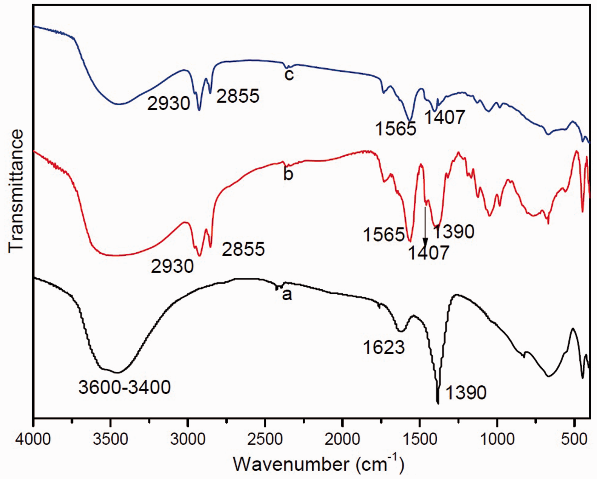
Particle size and morphology of LDH and RL-LDH
SEM images of pristine LDH, RL-LDH1, and RL-LDH2 samples are shown in Figure 5. Pristine LDH particles were mainly hexagonal platelets with sizes ranging from 100 nm to 150 nm (Figure 5(a)). RL-LDH1 and RL-LDH2 nanocomposite particles became aggregated, which was induced by RL introduction. However, the plate-shape morphology remained visible after RL intercalation. As seen from Figure 5(b) and (c), the particles of RL-LDH1 had higher aggregation extent than those of RL-LDH2.
Scanning electron micrographs of (a) LDH, (b) RL-LDH1, and (c) RL-LDH2.
TG-DSC analysis
The TG-DSC curves of pristine LDH, RL-LDH1, and RL-LDH2 samples are illustrated in Figure 6. The LDH was thermally decomposed through two steps: (1) desorption of physically adsorbed water and partial interlayer water between 293 K and 473 K with an endothermic peak at 385.5 K; (2) decomposition of nitrate anions and dehydration of brucite-like layers between 613 K and 853 K with an endothermic peak at 703.3 K. For RL-LDH1 (and RL-LDH2), the presence of a broad endothermic peak between 293 K and 423 K and the weight loss of 3.85% (and 5.42%) were related to water loss. The exothermic peak at ca. 452.8 K (and 453 K) was mainly induced by RL decomposition. Sharp exothermic peak at approximately 573.2 K (or 594 K) corresponded to the complete decomposition of species derived from RL, which implied that the chemical stability of RL was enhanced significantly in the confined region of RL-LDH2 galleries compared with that in RL-LDH1 galleries. The total weight losses of LDH, RL-LDH1 and RL-LDH2 were 46.68%, 65.24% and 67.62%, respectively.
Thermal gravimetry-differential scanning calorimetry curves of (a) LDH, (b) RL-LDH1, and (c) RL-LDH2.
Elemental analysis
Property of synthesized layered double hydroxide (LDH) and rhamnolipid (RL)-LDH.

The weight percentage of interlayer RL anion upon ICP data.
Specific surface areas
The specific surface areas of LDH, RL-LDH1, and RL-LDH2 were examined, and the results are also listed in Table 1. The specific surface areas of LDH, RL-LDH1, and RL-LDH2 were 39.39, 8.75, and 11.71 m2·g−1, respectively. The decrease in surface area could be due to the aggregation of RL-LDH plates (You et al., 2002) and stacking of RL anions in the interlayer spaces of LDH. In other words, the reduced surface area of RL-LDH nanocomposites demonstrated their strong hydrophobic property. A similar phenomenon was reported in LDH modified with DS− anions (Ruan et al., 2013). The specific surface area of RL-LDH1 was smaller than that of RL-LDH2 due to the higher aggregation extent of the former, which was in accordance with the results of SEM images.
Adsorption characterization
The adsorption isotherms of p-cresol on LDH, RL-LDH1, and RL-LDH2 at 293, 303, and 313 K are shown in Figure 7. The equilibrium adsorption capacities of p-cresol on both LDH and RL-LDH increased with the increasing initial concentration in the studied concentration range. Additionally, RL-LDH showed higher adsorption capacity for p-cresol than LDH although it has the smaller surface area, suggesting it’s adsorption behavior for p-cresol was unrelated to the surface area. The adsorption data in Figure 7 were analyzed using three isotherms, namely, Langmuir, Freundlich, and linear model.
The adsorption isotherms of p-cresol on LDH, RL-LDH1, and RL-LDH2 at varied temperatures.
The Langmuir isotherm is based on the basic assumption that adsorption occurs at specific homogeneous sites within the adsorbent, and the isotherm equation is described as follows ( Langmuir, 1916)

The Freundlich isotherm, assuming the presence of a heterogeneous surface with a non-uniform heat adsorption distribution on the surface, can be expressed as follows (Freundlich, 1906).

The linear model, indicating a partitioning adsorption mechanism, can be described simply as follows ( Gao et al., 2011)

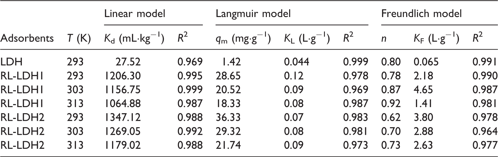
The parameters of the adsorption model for p-cresol at varied temperatures.
Effect of temperature on p-cresol adsorption
Because the linear model fitted the best to describe the adsorption of p-cresol on RL-LDH, the effect of temperature on p-cresol adsorption was discussed in detail based on the linear model. As seen from Figure 7 and Table 2, the equilibrium adsorption capacity for RL-LDH1 and RL-LDH2 decreased with increasing temperature, together with the Kd value decreased from 1206.30 to 1164.88 for RL-LDH1 and from 1347.12 to 1179.02 for RL-LDH2 with increasing temperature, suggesting that the interaction between p-cresol and RL-LDH nanoparticles was exothermic in nature.


Gibbs free energy, enthalpy, and entropy changes associated with p-cresol adsorption onto RL-LDH.

Comparison with other adsorbents available in the literature
Maximum adsorption capacities for p-cresol on various adsorbents.

As shown in Table 4, although RL-LDH had smaller adsorption capacity in comparison with commercial-activated alumina (Bakas et al., 2014), it exhibited higher adsorption capacity than some adsorbents such as the coconut shell-activated cha (Zhu and Kolar, 2014), pine wood-activated carbon (Das et al., 2013), swine manure char ( Fitzgerald et al., 2015). Most importantly, the obtained RL-LDH is a safe potential adsorbent for the removal of p-cresol due to its low toxicity and the RL-LDH nanocomposite (RL-LDH2) can be prepared through the facile delamination/reassembling process to effectively remove p-cresol.
Conclusions
RL-LDH nanocomposites were synthesized by ion exchange (RL-LDH1) and delamination/reassembling (RL-LDH2) method, respectively. The basal spacing of RL-LDH2 (d003 = 3.39 nm) was a little larger than that of RL-LDH1 (3.22 nm) while greatly larger than that of LDH (0.90 nm), revealing largely extended interlayer regions in RL-LDH2. The RL content of RL-LDH2 was higher than that of RL-LDH1. RL-LDH samples present higher adsorption capacities for p-cesol than that of the original LDH due to the strong hydrophobicity. The adsorption isotherms of p-cesol on RL-LDH1 and RL-LDH2 were well fitted by the linear model, implying a partitioning adsorption process. An adsolubilization mechanism model was proposed mainly involving hydrophobic interactions between p-cresol and the RL anions in the interlayer. The highest p-cresol adsorption capacity on RL-LDH2 can be related to the more loosely stacked interlayer RL anion arrangement. The adsorption capacity of RL-LDH2 was much better than some adsorbents reported. Overall, RL-LDH2 could be a safe potential adsorbent for removing p-cresol.
Footnotes
Declaration of Conflicting Interests
The author(s) declared no potential conflicts of interest with respect to the research, authorship, and/or publication of this article.
Funding
The author(s) disclosed receipt of the following financial support for the research, authorship, and/or publication of this article: This work was supported by the Natural Science Foundation of Shanxi Province of China (2013011040-8), Scientific and Technological Innovation Programs of Higher Education Institutions in Shanxi (2013159), National Training Programs of Innovation and Entrepreneurship for Undergraduates (201310122002), and Training Programs of Innovation and Entrepreneurship for Undergraduates in Shanxi (2014431).