
Abstract
Background:
Inflammatory response plays a pivotal role in the pathophysiology of depression. With this background, we planned this study to see if immune markers, interleukin 6 (IL-6), and erythrocyte sedimentation rate (ESR) are raised in subjects with major depressive disorder (MDD) and compare its level with healthy controls and first-degree relatives of subjects. We also explored if variation in the level of these markers was related to the severity of depression.
Methods:
This comparative observational study included 120 subjects, who were divided into three groups of 40 individuals each.
Group 1 was the MDD group, group 2 was the healthy first-degree relative group, and group 3 was the healthy control group. All the subjects were then made to undergo estimation of IL-6 (pg/mL) and ESR (mm/h) from our hospital biochemistry lab.
The patients in group 1 were additionally screened for the severity of depression.
Results:
The mean IL-6 and ESR measure within the MDD group was 9.20 ± 13.40 (pg/mL) and 5.90 ± 5.35 (mm/h), respectively. We found that the mean and median values of both IL-6 and ESR were highest in the MDD group as compared to other groups, which were statistically significant (p <0.05). A pairwise comparison found no significant difference in the IL-6 and ESR scores among the healthy controls and healthy first-degree relatives. The mean of IL-6 was highest in individuals with moderate severity of depression, while the mean of ESR was highest in individuals with severe depression.
Conclusion:
The immune markers, IL-6 and ESR, were significantly raised in the MDD group; however, the levels did not correlate significantly with the differential severity of the depressive disorder as per Hamilton depression rating scale (HAM-D) scores.
IL-6 and ESR are significantly raised in patients with MDD but are not associated with the severity of depression.Key Message:
Major depressive disorder (MDD) accounts for 1.84% of worldwide disability-adjusted life years. 1 The first-degree relatives of subjects with this illness embrace a thrice higher risk of MDD, and this disorder has approximately 35% heritability. 2 Studies indicate that gene expression patterns differ between depressed and nondepressed populations, which confer liability on the former population. 3 The changes in the neuroimmune axis, although they play an important part in the causation of this disorder, have been kept in the rear as compared to the classical neurotransmitter alteration. 4
The immune cells are largely derived from hematopoietic stem cells. 5 The microbial pathology or nontraumatic injury entails recruitment of the innate immune system. The pattern recognition receptors on antigen-presenting cells recognize the pathogen-associated molecular patterns (PAMPs) or danger-associated molecular patterns (DAMPs). 6 While the PAMPs are invoked as a result of microbial products, the facilitation of DAMPs occurs additively by physiological and psychological stressors. The DAMPs, along with the neurohormonal axis, modulate peripheral inflammation, forming a feedback loop known as an inflammatory reflex. 7
The exposure of the neuroimmune axis to psychological stressors causes the proliferation of neutrophils and monocytes. 8 The innate immune cells release cytokines such as IL-1 (interleukin) beta and IL-6. Various published studies in the past have shown the origin of depressive disorder as a result of neuroinflammation. 9 In a population of hepatitis C-afflicted patients, those undergoing long-term therapy by interferon-alpha developed depression. 10 One recent study investigated that a subset of the pediatric population with raised IL-6 levels was more susceptible to developing depression by early adulthood. 11
Sequenced treatment alternatives to relieve depression (STAR-D) trial has shown that 60–70% of the MDD subpopulation does not achieve complete remission from conventional antidepressants 12 This may reflect underlying disease mechanisms that are not ubiquitously treated by standard treatments. Hence, we might require a modified paradigm to achieve better therapeutic efficacy with the use of anti-inflammatory agents. 13 Tocilizumab, a humanized antibody to the IL-6 receptor, is garnering interest for its use in pharmacotherapy for affective disorders. 14 Other studies have examined nonsteroidal anti-inflammatory drugs as a treatment strategy for depression. However, the results have been mixed so far. 15 Ketamine, a novel antidepressant in the mitigation of treatment-resistant depression, has adjuvant anti-inflammatory action as it decreases levels of cytokines, including IL-6. 16 A study by Chavda et al. 17 noticed this trend in newly diagnosed MDD patients. Also, the same study noted significant differences in erythrocyte sedimentation rate (ESR) levels after 2 months of treatment with selective serotonin reuptake inhibitors (SSRIs). Abdel-Nasser et al. demonstrated a positive correlation of ESR measures with depressive symptoms in patients with rheumatoid arthritis (RA). 18 RA is also an inflammatory state, and its association with comorbid depression in this illness further corroborates the inflammatory hypothesis in depression.
Looking at the published literature, while most of the studies have found a relationship between IL-6 and depressive disorder, very few studies have explored this relationship with ESR. Also, most of the literature focuses on the comparison of immune markers among MDD and healthy control groups. None of the published studies, to our knowledge, have compared the heritability between the healthy first-degree relatives of patients suffering from MDD and healthy controls.
Hence, with this background, we planned this study to determine if immune markers, IL-6, and ESR are raised in subjects with depressive disorder. We also planned to see if the variation in the level of these markers was related to the severity of depression and showed any variation focusing upon healthy first-degree relatives of patients suffering from MDD and healthy controls.
Methods
This comparative observational study was conducted in the department of psychiatry of a tertiary care hospital for over 12 months (October 2022–October 2023) after obtaining permission from the institutional ethics committee. The sample size was calculated using Openepi software (a free web-based operating system program designed for public health) after consultation with an institute biostatistician. The significance level was kept at 0.05, and power (1-beta) was kept at 80%. The standard deviation (sigma) was kept at 1; the effect size was kept at 0.62, considering a prior study. 19 The sample size thus derived was 40 in each group.
A convenient sampling method was used.
Those found suitable for the study were included after written informed consent. We excluded patients who were taking antidepressants or other drugs regularly that affect ESR and/or IL-6, those suffering from any medical/ surgical comorbid illness, pregnant females, and those suffering from any other psychiatric illness.
We divided the subjects into three groups of 40 individuals each.
Group 1 was the MDD group, which consisted of patients who were diagnosed with MDD and were drug naïve or had a recurrent episode of MDD with a prior interepisodic recovery of 06 months. All the diagnoses were made as per Diagnostic and Statistical Manual of Mental Disorders, fifth edition (DSM-5) criteria after applying the Mini Internation Neuropsychiatric Interview (MINI).
Group 2 was the healthy first-degree relative group, which consisted of the first-degree relatives of the subject with MDD who accompanied them.
Group 3 was a healthy control group, which consisted of healthy subjects who were not related to MDD cases and did not have a family history of MDD among any first-generation members.
We applied MINI to subjects of both groups 2 and 3 to rule out any psychiatric illness (including MDD). We also ruled out any other medical and surgical illnesses based on the history and general and systemic examination of the subjects.
All the subjects were then made to undergo estimation of IL-6 (pg/mL) and ESR (mm/h) from our hospital biochemistry lab.
The specimen meant for IL-6 was collected in a yellow-topped vacutainer. The quantitative IL-6 level was determined via Beckmann Autoanalyzer DXI700, which uses the chemiluminescent immunoassay method (one-step immune enzymatic sandwich assay). The ESR samples were collected in purple-capped vacutainers containing ethylenediaminetetraacetic acid (EDTA). The samples were analyzed using a Roller 20LC autoanalyzer. This instrument measures sedimentation and aggregation by optical density and ESR by infrared microphotometer (950nm).
The hospital bore the cost of the test.
The patients in Group 1 were additionally screened for the severity of depression using the Hamilton depression rating scale (HAM-D)
Study Tools
Semi-structured proforma: A semi-structured proforma was used to collect demographic and clinical data.
Depression was assessed using the HAM-D, which is a 17-item clinician-administered depression assessment scale. Each behaviorally anchored item is rated on either a 3- or 5-point scale and summed to obtain the total score. A score of 8–16 indicates mild depression, 17–23 moderate depression, and a score of >24 severe depression. 20
Appropriate data was collected and tabulated and statistics were applied using Statistical analysis for the Social Sciences (SPSS) version 27 (BM Corp., Armonk, NY). 21 Descriptive statistics were used to summarise the data. The skewness and kurtosis of the variables within different groups were checked. The Shapiro–Wilk test was applied to the dataset. Once the data fulfilled three criteria of normality (skewness, kurtosis, and Shapiro–Wilk test), it was interpreted as a normal distribution. Where data was not normally distributed, nonparametric tests were applied. One-way analysis of variance (ANOVA) was used to make group comparisons in terms of age, and the chi-squared test was used to explore the association between “Group” and other variables. Posthoc pairwise tests for the Kruskal–Wallis test were performed using the Dunn Test method with Sidak correction. Significance was determined at p-value < 0.05.
Results
Table 1 shows the baseline characteristics of participants across the three groups. There was only a significant difference between the various groups in terms of the distribution of occupation (p = 0.0279).
Baseline Characteristics of Participants Across the Three Groups.
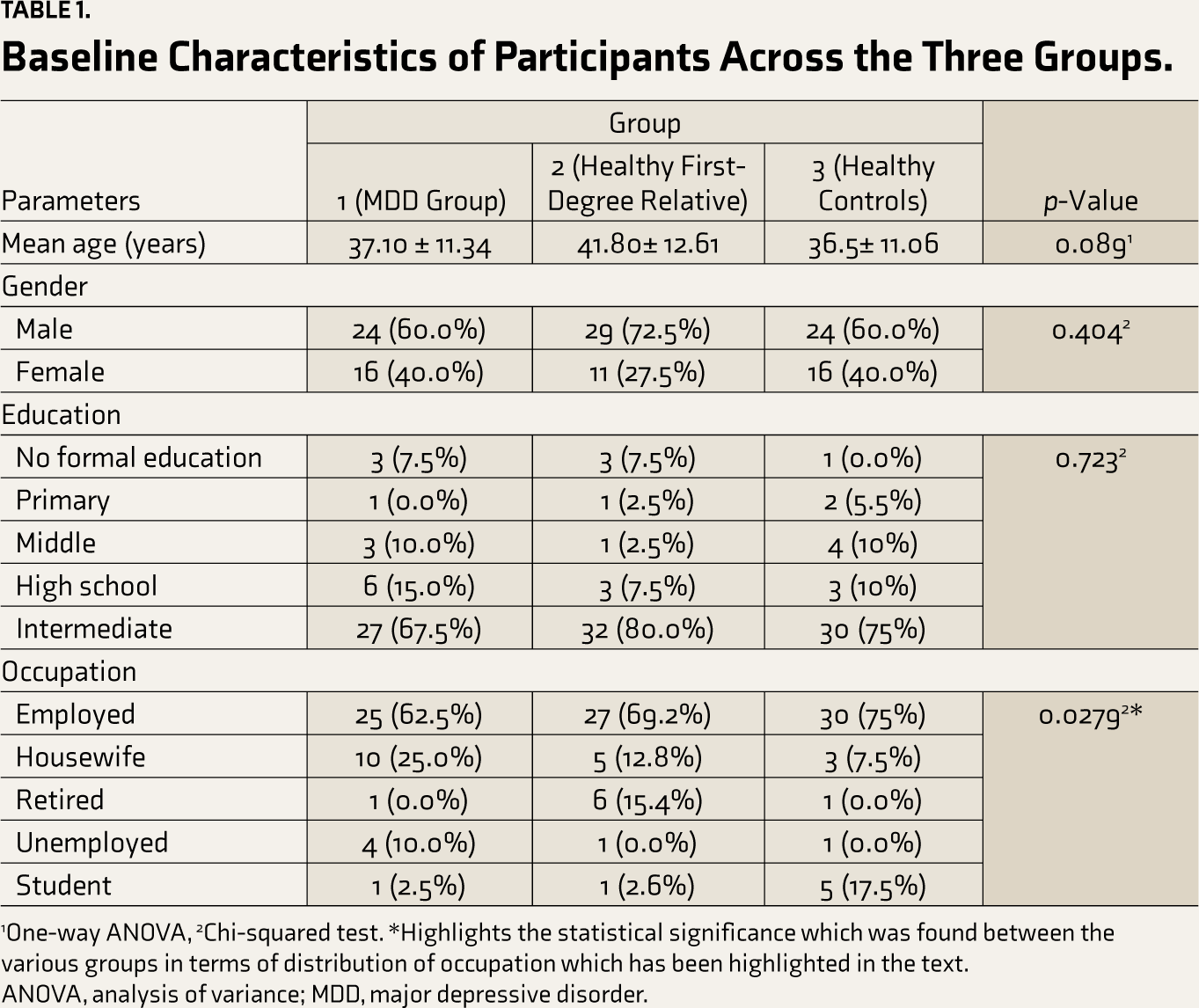
1One-way ANOVA, 2Chi-squared test. *Highlights the statistical significance which was found between the various groups in terms of distribution of occupation which has been highlighted in the text.
ANOVA, analysis of variance; MDD, major depressive disorder.
Out of the 40 individuals, as per the HAM-D scores, 15% were diagnosed with mild depression, 65% were diagnosed with moderate depression, and 20% were diagnosed as having severe depression. Two of them were diagnosed with MDD with psychotic features.
Table 2 shows the comparison of IL-6 values across various groups of subjects. We found that the mean and median value of IL-6 was highest in the MDD group as compared to the healthy first-degree relatives and healthy controls. On applying the Kruskal–Wallis test, this finding was statistically significant (p <0.001). However, looking at the pairwise comparison across various group subjects for IL-6, no significant difference was found among the healthy controls and healthy first-degree relative (p = 0.721). Similarly, we did a comparison of ESR values across various groups of subjects, as shown in Table 3. The mean and median values of ESR were highest in the MDD group as compared to other groups. On applying the Kruskal–Wallis test, this finding was statistically significant (p = 0.019). Looking at the pairwise comparison across various group subjects for ESR, no significant difference was found between the healthy controls and healthy first-degree relatives (p = 0.109).
Comparison of IL-6 Value Across Various Groups of Subjects.
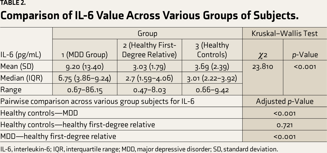
IL-6, interleukin-6; IQR, interquartile range; MDD, major depressive disorder; SD, standard deviation. Comparison of ESR Value Across Various Groups of Subjects.
Comparison of ESR Value Across Various Groups of Subjects.
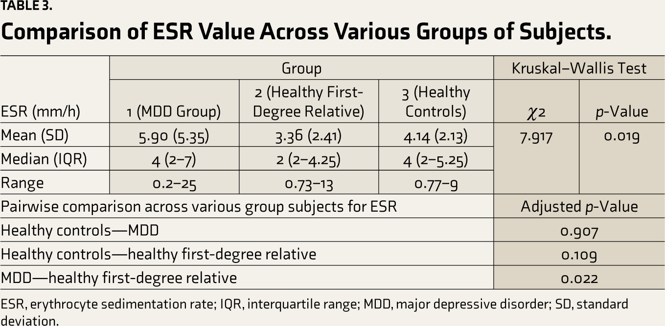
ESR, erythrocyte sedimentation rate; IQR, interquartile range; MDD, major depressive disorder; SD, standard deviation.
The association between the HAM-D scores and immune markers among the MDD group (group 1) is shown in Table 4. The mean IL-6 measure within the MDD cohort was calculated as 9.20 ± 13.40. The mean of IL-6 was highest in individuals with moderate severity of depression; however, on applying the Kruskal–Wallis test, there was no statistical significance (p = 0.113). Similarly, the mean ESR (mm/h) of the group was 5.90 ± 5.35, which was highest in individuals with severe depression, which, on applying the Kruskal–Wallis test, was not statistically significant (p = 0.057).
Comparison and Association Between the HAM-D Scores and Immune Markers (IL-6 and ESR) Among MDD Group.
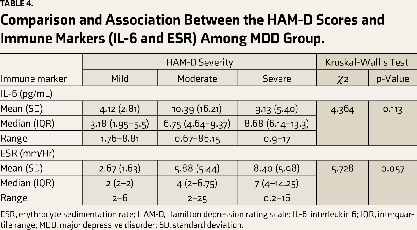
ESR, erythrocyte sedimentation rate; HAM-D, Hamilton depression rating scale; IL-6, interleukin 6; IQR, interquartile range; MDD, major depressive disorder; SD, standard deviation.
Discussion
This study examined the cross-sectional comparison of IL-6 measures across the three groups. We found higher values for IL-6 measures in patients with depression. The mean IL-6 value was at least 2–2.5 times higher than that of the other groups, which was also statistically significant. These results corroborate the existing research literature. A cumulative meta-analysis by Haapakoski et al discussed the plausible role of IL-6 in the pathology of depression, therapeutic response to antidepressants, the dose–response relationships, and as a predictor of future development of mood disorder. 22 While looking at the inflammatory profile of patients with treatment-resistant MDD having a chronic course and high severity, Krogh J et al. 23 inferred poor outcomes in those with higher levels of inflammatory markers, including IL-6
Another immune marker that we investigated was ESR. Limited studies or data are available so far that quote ESR levels as one of the parameters that affect MDD. In an experimental study by Chavda et al, the baseline comparison of newly diagnosed MDD patients with the control group reported increased levels of ESR, which decreased after treatment with SSRI. 17
We assumed that across the three groups, the measures of ESR and IL-6 would be highest among MDD patients, followed by first-degree relatives. The levels were assumed to be the lowest in the control group. In contrast to what we had expected, we found comparable ESR and IL-6 levels in the healthy first-degree relatives of MDD patients and the healthy control group. When comparing the mean ESR value, we found that the healthy control group (group 3) had a higher mean than the healthy first-degree relative group (group 2). Hence, we reach a plausible conclusion that the immune markers of depressive disorder are more of a state marker than a trait marker. While searching for published literature, we could not come across any study that has compared the level of immune-related genes among the first-degree relatives of cases suffering from depressive disorder and healthy controls. However, studies have demonstrated the role of immune-related genes in the pathophysiology of depression. A study by Gonzales et al 24 found out the role of interferon pathway in the pathophysiology of depressive disorder, especially focused upon immune-related gene USP18 (ubiquitin specific peptidase). Another study by Sforzini et al. 25 measured the expression of 16 immune-related candidate genes among the cases with depressive disorder versus a healthy control group, of which nine genes were differentially expressed across the groups. More studies in the future with specific target immune-related genes with a larger sample size might be able to uncover the potential heritability of this process and expand our horizon related to depression.
Another purpose of our study was to find a comparison and association between the HAM-D scores and the level of inflammatory markers. Through our findings, we could not establish a significant correlation between HAM-D severity and levels of IL-6. Similar results were found by Kofod J et al., 26 who conducted an open-label partially randomized trial on 90 outpatients with depression and found no correlation between any of the inflammatory markers with differential severity on the overall Montgomery–Åsberg Depression Rating Scale (MADRS). A study by Ryan 27 et al did not find any relationship between the inflammatory mediators (IL-1β, IL-6, IL-10, and tumor necrosis factor-alpha [TNF-α]) of depression and the HAM-D score. We also did not find a significant correlation between ESR and HAM-D scores.
Strengths
This study is the first of its kind from the northern part of India to explore the relationship of immune markers, especially ESR, with depressive disorder. Also, unlike previous literature, our study compared the levels of immune markers between the healthy first-degree relatives of patients suffering from MDD and healthy controls and found no significant association among them.
Limitations
Since ours was a comparative observational study done cross-sectionally, we did not follow up on cases with depressive disorder in the long term to see the changes in the level of immune markers following pharmacotherapy and/or psychotherapy. Since our study was a hospital-based one, we cannot comment on the generalizability of our results. Also, we did not do any structured evaluation to rule out MDD in the first-degree relatives of group 3 patients, and the medical and surgical exclusion of illness in groups 2 and 3 was done based on history from the subjects and general and systemic examination.
Conclusion
We conclude that the level of immune markers IL-6 and ESR was raised in subjects suffering from depression. Immune markers (IL-6 and ESR) levels do not correlate significantly with the differential severity of depressive disorder as per HAM-D scores. Also, neither of the immune markers showed significant differences when comparing the healthy first-degree relative and the healthy control group.
Footnotes
Declaration of Conflicting Interests
The authors declared no potential conflicts of interest with respect to the research, authorship and/or publication of this article.
Declaration Regarding the Use of Generative AI
We did not use artificial intelligence (AI) software to collect or analyze data, produce images or graphs, or write this article.
Ethical Approval
Ethics committee approval was taken via letter number-SRHU/HIMS/ETHICS/2022/350(Dated- 27/09/2022).
Funding
The authors received no financial support for the research, authorship, and/or publication of this article.
Informed Consent
Written informed consent was acquired from all participants.