
Abstract
A retrospective study was performed to determine the incidences of spontaneous findings in control laboratory New Zealand White (NZW) and Dutch Belted (DB) rabbits. Terminal body and organ weights data were also collected. A total of 2170 NZW (526 males/1644 females), 100 DB rabbits (50 animals per sex), aged 4- to 7-month-old were obtained from 158 non-clinical studies evaluated between 2013 and 2022. The NZW rabbits had greater mean terminal body weights than DB strain. Mixed cell infiltration in the lung was the most recorded finding in both strains, followed by pulmonary inflammation/mononuclear cell infiltration. Differentiation between pulmonary “infiltration”/“inflammation” remained challenging as interpretation of guidelines for diagnostic terminology may vary amongst pathologists. Other common findings included mineralization and basophilia of the renal tubules; hepatic/renal mononuclear cell infiltration, all more common in females. Cysts were commonly recorded, with high prevalence in the oviduct, thyroid gland, ovary in NZW strain, while uterine, pituitary gland, and thyroid gland cysts were the most identified in DB rabbits. Neoplasms and infectious etiologies were absent. Most of the animals were sexually mature. To our knowledge, this is the most recent comprehensive study of spontaneous lesions and organ weights in both rabbit strains and should facilitate the differentiation of spontaneous and induced lesions in safety studies.
Keywords
Introduction
Rabbits represent an excellent species in non-clinical drug development as they are considered the smallest and least expensive non-rodent animal models for reproductive, developmental, and general toxicology studies. The most common strain used in toxicity studies is the New Zealand White (NZW), but Dutch Belted (DB) rabbits are also recently considered a practical alternative for developmental toxicity testing, ocular studies and to investigate the effect of compounds on melanin development and binding. Differentiating drug-induced changes from spontaneous non–treatment-related findings is a challenge in the interpretation of histopathology results in preclinical toxicity studies in rabbits due to the paucity of published data. The variation in the anatomy of the rabbit, especially the digestive tract, when compared with the more common laboratory species, further complicates the evaluation of subtle morphologic changes in short-term studies. Both common and unusual spontaneous findings may occur in treated animals only, as there are a limited number of animals per group when compared to rodent studies. Therefore, compilation of historical databases of background lesions and organ weights from rabbit studies is very important. To the authors’ knowledge, reports of spontaneous lesions in rabbits are either outdated and/or take into account only NZW rabbits, with no data in DB rabbits. Publications of spontaneous findings in laboratory DB rabbits are lacking in the current literature. In addition, data of organ weights and terminal body weights in laboratory rabbits are outdated in the current literature. Therefore, the main aims of this study were to present and discuss the data set, terminal body and organ weights, and incidences of spontaneous findings of both laboratory NZW and DB rabbits from studies carried out at all Charles River Laboratories sites and to attempt a comparison of the findings between the two strains.
Materials and Methods
Animals
Tissue samples from a total of 2170 purpose-bred laboratory NZW rabbits (526 males and 1644 females) and 100 purpose-bred laboratory DB rabbits (50 animals per sex) were obtained from control groups of 158 non-clinical toxicological studies evaluated between 2013 and 2022. The animals originated from accredited suppliers (Envigo RMS LLC, UK and Charles River Laboratories, Canada). Their age ranged from 4 to 7 months at the start of the study and with body weight ranging from 2 to 6 kg. All control rabbits in the study were from groups which were untreated or where an appropriate vehicle was given. They were allowed to acclimatize for a minimum period of 20 days prior to dosing. Several routes of administration of the vehicles were applied and included oral gavage, intra-muscular, intra-venous, intra-arterial, subcutaneous, ocular (topical) instillation and intra-ocular (intra-vitreal and intra-cameral), intra-dermal, intra-vaginal, intra-articular, and intra-nasal routes. The total number of studies for each route of administration used in both NZW and DB rabbits is shown in Table 1. In DB rabbits, few routes of administration (ocular topical instillation, intra-vitreal, intra-nasal, and intra-cameral) were implemented.
Number of studies per route of administration in control animals in both strains (New Zealand White and Dutch Belted Rabbits).
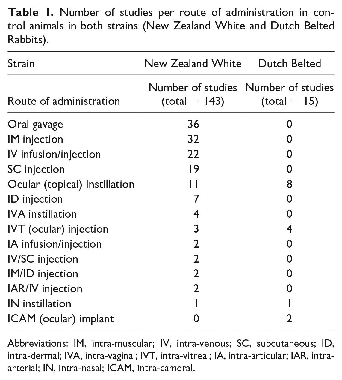
Abbreviations: IM, intra-muscular; IV, intra-venous; SC, subcutaneous; ID, intra-dermal; IVA, intra-vaginal; IVT, intra-vitreal; IA, intra-articular; IAR, intra-arterial; IN, intra-nasal; ICAM, intra-cameral.
Animals were identified by a subcutaneously implanted electronic identification chip (localized in the interscapular or gluteal region) or ear tag. Rabbits were maintained as an outbred colony, housed in HEPA (High-Efficiency Particulate Absorbing)-filtered barrier rooms and bred via a pair-mating system. They were housed singly (for both males and females) or, occasionally, group-housed (females only) in appropriately sized stainless-steel cages with a Techniplast “Noryl” dual-level interior, a perforated floor and a suspended tray containing absorbent paper, corncob bedding (Pel-O’Cobs) or other suitable material. Cage identification followed a color-coded cage card with appropriate legal statutory requirements. Dumbbells, bunny blocks, chains, and/or Timothy cubes/irradiated hay were supplied for cage enrichment, and resting pads were provided as needed. Objects for chewing and devices for hiding in were provided with a certificate of analysis for significant contaminants. An analytical certificate for each batch of chewing objects and hiding devices used was retained at the Testing Facility. The animals were given a period of exercise in a separate floor pen (a minimum of 30 minutes, and no longer than 45 minutes) up to 3 times per week. There was automatic control of temperature and humidity, target ranges being respectively 16°C to 22°C ± 2°C and 30% to 70% ± 20%, with a minimum of 10 air-changes per hour. There was automatic control of light cycle, with light hours normally being 07:00 to 19:00 hours and dark hours being 19:00 to 07:00 hours. Each cage was supplied with an automatic watering valve and stainless-steel food hopper. The animals were fed ad libitum with pellets with the following diets: (1) Teklad Global Rabbit diet (pellets supplied by Harlan Teklad); or (2) PMI Nutrition International, LLC Certified Rabbit LabDiet 5322; or (3) Lab Diet Certified High Fiber Rabbit Diet 5325. The animals were also offered hay 3 times per week and a small portion of a limited selection of fresh fruits and/or vegetables at least twice a week. They had ad libitum access to domestic mains quality water supplied through an automatic watering system. The animals had received an appropriate veterinary care and examination. Animals supplied were from colonies demonstrated to be free of organisms listed by the European Federation of Laboratory Animal Science Associations (FELASA). Rabbit colonies were screened quarterly by the Polymerase Chain Reaction (PCR) method, Multiplexed Fluorometric ImmunoAssay (MFIA), agglutination, culture, direct examination, or serology for viruses, bacteria, and some parasites and fungi in agreement with the FELASA working group recommendations. 14 All the rabbits were kept under a VAF/Plus (Virus Antibody Free) and SPF (Specific Pathogen-Free) health status and the colonies were tested for, and free of Rabbit Hemorrhagic Disease (RHDV), Rabbit Rotavirus, Parainfluenza virus types 1, 2, and 5 (PIV1, PIV2, PIV5), Lymphocytic Choriomeningitis virus (LCMV), Reovirus, adenovirus, coronavirus, and Myxomatosis virus/Leporipoxvirus; Tyzzer Disease (Clostridium piliforme), Treponema cuniculi and Treponema paraluiscuniculi, Bordetella bronchiseptica, Pasteurella multocida (and other pasteurellaceae), Pseudomonas aeruginosa, Lawsonia sp., salmonellosis, Escherichia coli (enteropathogenic strains), Filobacterium rodentium (CAR Bacillus), Helicobacter species, and Staphylococcus aureus; dermatophytes, Passalurus ambiguus, Pneumocystis oryctolagi, pinworms, Encephalitozoon cuniculi, Cheyletiella parasitovorax, Leporacarus gibbus, Psoroptes cuniculi, and other ectoparasites and helminths. Screening for Eimeria species was not consistently performed in the colonies. All studies were conducted in accordance with the UK Animals (Scientific Procedures) Act 1986, which conforms to the European Convention for the Protection of Vertebrate Animals Used for Experimental and Other Scientific Purposes (Strasbourg, Council of Europe) (for studies conducted in the United Kingdom) and the USDA Animal Welfare Act (9 CFR, Parts 1, 2, and 3) (for studies conducted in the United States and Canada).
Pathological Examination
Rabbits were euthanized by intra-venous injection with sodium pentobarbitone followed by exsanguination. Tissues were trimmed to remove as much fat as possible, damp-dried on absorbent paper, and weighed with a calibrated balance. Any organ weight found to be inconsistent or flagged as out of range was reweighed where applicable. Paired organs were weighed together, unless lesions were present, in which case they were weighed individually. Tissues were preserved in 10% neutral buffered formalin, embedded in paraffin wax, sectioned to a 4 to 5 mm thickness, and stained with hematoxylin and eosin. They were examined by a board-certified veterinary pathologist or a veterinary pathologist with experience in laboratory animal toxicologic pathology and were entered directly into a computerized database (Instem Provantis). For each histological finding, prevalence and reference range were calculated, per each sex and strain. The reference range is defined by the lower and upper reference value and was calculated as the lowest and highest percentage found on a study for each finding.
Study Design
Data were collected retrospectively from control groups of NZW and DB rabbit studies evaluated over a period of ten years (2013-2022). Data were available from 158 controlled studies, with five to twenty-two animals per sex per study, giving a total of 2270 control animals (2170 NZW rabbits and 100 DB rabbits). Studies incorporated into the present investigation were selected based on the following criteria:
(a) Male and female animals from both strains of rabbits with a minimum of five animals per sex per group.
(b) At least one control or untreated group.
(c) GLP compliant toxicological studies.
Male and female NZW rabbits from the selected studies were aged at necropsy from 24 to 28 weeks for organ and body weight measurements and between 13 and 69 weeks for histopathology evaluation. Male and female DB rabbits selected for the studies to be included in the investigation were aged at necropsy from 21 to 32 weeks for organ and body weight measurements and between 18 and 65 weeks for the histopathological findings. A narrow age range was selected for organ weight data, in order to avoid the range of values being too wide.
Results
Terminal Body and Organ Weight findings
The results showing the mean values, standard deviations, and ranges of absolute body and organ weights are outlined in Table 2 for NZW rabbits and in Table 3 for DB rabbits. Terminal body weight differed between the two strains selected for a similar age range. Male and female NZW rabbits had a greater mean terminal body weight (3.3 kg ± 0.2 kg and 3.6 kg ± 0.3 kg, respectively) compared to male and female DB rabbits (1.8 kg ± 0.1 kg and 1.9 kg ± 0.1 kg, respectively). Females in both strains presented a slightly higher terminal body weight compared to males. Overall, NZW rabbits had greater mean values of organ weights compared to DB rabbits, except for the lung in female DB rabbits which had slightly higher weight (11.9 g ± 3.527 g) compared to NZW female rabbits (10.8 g ± 1.396 g). In both NZW and DB rabbits, males showed a greater mean value of adrenal gland weight (0.338 g ± 0.078 g in DB and 0.356 g ± 0.101 g in NZW rabbits) and heart weight (4.777 g ± 0.496 g in DB and 7.106 g ± 0.812 g in NZW rabbits) compared to females. Male NZW rabbits showed higher mean values of kidney weight (15.829 g ± 1.496 g), liver weight (82.013 g ± 18.952 g) and lung weight (11.342 g ± 1.475 g) compared to females (respectively 14.683 g ± 1.596 g; 72.412 g ± 14.008 g and 10.801 g ± 1.396 g); however, there were no significant differences in the same organ weight mean values in the DB strain.
New Zealand White rabbit—absolute terminal body weight (kg) and absolute organ weight (g).
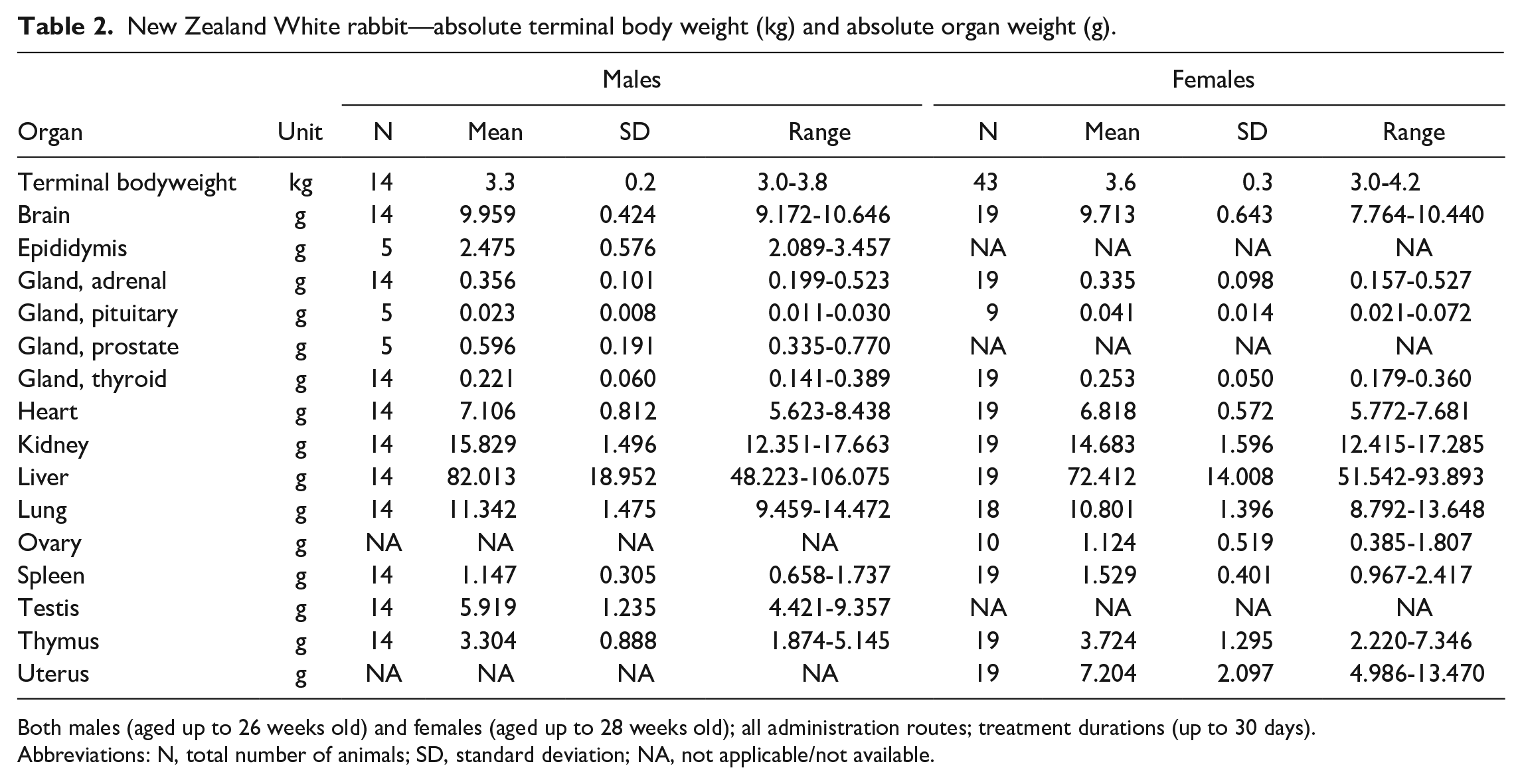
Both males (aged up to 26 weeks old) and females (aged up to 28 weeks old); all administration routes; treatment durations (up to 30 days).
Abbreviations: N, total number of animals; SD, standard deviation; NA, not applicable/not available.
Dutch Belted rabbit—absolute terminal body weight (kg) and absolute organ weight (g).
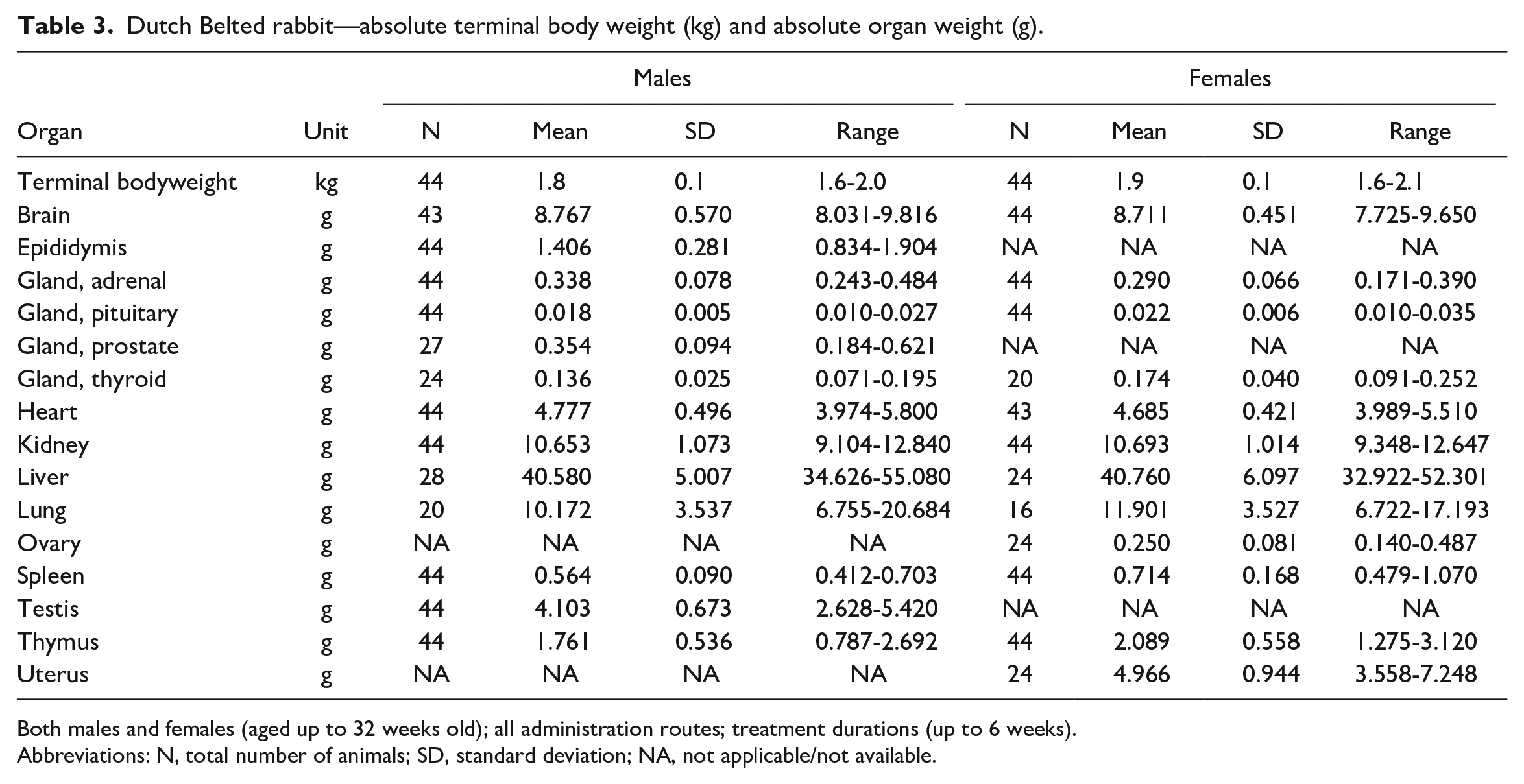
Both males and females (aged up to 32 weeks old); all administration routes; treatment durations (up to 6 weeks).
Abbreviations: N, total number of animals; SD, standard deviation; NA, not applicable/not available.
In both NZW and DB rabbits, females showed greater mean values of pituitary gland weight (0.022 g ± 0.006 g in DB and 0.041 g ± 0.014 g in NZW rabbits), thyroid gland weight (0.174 g ± 0.040 g in DB and 0.253 g ± 0.050 g in NZW rabbits), spleen weight (0.714 g ± 0.168 g in DB and 1.529 g ± 0.401 g in NZW rabbits), and thymus weight (2.089 g ± 0.558 g in DB and 3.724 g ± 1.295 g in NZW rabbits) compared to males. No other significant strain or sex organ weight differences were noted.
Common Histopathological Findings
The results showing the most common histopathological findings encountered in the 2170 control NZW rabbits and 100 DB rabbits evaluated at our laboratory are outlined in Tables 4 to 12. Inflammatory mixed cell infiltration in the lung was the most recorded finding in both NZW (19.0% females/12.0% males) and DB (38.0% females/22.0% males) rabbits. Other common lesions were mineralization of the renal tubules (NZW = 16.3% females/14.6% males), renal tubular basophilia (NZW = 15.1% females/11.4% males; DB = 16.0% females/10.0% males), hepatic mononuclear cell infiltrations (NZW = 10.5% females/7.1% males) and renal mononuclear cell infiltrations (DB = 14.0% females/10.0% males). All of these findings were more commonly identified in females. Inflammation and inflammatory cell infiltrates were the most recorded histopathological findings, and differentiation between “infiltration” and “inflammation” was defined by accumulation of immune cells and the presence or absence of other histological changes typical of inflammation. 15 The term “infiltration” was used when there was an aggregate/clump of inflammatory cells without further changes suggesting inflammation. The term “inflammation” was applied when the accumulation of inflammatory cells was accompanied by extravasated erythrocytes (hemorrhages), microvascular exudation of plasma fluids and proteins (edema), accumulation of fibrillar beaded eosinophilic extracellular material (fibrin) and/or evidence of tissue destruction (necrosis), and granulation tissue and/or fibrosis. 15 Overall, inflammation was mostly encountered in the lung of male NZW rabbits (5.7%) and female DB rabbits (14.0%). Classification of inflammatory changes in the lung remains, however, challenging and diagnostic terminology often drifts among pathologists. Common locations for mononuclear infiltrations in NZW rabbits in this study included the liver, eyelid, lung, nasal cavity, vagina, thyroid gland, kidney, heart, and lacrimal gland. Cysts were commonly recorded, with high prevalence in the oviduct (11.0%), thyroid gland (10.5% females/9.7% males) and ovary (1.4%) in NZW rabbits, while uterine (6.0%), pituitary gland (2.0% females/6.0% males), and thyroid gland (6.0% females/0.0% males) cysts were the most identified in DB strain. Neoplasms and infectious etiologies were not found in any of the rabbits in this study. Bacteria were recorded only in a female NZW rabbit in the kidney and in the spinal cord, but with no associated tissue reaction and so considered contaminant. Most of the animals in this study were sexually mature, based on histological criteria.
Cardiovascular and respiratory systems.
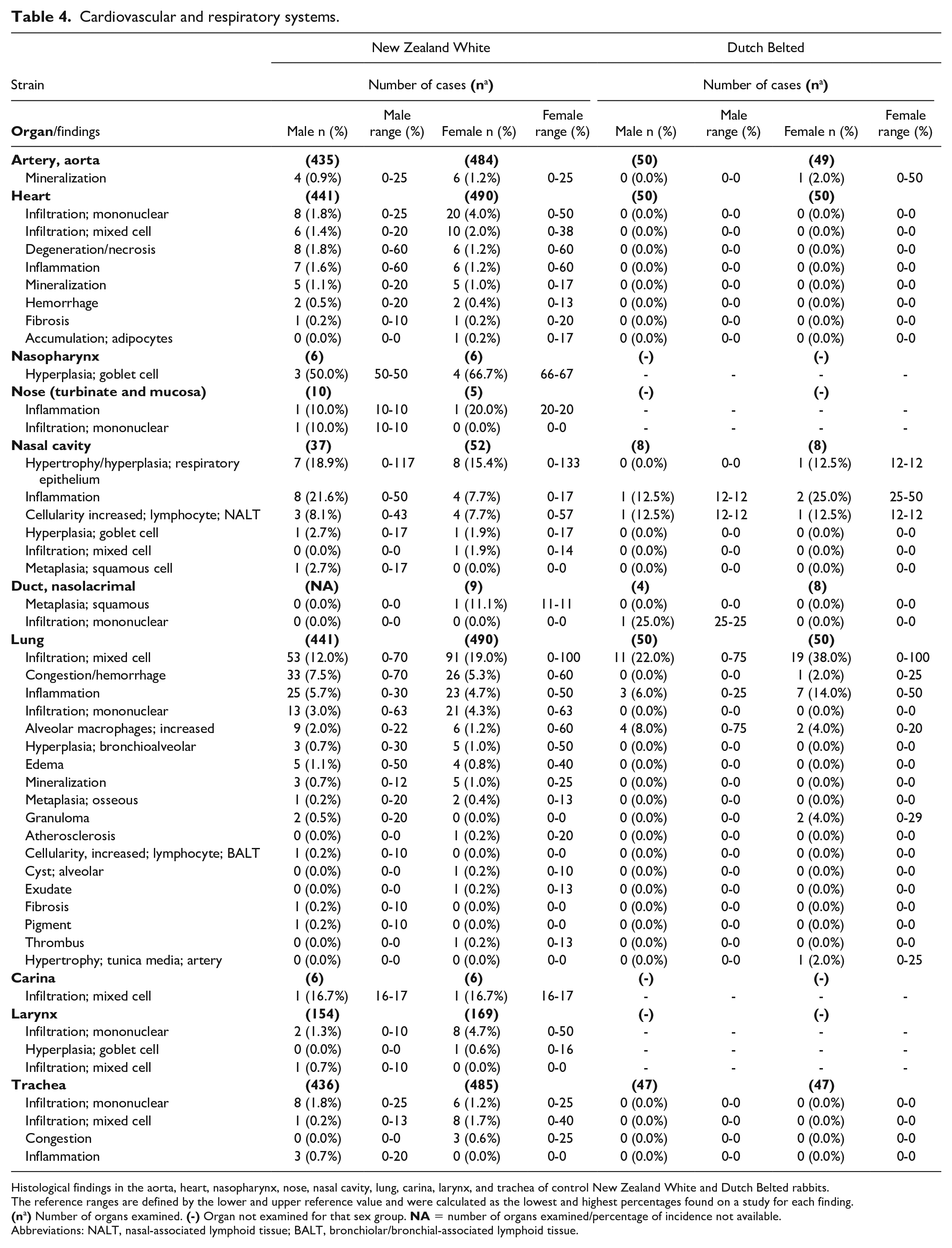
Histological findings in the aorta, heart, nasopharynx, nose, nasal cavity, lung, carina, larynx, and trachea of control New Zealand White and Dutch Belted rabbits.
The reference ranges are defined by the lower and upper reference value and were calculated as the lowest and highest percentages found on a study for each finding.
Abbreviations: NALT, nasal-associated lymphoid tissue; BALT, bronchiolar/bronchial-associated lymphoid tissue.
Upper digestive tract, gastrointestinal, exocrine pancreas, and hepatobiliary systems.
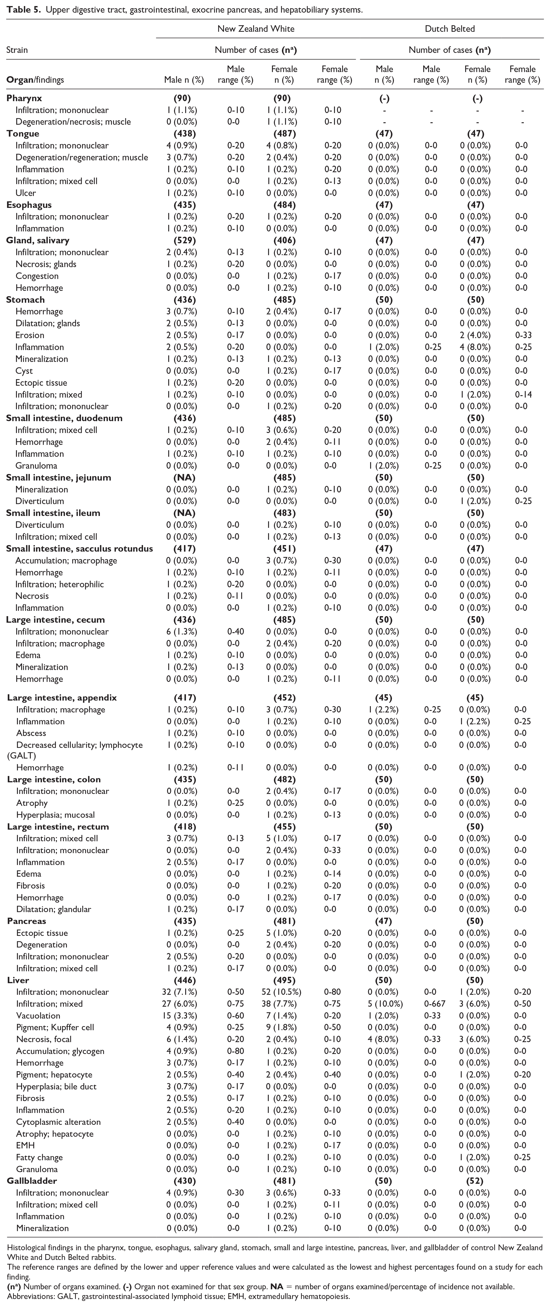
Histological findings in the pharynx, tongue, esophagus, salivary gland, stomach, small and large intestine, pancreas, liver, and gallbladder of control New Zealand White and Dutch Belted rabbits.
The reference ranges are defined by the lower and upper reference values and were calculated as the lowest and highest percentages found on a study for each finding.
Abbreviations: GALT, gastrointestinal-associated lymphoid tissue; EMH, extramedullary hematopoiesis.
Urogenital systems.
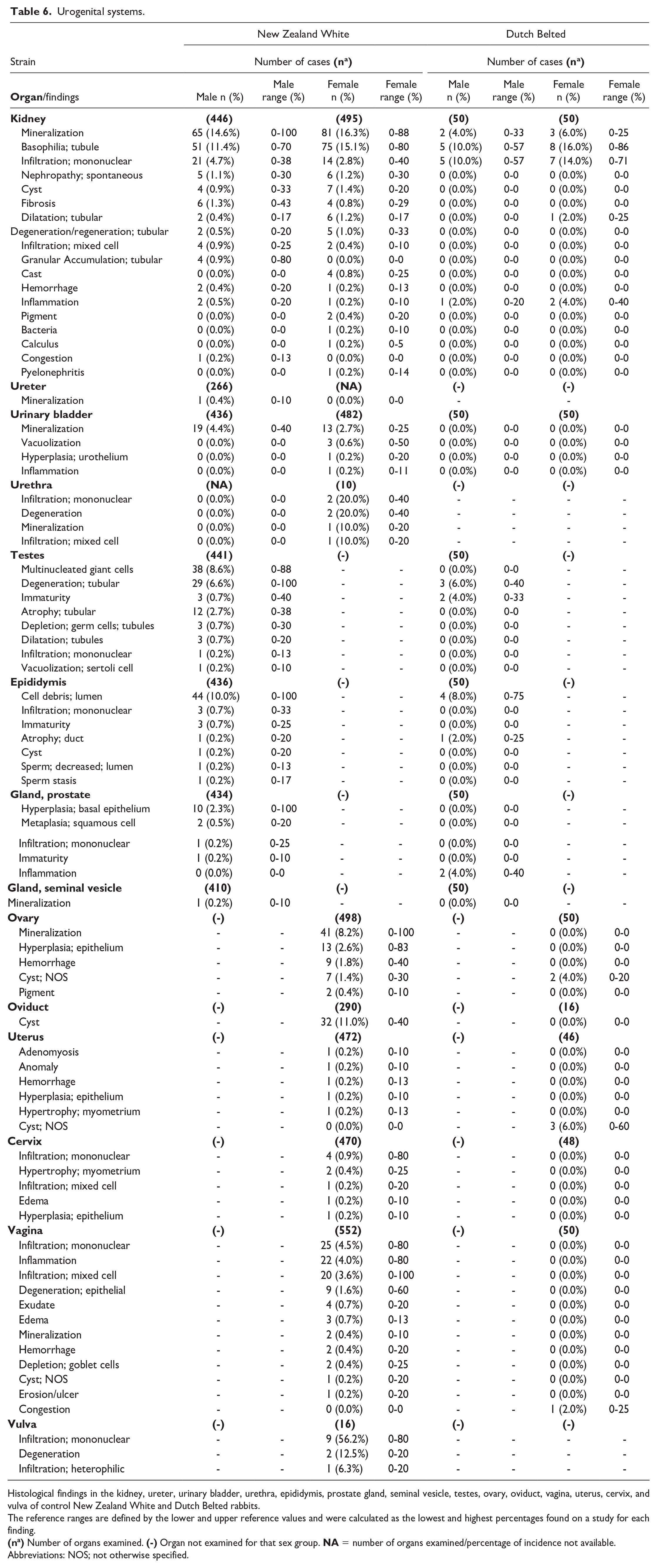
Histological findings in the kidney, ureter, urinary bladder, urethra, epididymis, prostate gland, seminal vesicle, testes, ovary, oviduct, vagina, uterus, cervix, and vulva of control New Zealand White and Dutch Belted rabbits.
The reference ranges are defined by the lower and upper reference values and were calculated as the lowest and highest percentages found on a study for each finding.
Abbreviations: NOS; not otherwise specified.
Endocrine system.
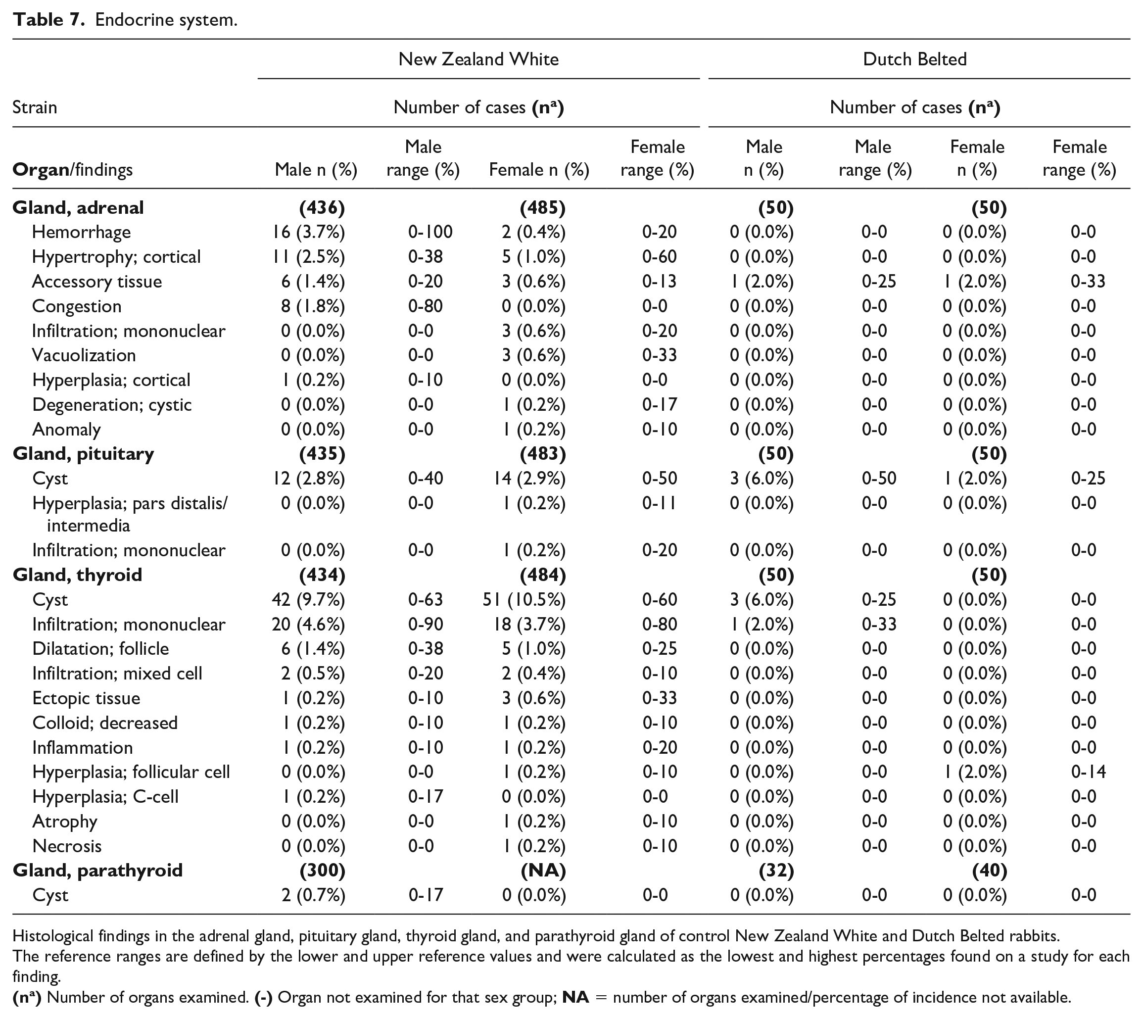
Histological findings in the adrenal gland, pituitary gland, thyroid gland, and parathyroid gland of control New Zealand White and Dutch Belted rabbits.
The reference ranges are defined by the lower and upper reference values and were calculated as the lowest and highest percentages found on a study for each finding.
Hematopoietic system.
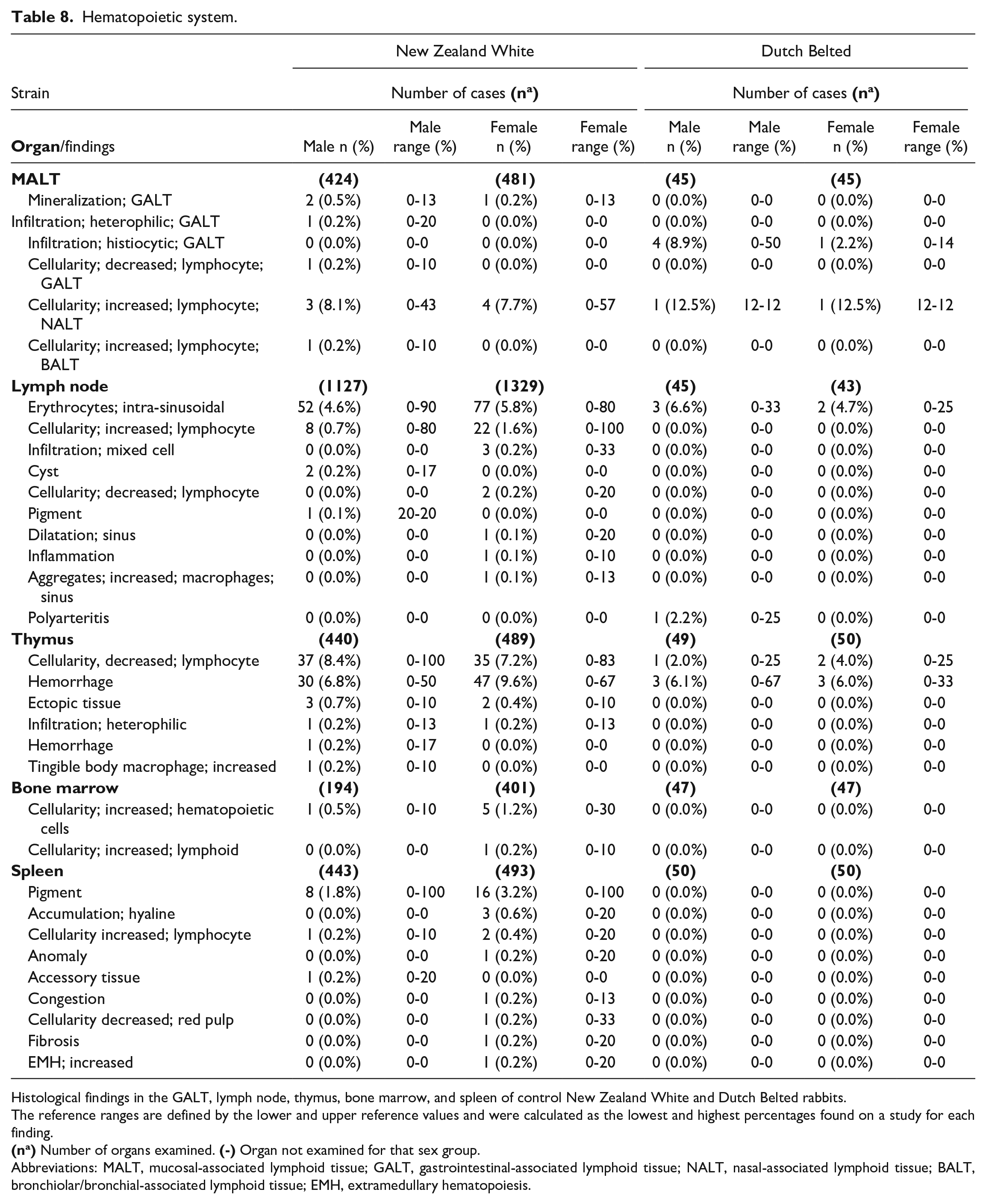
Histological findings in the GALT, lymph node, thymus, bone marrow, and spleen of control New Zealand White and Dutch Belted rabbits.
The reference ranges are defined by the lower and upper reference values and were calculated as the lowest and highest percentages found on a study for each finding.
Abbreviations: MALT, mucosal-associated lymphoid tissue; GALT, gastrointestinal-associated lymphoid tissue; NALT, nasal-associated lymphoid tissue; BALT, bronchiolar/bronchial-associated lymphoid tissue; EMH, extramedullary hematopoiesis.
Integumentary system and mammary gland.
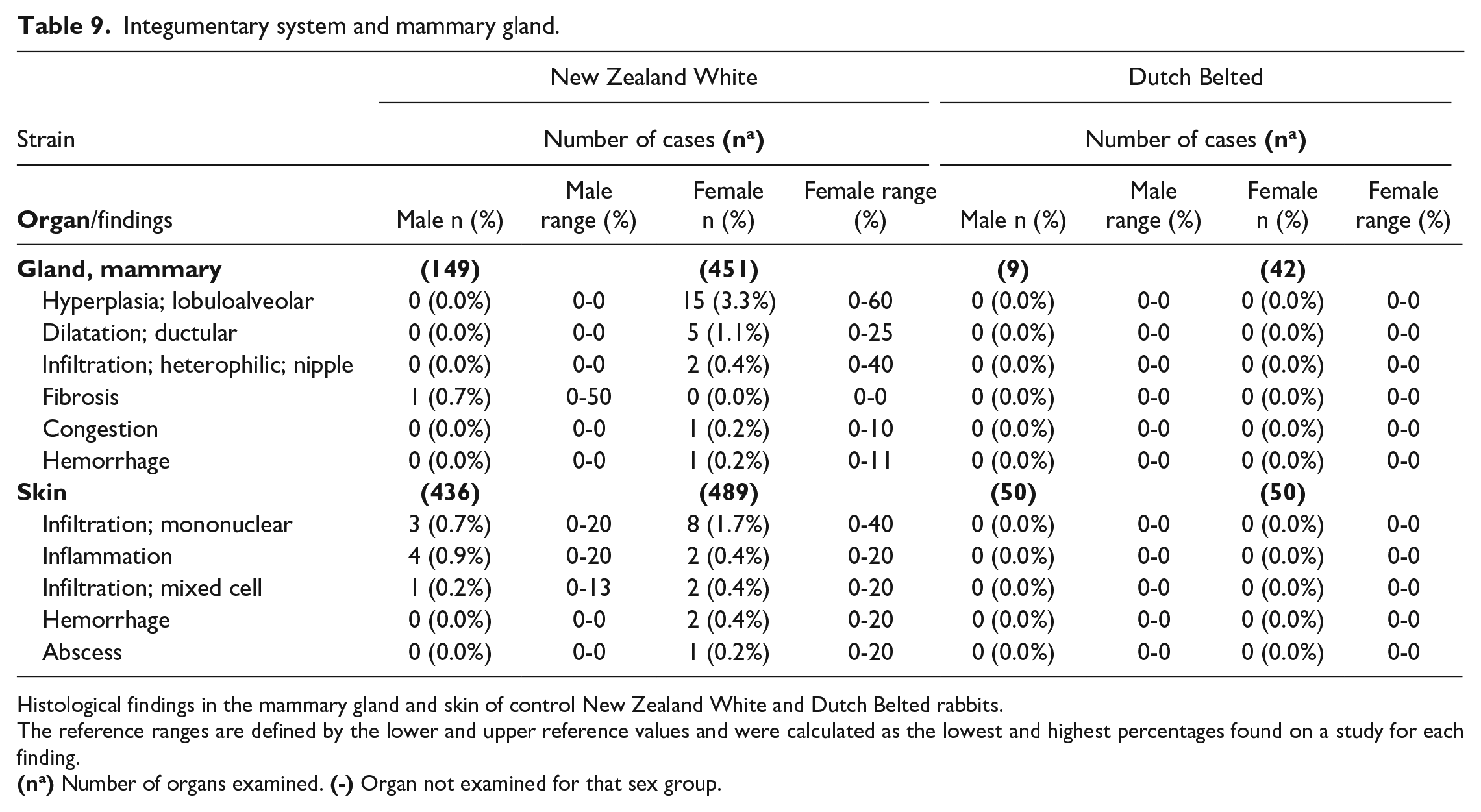
Histological findings in the mammary gland and skin of control New Zealand White and Dutch Belted rabbits.
The reference ranges are defined by the lower and upper reference values and were calculated as the lowest and highest percentages found on a study for each finding.
Musculoskeletal system.
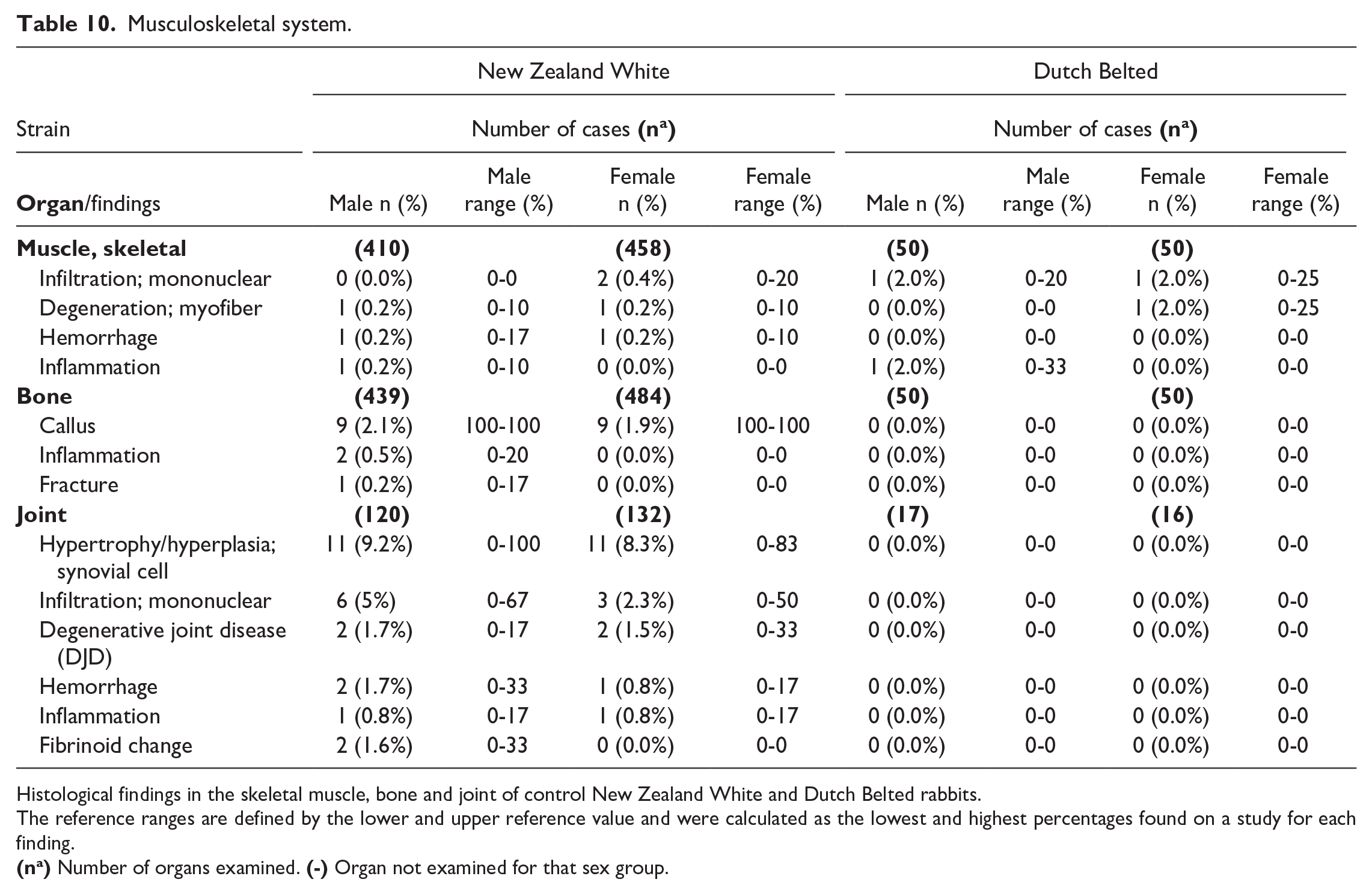
Histological findings in the skeletal muscle, bone and joint of control New Zealand White and Dutch Belted rabbits.
The reference ranges are defined by the lower and upper reference value and were calculated as the lowest and highest percentages found on a study for each finding.
Ocular system. a
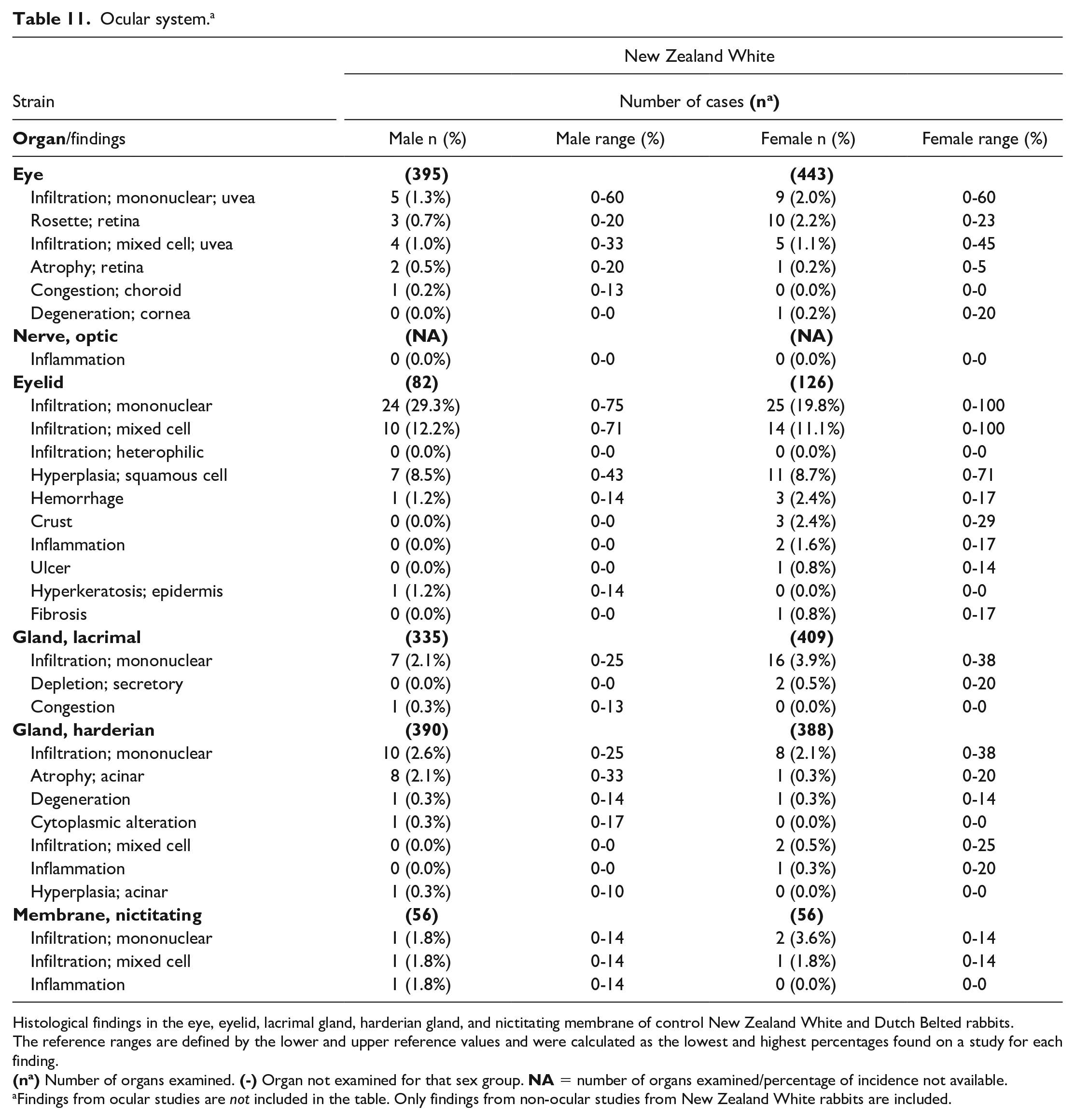
Histological findings in the eye, eyelid, lacrimal gland, harderian gland, and nictitating membrane of control New Zealand White and Dutch Belted rabbits.
The reference ranges are defined by the lower and upper reference values and were calculated as the lowest and highest percentages found on a study for each finding.
Findings from ocular studies are not included in the table. Only findings from non-ocular studies from New Zealand White rabbits are included.
Nervous system.
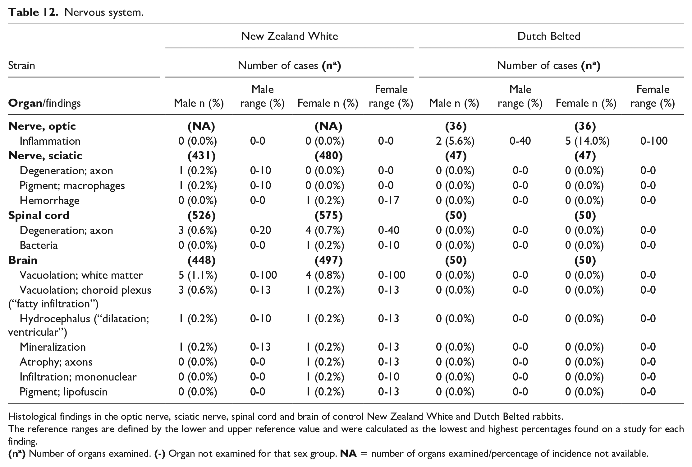
Histological findings in the optic nerve, sciatic nerve, spinal cord and brain of control New Zealand White and Dutch Belted rabbits.
The reference ranges are defined by the lower and upper reference value and were calculated as the lowest and highest percentages found on a study for each finding.
Findings Per Organ Systems
Respiratory and cardiovascular system
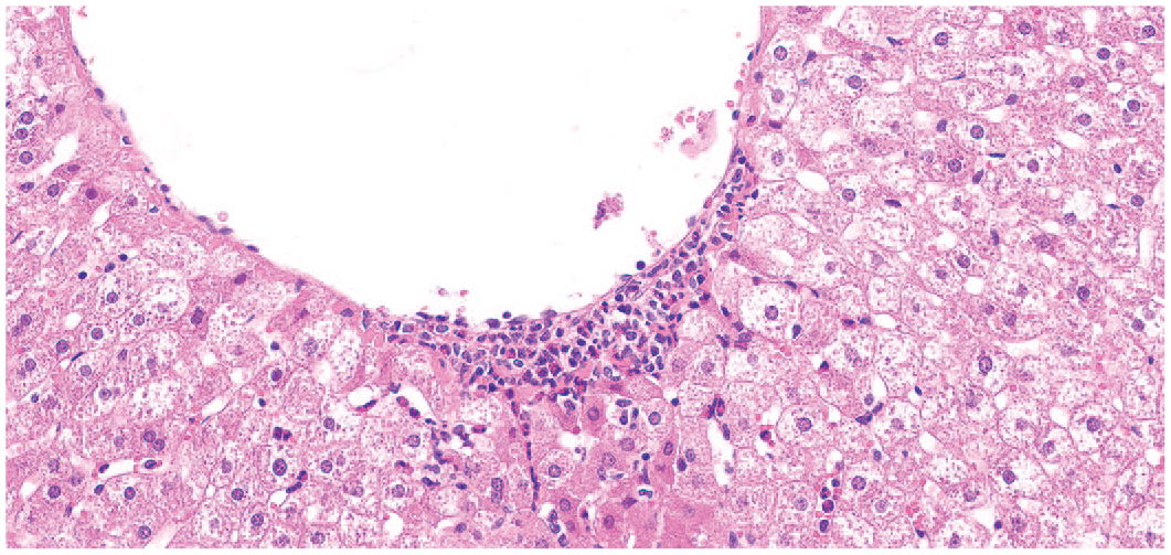
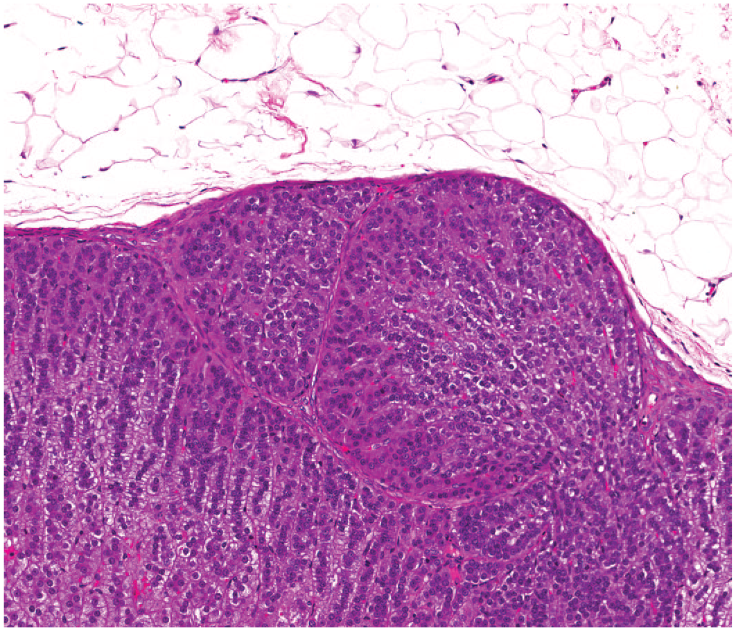
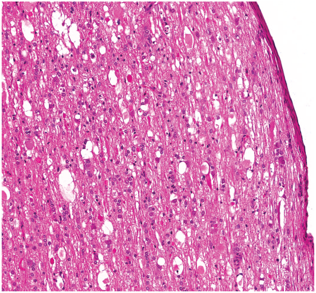
The lung was the most affected organ, and there was at least one lung finding in every study evaluated. The most common findings in the lung and cardiovascular system are documented in Table 4. In both strains, mixed cell infiltration in the alveoli was the most common finding (Figure 1) and mostly identified in females in both NZW (19% females/12% males) and DB (38% females/22% males) rabbits. In NZW rabbits, pulmonary agonal congestion/hemorrhage (7.5% females/5.3% males), and alveolar inflammation (either mixed or mononuclear cell) (5.7% males/4.7% females) were the second most common findings in the lung with similar incidences in both males and females. In DB rabbits, the second most common finding was inflammation commonly seen in females (14% females; 6.0% males). Mononuclear inflammatory infiltrates were the most common findings in the larynx (4.7% females/1.3% males) and trachea (1.8% males/1.2% females) of NZW rabbits and with no striking sex predilection. Interestingly, no histological changes were seen in the trachea of control DB rabbits. In the nasal cavity, the most common finding in NZW rabbit was hyperplasia/hypertrophy of the respiratory epithelium (18.9% males/15.4% females), while no relevant findings were present in DB rabbits. The nasopharynx, nose, carina, and larynx were only examined in NZW rabbits, and only a limited number were sampled.
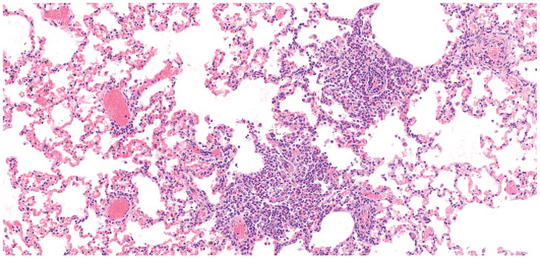
Lung. Infiltration; mixed. Alveoli are multifocally expanded by a mixed population of heterophils, lymphocytes and alveolar macrophages. NZW rabbit. Female. Hematoxylin and eosin.
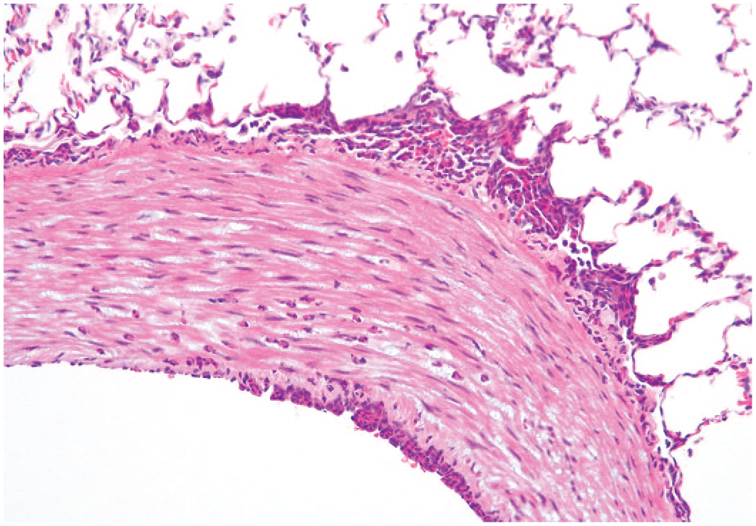
Compared with the lung, incidences of spontaneous findings in the cardiovascular system of NZW rabbits were less numerous. The most common findings were recorded in the heart, with the most common histological change being mononuclear cell infiltration (Figure 2), more prevalent in NZW females (4.0% females/1.8% males). Myocarditis (inflammation) was only identified in 1.6% of NZW male and 1.2% of NZW female hearts examined. Other less common changes in the heart of NZW rabbits were degeneration/necrosis and myocardial mineralization (1.8% males/1.2% females), which showed no sex predilection. Interestingly, no histological changes were present in the 100 examined hearts of DB control male and female rabbits. The only histological change identified in the aorta of NZW rabbits was mineralization, most commonly seen in females (1.2% females/0.9% males). Only one case of tunica media mineralization was identified in a female DB rabbit (Figure 3).
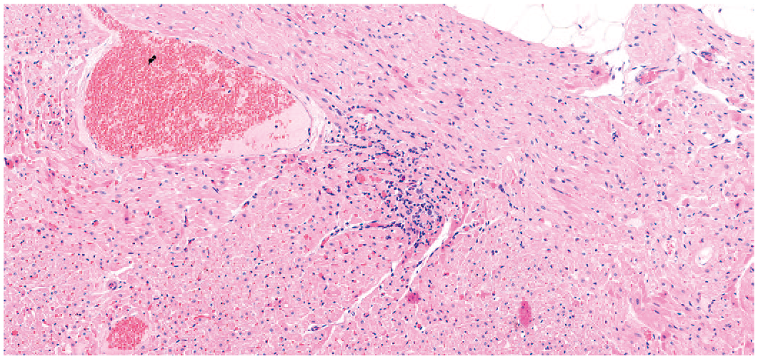
Heart. Infiltration; mononuclear cell. The interstitium is minimally expanded by multifocal mononuclear cells (lymphocytes). NZW rabbit. Male. Hematoxylin and eosin.
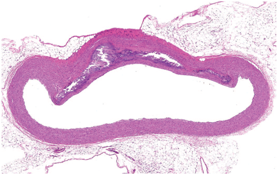
Aorta. Mineralization. The tunica media is expanded by moderate amount of amorphous basophilic extracellular material. DB rabbit. Sex not recorded when image taken. Hematoxylin and eosin.
Upper digestive tract, gastrointestinal, exocrine pancreas, and hepatobiliary system
Common lesions encountered in the upper digestive tract, salivary gland, gastrointestinal tract, pancreas, liver, and gallbladder are shown in Table 5. Compared to other organ systems, the gastrointestinal tract was the one with the least common lesions identified. Mononuclear cell infiltrations in the pharynx, tongue, cecum, and rectum were among the most common lesions present in NZW rabbits, with no clear sex predilection. In the stomach of NZW rabbits, submucosal hemorrhages were the most common finding followed by dilatation of the gastric glands, inflammatory foci and mucosal erosion, although a clear sex predilection was not identified. In the DB strain, gastric inflammation (gastritis) was the most common lesion encountered in the gastrointestinal tract, mostly prevalent in females (8.0% females/2.0% males), followed by mucosal erosions and mixed cell mucosal infiltration. In the DB strain, the pharynx was not examined. In the remaining gastrointestinal tract of the DB strain, the only lesions identified were in the duodenum (one male with a submucosal granuloma), jejunum (one female with diverticulum), and appendix (one male with macrophage accumulation and one female with inflammatory foci in the mucosal lamina propria). No infectious etiologies were identified histologically in any part of the gastrointestinal system and upper digestive tract. The exocrine pancreas was examined in both strains but only NZW rabbits presented histological changes with the most common lesion being ectopic splenic tissue (Figure 4), most common in females (1.0% females/0.2% males) followed by pancreatic acinar degeneration and inflammatory infiltrates expanding the periacinar and periductular interstitium.
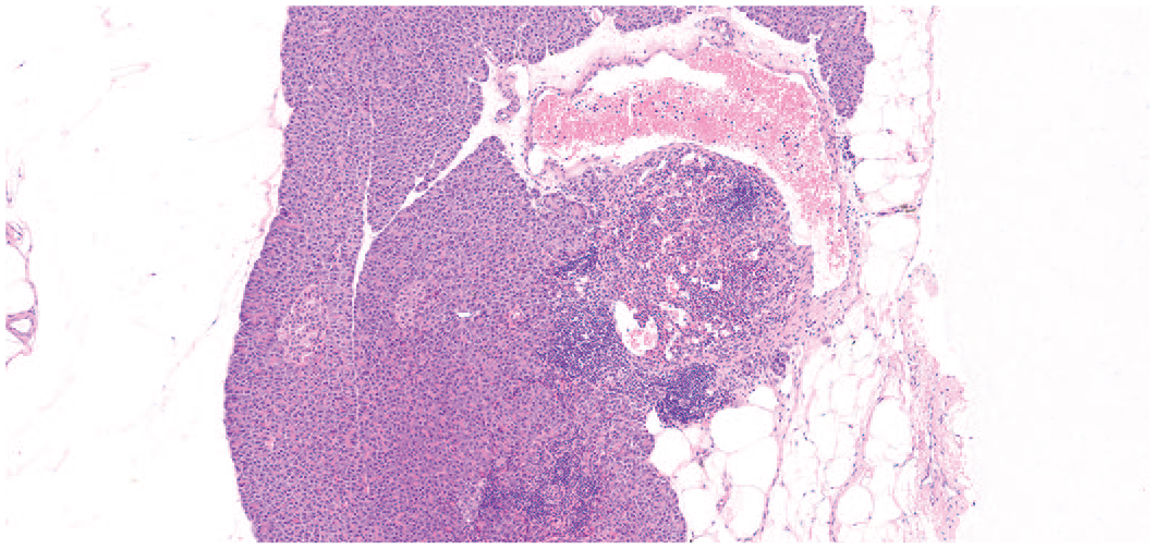
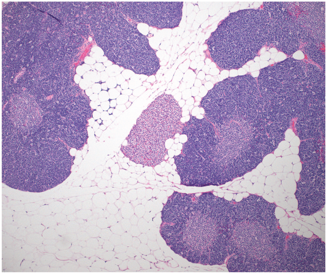
Pancreas. Ectopic tissue; spleen. Pancreatic acini are surrounded by a well-demarcated nodule of normal splenic tissue characterized by both white and red pulp. There is an accompanying mononuclear inflammatory cell (lymphocytic) infiltrate surrounding the accessory spleen. NZW rabbit. Female. Hematoxylin and eosin.
In the liver, foci of inflammatory mononuclear cell infiltrates (mostly lymphoplasmacytic) and mixed cells (viable heterophils and lymphocytes) within portal to periportal areas and centrilobular areas (Figure 5) were the most common findings recorded in NZW rabbits. Both infiltrates in NZW rabbits were slightly more common in females (mononuclear cell infiltrates: 10.5% females/7.1% males, and mixed cell infiltrates: 7.7% females/6.0% males). Inflammation of the liver was less prevalent in NZW rabbits and with similar prevalence in females and males. In the DB strain, mixed cell infiltrations were the most common finding and slightly more prevalent in males (10.0%) than females (6.0%). Foci of hepatocellular necrosis (Figure 6) were seen in both strains. Other less frequent changes seen in NZW rabbits were pigment accumulation in macrophages and hepatocytes, glycogen accumulation, bile duct hyperplasia, and portal to periportal accumulation of collagen with similar incidence in both males and females. In the DB strain, other less frequent hepatic changes included vacuolation (seen only in a male), hepatocellular pigment, and fatty change (seen only in a female). The gallbladder was examined in both strains, but only NZW rabbits presented histological changes with the most frequent being mononuclear infiltration (0.9% males/0.6% females) expanding the mucosa.
Liver. Infiltration; mixed cell, centrilobular. A focal infiltration of mixed inflammatory cells (viable heterophils and lymphocytes) surrounds the central vein. Hepatocytes show minimal cytoplasmic rarefaction (glycogen accumulation), but otherwise they are unremarkable. NZW rabbit. Female. Hematoxylin and eosin.
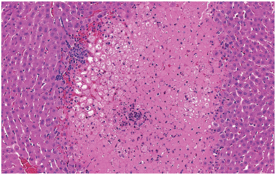
Liver. Necrosis. There is a focal, well-demarcated, hepatic area characterized by loss of differential staining and retention of architecture and hepatocytes show increased cytoplasmic eosinophilia, nuclear fading, and karyolysis. Mixed cell inflammatory foci are also present. DB rabbit. Sex not recorded when image taken. Hematoxylin and eosin.
Urogenital system
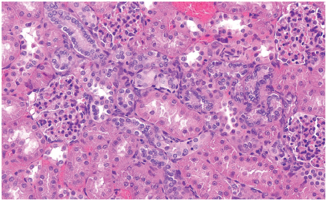
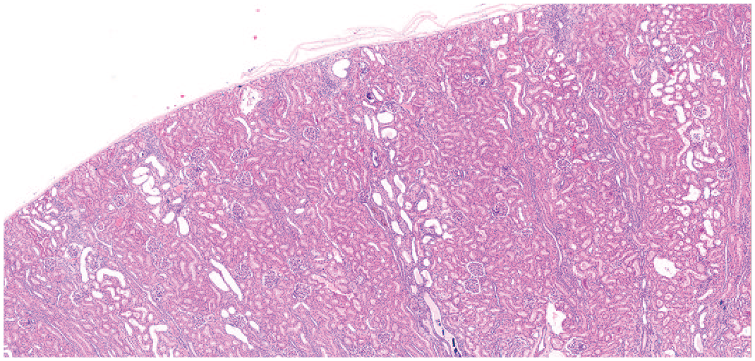
Common lesions encountered in the urogenital system are outlined in Table 6. The most common findings in NZW and DB rabbits were identified in the kidney. Tubular renal mineralization of the proximal and medullary tubules (Figure 7) and tubular basophilia (Figure 8) were the most recorded lesions in NZW rabbits followed by cortical interstitial mononuclear cell infiltration, most commonly seen in males (4.7%). In females, tubular mineralization (16.3%) and basophilia (15.1%) were more prevalent than in males (respectively 14.6% and 11.4%).
Kidney. Mineralization; proximal tubule. Focally, the proximal tubule is mildly dilated, and the lumen expanded by extracellular amorphous basophilic material (mineralization) which compresses the mildly vacuolated tubular epithelium. NZW rabbit. Female. Hematoxylin and eosin.
Kidney. Basophilia; proximal tubules. The proximal tubules multifocally show increased tinctorial difference (basophilia) and nuclear crowding compared with the surrounding unremarkable proximal tubules. NZW rabbit. Male. Hematoxylin and eosin.
Other less common renal changes present in the NZW rabbits were nephropathy, renal cysts, interstitial renal fibrosis, proximal tubular dilatation, foci of tubular degeneration and regeneration, mixed cell infiltration, tubular casts (only seen in females), and pigmented tubules. Nephropathy was seen in both males and females with similar prevalence. The histological findings of “chronic nephropathy” (Figure 9) were generally recorded separately (basophilia of the tubules, dilated/cystic tubules, pigmented tubules, interstitial inflammatory cell infiltrate), and the term nephropathy was only used when at least three of the aforementioned components were present. 2 Tubular basophilia and interstitial mononuclear cell infiltration were the most identified changes in the DB strain and were more common in females (respectively 16.0% and 14.0%) than in males (both 10.0%). Mineralization of the tubules was the second most common change in DB and with similar incidence between males and females. Apart from renal inflammation (identified in a male) and tubular dilatation (identified in a female), there were no other histological changes identified in the kidney of the DB strain. In the ureter and urinary bladder, the lesions most identified in NZW rabbits were foci of mineralization (commonly located within the mucosa), whereas foci of mononuclear inflammatory infiltrates predominate in the urethra of the NZW strain. Mineralization of the urinary bladder was slightly more common in males (4.4%) than females (2.7%). The ureter and urethra were not examined in the DB strain, and no histological changes were seen in the urinary bladder of DB rabbits.
Kidney. Nephropathy. Multifocally, the renal proximal tubules are mildly dilated (dilated/cystic tubules) and are surrounded by basophilic tubules, occasionally showing intra-cytoplasmic brown to yellow pigment (at the center of the image) and admixed with multifocal interstitial mononuclear inflammatory cell infiltrate. A few foci of tubular mineralization are also present. NZW rabbit. Female. Hematoxylin and eosin.
Within the reproductive systems, the testes and ovaries were the organs with most recorded changes.
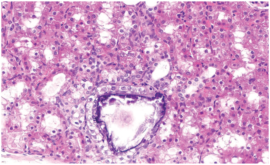
In the testes, the most commonly identified lesion in NZW rabbits was accumulation of multinucleated giant cell (8.6%) within the lumen of the seminiferous tubules and tubular degeneration (6.6%). A few testes (2.7%) presented a proportion of tubules that had lost all germ cells (tubular atrophy) or/and showing tubular dilatation (Figure 10). Degeneration of the tubules (6.0%) was the only histological change identified in the DB strain. In the females, ovarian mineralization (Figure 11) was the most frequent histological lesion identified in NZW rabbits (8.2%) followed by ovarian epithelial capsular hyperplasia (2.6%), hemorrhages (1.8% each), ovarian cysts (1.4%) and pigment deposition (0.4%). Ovarian cysts (4.0%) were the only recorded lesion in the DB strain. Cysts were common in the oviduct (11%) of NZW rabbits but were not identified in the DB strain. Changes in the uterus in NZW rabbits were minimal but no histological findings were present in the DB strain. In males, the most common epididymal change in both DB and NZW rabbits was the presence of tubular luminal debris (respectively 8.0% and 10%). Hyperplasia of the prostatic glandular acini (2.3%) was the most common recorded prostatic change in the NZW rabbits, while the only histological lesion in the prostate of DB was inflammation (prostatitis) (4.0%). Spontaneous focal keratinized squamous metaplasia of the prostate and proprostate epithelium (Figure 12) was identified only in NZW rabbits (0.5%). Seminal vesicles were examined in both strains but only NZW rabbits presented histological changes consistent with mineralization (0.2%). The majority of rabbits in this study were sexually mature with only 3 out of the 441 NZW testes (0.7%) and 2 out of 50 DB testes (4.0%) recorded as immature histologically.
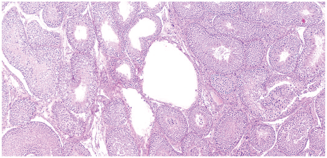
Testes. Tubular dilatation. Focally, seminiferous tubules show increased luminal diameter (dilatation) and lined by sertoli cells and sparse germ cells. NZW rabbit. Male. Hematoxylin and eosin.
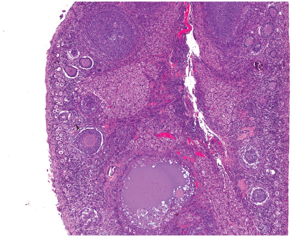
Ovary. Mineralization. There are multiple foci of basophilic concretions (mineralization) surrounded by primary and primordial follicles. NZW rabbit. Female. Hematoxylin and eosin.
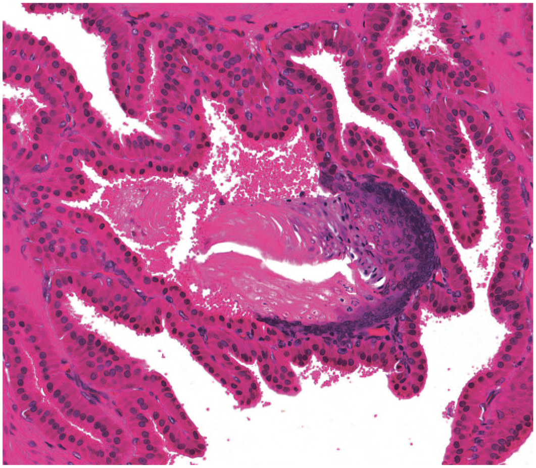
Prostate. Squamous metaplasia. There is gradual squamous differentiation of the prostatic epithelium, showing transformation from a basilar nucleated epithelial layer to an anucleate squamous layer toward the lumen. There is evidence of superficial keratinization and sloughing of cornified debris into the gland alveolar lumen; the squamous metaplastic focus is located within the gland epithelium and minimally extended into the underlying lamina propria. NZW rabbit. Male. Hematoxylin and eosin.
Endocrine system
Common lesions encountered in the endocrine system are shown in Table 7. Common lesions identified in the adrenal glands of NZW rabbits were hemorrhages and diffuse adrenal cortical hypertrophy, both more common in males (respectively 3.7% and 2.5%) than females (respectively 0.4% and 1.0%). Accessory adrenal gland (ectopic) tissue (Figure 13) was the only lesion recorded in the adrenal gland of a male and a female DB rabbit. Cysts in the thyroid gland and pituitary gland were the most common histological changes recorded in both NZW and DB rabbits, and with similar prevalence in both males and females. Mononuclear cell infiltration was the second most common finding in the thyroid gland in NZW rabbits. Other findings in the thyroid gland of NZW rabbits were follicular dilatation, thymic ectopic tissue, and mixed cell infiltration. In the thyroid gland of the DB strain, the only findings recorded in addition to cysts were mononuclear infiltration (in a male) and follicular hyperplasia (in a female). Parathyroid cysts were the only lesion present in the parathyroid glands and only detected in NZW rabbits.
Adrenal gland. Accessory tissue. There is a focus of unremarkable adrenal cortical tissue located external to the adrenal gland section and in contact with the capsule at one pole of the adrenal gland. The accessory tissue is composed of unremarkable cortical zona glomerulosa and zona fasciculata. NZW rabbit. Female. Hematoxylin and eosin.
Hematopoietic system
Common lesions of the hematopoietic system are listed in Table 8. Foci of mineralization were the most common lesion recorded in the GALT (Gastrointestinal-Associated Lymphoid tissue) of NZW rabbits followed by minimal heterophilic infiltration expanding the sinuses. Histiocytic cell infiltration was the only finding identified in the GALT of DB males (8.9% males/2.2% females), and the same change was not present in the NZW rabbits. Several lymph nodes were examined in NZW rabbits, and the findings were merged as they were similar within the individual lymph nodes. The lymph nodes examined in NZW rabbits were the axillary, iliac, inguinal, lumbar, mandibular, mesenteric, parotid, renal, and sacral lymph node. In DB rabbits, only the mandibular lymph node was examined. Sinus erythrocytosis (eg, intra-sinusoidal erythrocytes) was the most common finding in the lymph nodes of both strains, and most prevalent in females NZW rabbits (5.8% females/4.6% males) and in male DB rabbits (6.6% males/4.7% females). The second most common change in the lymph nodes of NZW rabbits was increased lymphoid cellularity of the germinal centers and paracortex (suggesting lymphoid follicular hyperplasia), most common in females (1.6% females/0.7% males). In addition, in the lymph node of one male DB rabbit, there was a case of polyarteritis (Figure 14). Polyarteritis was not seen in NZW rabbits.
Artery, Lung. Inflammation. Multifocally, there is accumulation of viable heterophils which infiltrate the tunica intima and replace the degenerate endothelium and reach the tunica media and adventitia. The smooth muscle myofibers of the tunica media are multifocally separated by increased clear spaces (edema) and previously described heterophils. NZW rabbit. Male. Hematoxylin and eosin.
The most common histological findings in the thymus of NZW rabbits were decreased lymphoid cellularity, showing similar prevalence in male and females, and medullary hemorrhages. Both changes were the only ones identified in the thymus of the DB strain. Other common thymic changes were ectopic tissue (Figure 15) and heterophilic infiltration. Figure 15 is from a control rabbit from a general toxicology study which was not included in the Table 8. In chronic studies, the terminology “decreased lymphocyte cellularity” occasionally overlapped with “atrophy” and “involution” and distinguishing them was challenging. Bone marrow was evaluated in both strains, but only NZW rabbits showed findings, with the most common change being increased hematopoietic cells either myeloid or erythroid. In NZW rabbits, the most common finding in the spleen was the presence of abundant intra-cellular (intra-histiocytic) and extracellular pigment (most likely consistent with hemosiderin), which was more commonly identified in females (3.2% females/1.8% males) and was associated with a few cases of hyaline accumulation, and increased cellularity of the white pulp (suggesting reactivity). The spleen of DB rabbits showed no findings.
Thymus. Ectopic tissue; parathyroid. Focally, within the adipose tissue surrounding the thymus, there is a focal, well-demarcated focus of unremarkable parathyroid tissue (ectopia). DB rabbit. Sex not recorded when image taken. Hematoxylin and eosin.
Integumentary system and Mammary gland
Common lesions encountered in the integumentary system and mammary gland are shown in Table 9. Mammary gland tissue and skin tissue were examined in both strains but only the NZW rabbits showed histological findings. Mammary gland hyperplasia (with increased secretory activity) was only identified in females (3.3% females) and was the most common histological finding in the female mammary gland, followed by ductular glandular dilatation (1.1%) and foci of inflammatory heterophilic infiltration (0.4%). The only histological change identified in male mammary glands (but not in the female) was fibroplasia, as seen in one animal.
The routine skin section examined was associated with mammary gland tissue. The most common histological finding in NZW rabbits was the presence of foci of mononuclear inflammatory infiltrates, more commonly present in females (1.7% females/0.7% males), followed by inflammation and mixed inflammatory infiltrates located within the dermis. There were other histological changes only in the NZW strain (in studies with subcutaneous and dermal injections—not listed in Table 9) in the skin of the vehicle administration sites: perivascular hemorrhage, arterial and/or venous endothelial degeneration, crusts, epidermal erosions, accumulation of pigmented (hemosiderin-laden) macrophages, mixed cell inflammatory infiltrates, and myofiber degeneration, which were all considered to be procedural-related rather than a reaction to the vehicle.
Musculoskeletal system
Common lesions encountered in the musculoskeletal system are shown in Table 10. Mononuclear infiltration was the most common finding in the skeletal muscles of NZW rabbits and only identified in females (0.4%). The latter was also the most common finding identified in the DB strain in both males and females (2.0% both). Degeneration of myofibers (Figure 16) was the second most common histological finding in NZW rabbits and showed the same prevalence in both males and females (0.2% both). Femoral and ulnar bones were examined in NZW rabbits. In NZW rabbits, callus formation in the ulnar bone was the most common change. This was followed by mixed inflammation (0.5%) of the femur (present only in males) and a male presenting a femoral fracture (0.2%), secondary to trauma. The femorotibial joint of NZW rabbits was characterized by similar prevalence in both males and females of synovial hyperplasia and hypertrophy followed by mononuclear cell synovial inflammatory infiltrates and degenerative joint disease. Only the femoral bone was examined in DB rabbits, and no findings were identified in the bone and femorotibial joint.
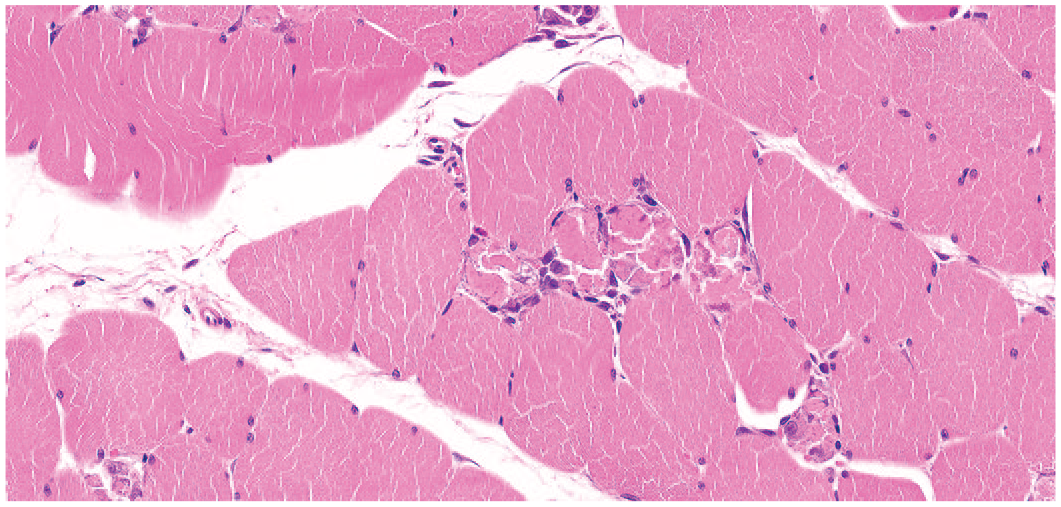
Skeletal muscle. Necrosis and degeneration, myofiber. Multifocally, myofibers are fragmented and show loss of cellular details, cytoplasmic hypereosinophilia and karyolysis. The affected myofibers are surrounded by minimal mononuclear cell infiltration. NZW rabbit. Male. Hematoxylin and eosin.
Ocular system
Common lesions encountered in the ocular system (eyes and annexa) are shown in Table 11. Findings from ocular studies are not included in the table. Only findings from non-ocular studies from NZW rabbits are included. A majority of studies from DB rabbits are ocular studies, and therefore, were not included in the table. The eye was the organ with the most identified histological changes with the most common being mononuclear infiltration in the choroid, cornea, and limbus (2.0% females/1.3% males) and retinal rosettes (2.2% females/0.7% males), both more common in females than males. A less common change was retinal atrophy (0.5% males/0.2% females) (Figure 17). In the eyelid, mononuclear and mixed inflammatory infiltrations were common findings in NZW strain and with no sex predilection. Mononuclear infiltration was the most recorded finding in NZW rabbits in the lacrimal gland (3.9% females/2.1% males) and harderian gland (2.6% males/2.1% females). Harderian glands in NZW rabbits were characterized by the highest prevalence of mononuclear inflammatory infiltrate followed by acinar atrophy and degeneration, cytoplasmic alteration, and mixed cell infiltration. There were other histological changes from ocular studies in both NZW and DB strains identified in the eye (not listed in Table 11) consistent with needle tract lesions, lens degeneration, and corneal erosion which were considered procedural-related. In ocular studies, findings identified only in the DB strain were retinal detachment, iris adhesions, discontinuation of the Descemet membrane, fibroplasia of the limbus, retinal degeneration, displacement of the photoreceptor nuclei, and corneal hypertrophy which were also considered procedural-related.
Eye, retina. Atrophy. Diffusely the retina is half the normal thickness with loss of the outer plexiform, outer nuclear, and photoreceptor layers. NZW rabbit. Male. Hematoxylin and eosin.
Nervous system and ear
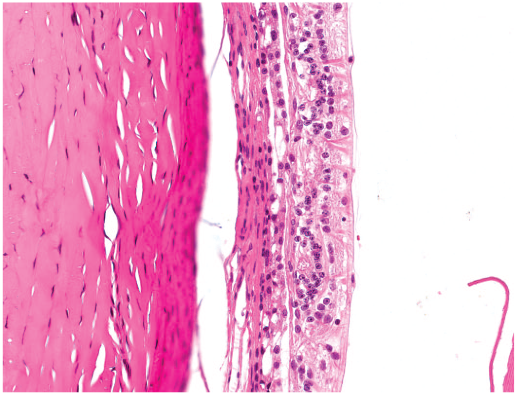
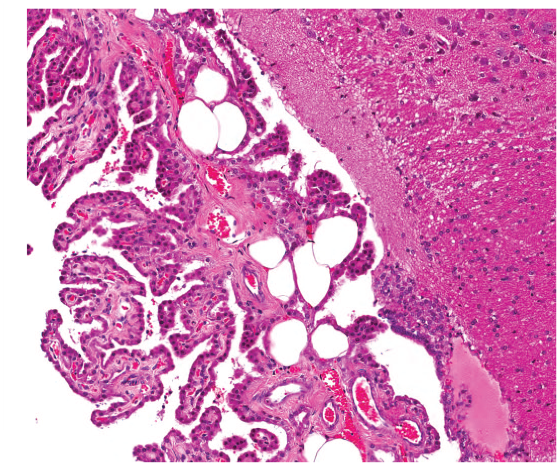
Common lesions encountered in the nervous system and ear are shown in Table 12. The most common histological finding in the brain of NZW rabbit was vacuolation of the choroid plexus followed by foci of fatty infiltration of the choroid plexus (Figure 18), ventricular dilatation (Figures 19 and 20), and foci of mineralization, all showing no clear sex predilection. The most common change in the spinal cord of NZW rabbits was axonal (“Wallerian”) degeneration (Figure 21), whereas only minimal changes were seen in the sciatic nerve. No histological changes were identified in the brain, spinal cord, and sciatic nerve of DB rabbits. The optic nerve was the only nervous tissue in the DB strain with histological findings consisting of inflammation (mostly mononuclear), with slightly more prevalence in females (14.0% females/5.6% males) No histological changes were identified in the optic nerve of NZW rabbits.
Brain, choroid plexus. Vacuolation (“Fatty infiltration”). Multifocally, in the connective tissue underlying the ependymal cells, there are round, discrete, and large vacuoles. NZW rabbit. Male. Hematoxylin and eosin.
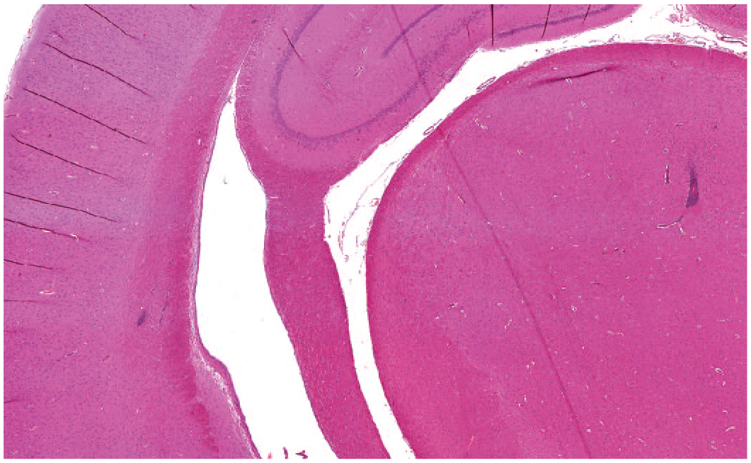
Brain. Hydrocephalus (“Dilatation, ventricles”); infiltration, mononuclear. Diffusely there is mild dilatation of the third ventricle at the level of the diencephalon. Multifocally, within the gray matter, small caliber vessels are surrounded by a mononuclear cell infiltration forming perivascular cuffs. NZW rabbit. Female. Hematoxylin and eosin.
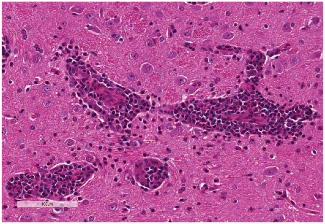
Brain. Infiltration, mononuclear; perivascular. Higher magnification of perivascular mononuclear cuff as described in Figure 19. NZW rabbit. Female. Hematoxylin and eosin.
Spinal cord. Degeneration, axonal. Myelin sheaths are dilated up to 2 times normal and contain glassy, eosinophilic, swollen axons (spheroids), or fragmented myelin debris (ellipsoids) admixed with single macrophages (gitter cells). NZW rabbit. Female. Hematoxylin and eosin.
Discussion
The purpose of this study was to report the spontaneous background lesions and terminal body and organ weights in laboratory NZW and DB rabbits. This report of the Charles River Laboratories data outlines the most common lesions and presents some of the interesting histological findings (Figures 1-20). Reports of spontaneous lesions in rabbits of this kind are either quoting outdated studies,2,7 occasional single tissue reports,8,12,19 or case reports from pet animals. To the authors’ knowledge, the most updated review of spontaneous lesions in rabbits considered only NZW strain, 5 and reports of spontaneous lesions in DB rabbits are lacking in the current literature. Data of organ weights and terminal body weights in laboratory rabbits are outdated in the current literature. In recent years, rabbits have been shown to have advantages as a non-rodent model for developmental and toxicity studies as they are considered the smallest and least expensive animal models in which most, if not all, of the reproductive and toxicologic endpoints can be measured and evaluated in order to translate them to larger animals and humans. 9 Recently, rabbits have been explored as a non-rodent species for general toxicology studies, particularly given supply issues of routine non-rodent species affecting study lead times. 20 Historically used in vaccine, cosmetic, and medical device testing, nowadays rabbits are currently implemented in the pharmaceutical industry for embryo-fetal development studies, ophthalmic therapeutics, some medical devices and implants, and vaccines. 20 The most common strain used in toxicity studies is the NZW. Based on a relatively recent study, 21 however, the DB rabbit is also considered to be a practical alternative to the NZW strain for developmental and general toxicity testing. The main advantages of using the DB rabbit are their size as the DB strain would require approximately 40% less drug than studies conducted with NZW rabbits. 21 In addition, because of their pigmentation, they offer the ability to test the potential effects of various compounds on melanocyte development and to evaluate drugs that can bind to melanin and pigmented retinal epithelium. 21 The DB rabbit might also represent a better choice than the NZW rabbit to study wound healing modulation following filtration surgery in the eyes as a model for glaucoma surgery. 17
This study presents terminal body and organ weight ranges and spontaneous lesions occurring in young laboratory NZW and DB rabbits from control groups in studies evaluated over the last ten years at Charles River Laboratories. The findings in this study were generally consistent with the incidence and occurrence of similar lesions described in the INHAND paper. 3 Overall, male and female NZW showed a greater mean terminal body weight than the DB strain, and this is consistent with the literature, 21 as DB rabbits tend to be smaller than NZW rabbits. Terminal body weight and organ weight data are very limited in the literature and often old publications; 4 however, no significant changes from this study were identified.
Histologically, the most recorded lesions in NZW and DB rabbits in this data set were inflammatory cell foci in the lung (mixed cell infiltration), most common in females. The results in NZW rabbits are consistent with the most recent review in this strain. 5 Inflammation was also commonly encountered in the lung in NZW rabbits and DB rabbits. Classification of inflammatory changes in the lung can be challenging and diagnostic terminology and criteria to differentiate “infiltration” from “inflammation” according to agreed guidelines 15 often drifts among pathologists. Generally, two types of inflammatory changes seemed to emerge in rabbits—chronic interstitial inflammation (expansion of the septa and alveoli of mostly mononuclear inflammatory cells accompanied by interstitial and septal fibrosis and/or pneumocyte type II hyperplasia) and bronchopneumonia (acute to subacute inflammation of both bronchi, bronchioles, and alveoli, including aspiration pneumonia). Bronchopneumonia types of inflammatory changes and focal granulomas were sometimes related to the housing of the NZW Rabbits, such as due to inhaled dust particles or hair shafts inadvertently injected when blood sampling 7 or to the type of study (thrombi/emboli in continuous intra-venous infusion studies). Increased alveolar macrophages were often seen in the lungs of both strains and showed no particular distribution pattern, peripherally placed just beneath the pleura, or at the bronchoalveolar junction, as described in the literature. 17 Increased diffusely distributed macrophages was also reported to occur in DB rabbits that relatively frequently show cardiomyopathy of unclear origin; 3 however, in this study, the heart of the DB strain showed no histological changes. Epithelial hyperplasia/hypertrophy of the nasal cavity and nasal-associated lymphoid tissue (NALT) hyperplasia were also common changes present in the respiratory tract of this study. Mononuclear inflammation in the nasal mucosa (rhinitis) and extending into the nasal cavity is often described associated with hyperplastic and hypertrophic changes of the nasal epithelium and with secondary reactive NALT hyperplasia. This is a common spontaneous condition in rabbits as they are animals with obligate nasal respiration, and this predisposes to local inflammation. 1
The kidney was the second organ with the most findings identified in both strains. Foci of mineralization were commonly seen in the urinary tract of NZW rabbits, particular the urinary bladder and renal tubules, consistent with a recent review of NZW rabbits. 5 Mineralization of the renal tubules in NZW rabbits was the most common degenerative change in this strain, and more frequent in females. This is a common spontaneous change in rabbits. 3 Although calcium absorption and metabolism in the rabbit are poorly understood, mineralization of the urinary tract is thought to be related to higher ionized blood calcium in the serum. 18 Rabbits normally have higher blood calcium than other laboratory animal species and are predisposed to cystic, urethral, ureteral, and renal calculi. Rabbits excrete 45% to 60% excess calcium through the urine as calcium carbonate and so mineralized foci are commonly seen throughout the urinary tract. 18 Rabbits are also particularly sensitive to high dietary levels of vitamin D as excess can induce mineralization of various tissues including the kidney, where calcium deposits can be observed in the tubular epithelium and the glomerular or tubular basement membranes. The pathogenesis is linked to vitamin D-induced increased intestinal absorption of calcium and increased tubular resorption. 23 In this study, mineralization was commonly seen in the proximal tubules. Basophilia of the renal tubules alone was also often identified in both strains, more commonly in females than males. This change may also be described as “nephropathy” when in combination with dilated cystic tubules, pigmented tubules, and interstitial inflammation. 13 Only a few cases of nephropathy were identified in this study. This spontaneous nephropathy syndrome is occasionally seen in clinically normal/healthy NZW rabbits from colonies free from E. cuniculi. 3
The liver also presented frequent changes in NZW rabbits, such as inflammatory infiltrates (most commonly in females) and hepatocyte vacuolation. Lymphocytic cell infiltration into the periportal area of the liver is a quite common change2,5 and is often not associated with a clear etiology. Lymphocytic infiltration increases with age and is more common in female rabbits, 2 consistent with the findings in this study. Inflammation of the liver was not a common finding in either strain in this study. Hepatocellular vacuolation (most likely glycogen) was the third most frequent change in the liver, believed to be influenced by variations in fasting and timing of euthanasia. 2
Mononuclear infiltrations (lymphocytes, plasma cells, and macrophages) were also commonly recorded in the eyelid, eye, lung, nasal cavity, liver, vagina, thyroid gland, kidney, heart, and lacrimal gland. Mononuclear cell infiltrations in these locations are spontaneous background changes, well described in the current literature, and often a clear cause of these infiltrations is not well-established histologically.2,5 Mononuclear infiltration (predominantly lymphocytic) was often seen in the thyroid gland of NZW rabbits but recorded only once in a male DB rabbit. Inflammation of the thyroid gland was rarely recorded. Lymphocytic thyroiditis has been described as an autoimmune disease with a polygenic inheritance in NZW rabbits. 13 It was in most cases associated with prominence of C-cell nests between follicles which in some cases were recorded as separate entities.
Common ocular findings in NZW rabbits on non-ocular studies were mononuclear infiltration in the choroid, cornea, and limbus, consistent with a recent review, 5 and retinal rosettes, particularly in females. The latter is in accordance with the recent literature 12 which found retinal folds and rosettes to be the most common retinal finding. In this study, retinal dysplasia, a focal area of retinal disorganization with infiltration of retinal pigment epithelial cells and occasionally vacuolation, was not recorded. A clear cause of retinal rosette is not identified in rabbits; however, in rats, this is thought to be related to non-specific response to diverse stimuli that affect retinal cell differentiation during development. 11 Infectious etiologies were not present in this study. There were other histological changes from ocular studies in both NZW and DB strains (not listed in Table 11) consistent with needle tract lesions, lens degeneration, and corneal erosion which were considered procedural-related. In ocular studies, findings only identified in the DB strain were likely secondary to ocular procedures. Retinal detachment has been previously described in the DB strain due to injection of distilled water into the vortex vein 10 so this finding in our rabbits may be due to injection of the vehicle.
Increased cellular debris in the epididymis, multinucleated giant cells, and tubular degeneration of the testes were often seen in rabbits in both strains. The results in NZW rabbits are similar to a recent publication on NZW rabbits. 5 Increased epididymal cellular debris was a frequent finding, and this has also been previously reported as being common in rabbits, 3 although a clear cause of this change is not well established. The presence of multinucleated giant cells in the lumen of the testicular seminiferous tubules is also a common finding reported in rabbits5,3,16 and is thought to be related to stress associated with handling or environmental factors. 16 Multinucleated giant cells and degenerative changes were also investigated in Himalayan rabbits and considered a remnant of previous seasonal and possibly also social testicular regression, often seen in wild living animals. 22
Cysts were often identified in the female reproductive tract in this study, consistent with a previous publication.2,5 Ovarian cysts are common spontaneous findings in domestic species and are described as Graafian follicle cysts, luteinized cysts, or ovarian epithelial cysts, cystic rete ovarii, the latter being relatively rare in rabbits. 15 A less common finding was ovarian capsular epithelial hyperplasia, although this likely reflected normal ovarian surface epithelium which in rabbits form structures resembling ovarian papillomas. The ovarian mesothelium (surface epithelium) forms short, broad papilla, or papillae with slender villous processes. 3 As expected in young animals, neoplastic lesions were never recorded in any organ.
In conclusion, rabbits represent a suitable non-rodent species for routine studies in nonclinical drug development and, as such, there is a need to gather historical control data set. Unlike current published Historical Control Data (HCD) papers, this study presents a comprehensive set of background body weights, organ weights, and histopathology findings in both NZW and DB rabbits which should facilitate the differentiation of spontaneous and induced lesions in toxicological safety studies in both strains of rabbits.
Footnotes
Acknowledgements
The authors thank Typhaine Lejeune for providing the histological images in Dutch Belted rabbits; Sylvie Wise for the information provided from the Canadian site of Charles River; Bevin Zimmerman for the information provided from the American sites of Charles River; and Chereen Collymore for her help in providing the rabbit documentation for cages.
Declaration of Conflicting Interests
The author(s) declared no potential conflicts of interest with respect to the research, authorship, and/or publication of this article.
Funding
The author(s) received no financial support for the research, authorship, and/or publication of this article.