
Abstract
The authors performed a retrospective study to determine the incidences and range of spontaneous pathology findings in the lymphoid and haemopoietic systems of control Charles River CD-1 mice (Crl: CD-1(ICR) BR). Data was collected from 2,560 mice from control dose groups (104-week and 80-week carcinogenicity studies; 13-week studies), from regulatory studies evaluated at the authors’ laboratory between 2005 and 2010. Lesions of the lymphoid and hematopoietic systems were uncommon in 13-week studies but were of high incidence in the carcinogenicity studies (80- or 104-week duration). The most common finding overall was lymphoid hyperplasia within the spleen, thymus, and lymph nodes. The finding of benign lymphoid hyperplasia of the thymus is unusual in other mouse strains. The most common cause of death in the carcinogenicity studies was lymphoma. It is hoped that the results presented here will provide a useful database of incidental pathology findings in CD-1 mice on carcinogenicity studies.
Introduction
CD-1 mice are the most common strain of mouse used for chronic studies in Europe. However, despite their common usage, specific information on the incidence of spontaneous background pathology findings in the lymphoid and haemopoietic system is not readily available. Most publications are based on findings in B6C3F1, C57BL/6, BALB/c (Frith et al. 2001; Ward et al. 1999) or transgenic mice (Mahler et al. 1998). Since some of these incidental findings may resemble drug-induced lesions and would thus hinder the interpretation of pathology results, it is important for the pathologist to have available some reference material such as a historical control database with incidences of spontaneous findings in this strain.
The main aims of this study were to offer the study director, reviewing toxicologist, and/or study pathologist the range and incidences of the most common spontaneous lesions of the lymphoid and hematopoietic systems in control CD-1 mice (Crl: CD-1(ICR) BR) from studies carried out at Charles River Edinburgh.
Materials and Methods
Animals
Tissue samples from a total of 2,560 CD-1 mice (1,658 from eight 104-week studies, 608 from five 80-week studies, and 294 from fourteen 13-week studies) were obtained from control groups of preclinical toxicological studies evaluated between 2005 and 2010. The animals were purpose-bred for laboratory use and came from Charles River European suppliers (Charles River UK Ltd., Margate, Kent). All control animals incorporated into the study were obtained from groups of animals that had been sham dosed with an appropriate vehicle. Control groups comprised 10 to 60 mice, depending on study duration.
All animals were between 19 and 33 g at the start of each study. Males and females were housed separately in groups of 3 animals per cage. The temperature and humidity were automatically controlled at 19–23oC and 40–70%, respectively, with a minimum of 15 air changes per hour. An automatic light cycle of 0700–1900 (12-hour cycle) was maintained. Animals were fed an ad libitum commercial rodent diet (Rat and Mouse [modified] No. 1 Diet SQC Expanded, Special Diet Service Ltd, 1 Stepfield, Witham, Essex, UK). Wooden chew-sticks and play tunnels were also offered to all animals for environmental enrichment.
All studies were conducted in accordance with the UK Animals (Scientific Procedures) Act 1986, which conforms to the European Convention for the Protection of Vertebrate Animals Used for Experimental and Other Scientific Purposes (Strasbourg, Council of Europe).
Pathological Evaluation
Animals were humanely euthanized by a rising concentration of carbon dioxide and exsanguinated via femoral veins. A detailed necropsy was performed under the supervision of a veterinary pathologist who was either present throughout the necropsy sessions or remained on call after observing the commencement of the necropsy. Tissues were preserved in 10% neutral buffered formalin, embedded in paraffin wax, sectioned to a 4–5 µm thickness, and stained with hematoxylin and eosin. Data from all studies were recorded by direct computer entry by the study pathologist using PLACES 2000 (Instem, Apoloco Limited Systems, Plymouth Meeting, PA, USA). Proliferative changes were classified wherever possible according to published criteria (Frith et al. 2001; Ward et al. 1999). Each study was subjected to an internal peer review and all data reviewed by the Quality Assurance Department at Charles River’s Edinburgh facility prior to release of the final pathologist’s report. Data presented here are from untreated control groups only. All unscheduled sacrifice animals from the untreated control groups are included in this data.
Study Design
Data were collected retrospectively from control groups of CD-1 mouse studies evaluated over a period of 5 years (2005–2010). From this pool of information, studies to be incorporated into the present investigation were selected based on the following criteria:
At least one control or untreated group (controlled studies) and
Good Laboratory Practice (GLP)–compliant toxicological studies with evaluation of a full tissue list.
Study materials including histological incidence tables, individual animal data listings, and a few selected glass slides were retrieved from the archives and analyzed for pathology findings under each body and organ system.
Results
Tables 1 through 4 present the spontaneous histopathological findings recorded in 104-, 80-, and 13-week studies at our facility.
Histological findings of the haematopoietic system in CD-1 mice: Thymus findings.
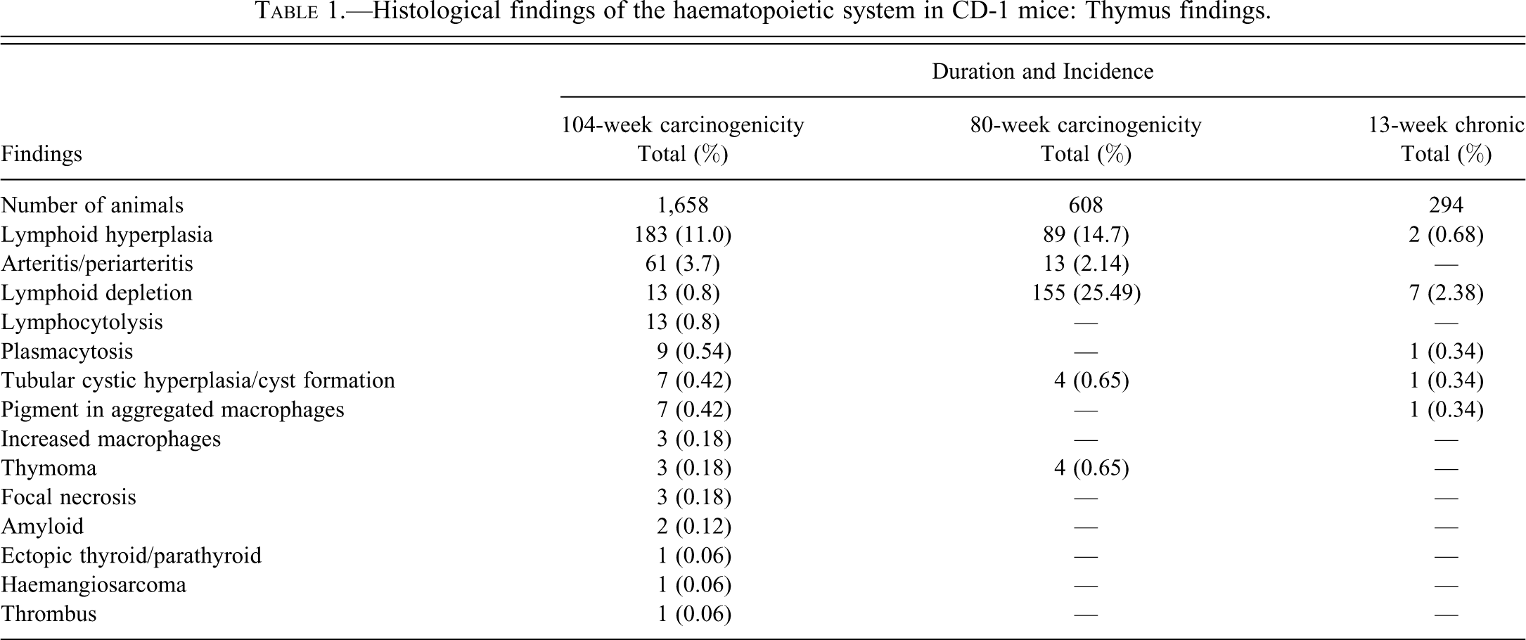
Thymus (Table 1)
Lymphoid hyperplasia was the most common finding of the thymus in the 80- and 104-week carcinogenicity studies. This change was also present to a lesser extent in the 13-week studies. At necropsy an affected thymus may appear enlarged. Lymphoid hyperplasia usually occurs in thymuses that have undergone physiological involution, and is focal or diffuse. It comprises apparently normal thymic tissues within an atrophic thymus. There may be atypical T-cell hyperplasia, B-cell aggregates, or germinal centre formation. Although lymphoid elements may be more enlarged in females (hormonally mediated) in comparison to the epithelial components, a normal medullary/cortex lobular pattern is preserved. This lesion should be differentiated from benign thymoma. These hyperplastic lesions do not progress to neoplasia. Lymphoid hyperplasia is an aging change that is more common in mice older than 60 weeks. Although more common in females, it can occur in both sexes. “Lymphoid overspill” is seen as sheets of non-neoplastic mature lymphocytes present perivascularly and within liver, mediastinum, periovarian tissues, periuterine tissues, renal pelvis, and lacrimal and salivary glands.
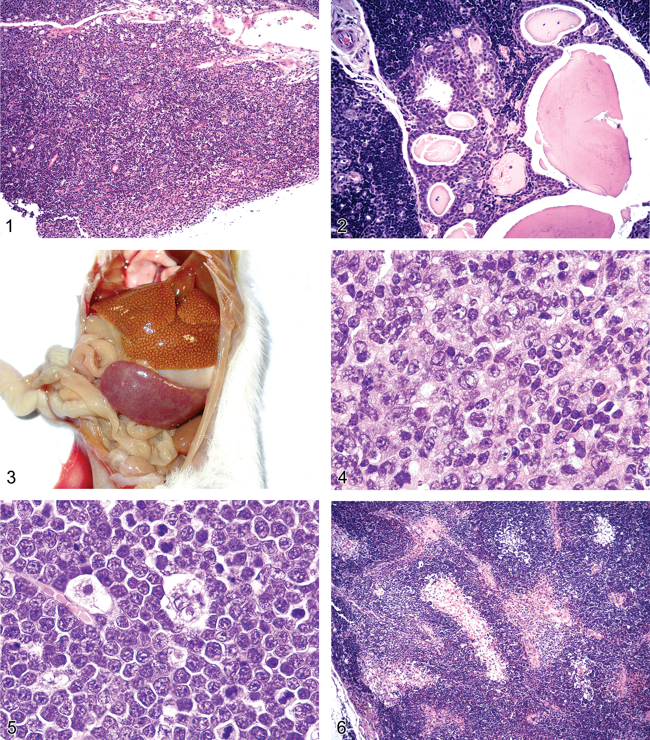
Lymphoid depletion (involution) (Figure 1) was recorded in the thymus of animals from both 80- and 104-week duration carcinogenicity studies and from 13-week chronic studies. Depletion affects both cortical and medullary elements, and the incidence increases with age (physiological involution). Males are more severely affected than females. Adipose infiltration of connective tissue and septae often accompanies lymphoid depletion. The medullary epithelial elements form cords/ribbons and the lesion may progress to focal/diffuse cystic hyperplasia. Cystic structures are lined by cuboidal or columnar cells, which are occasionally ciliated and contain some secretory goblet cells and are filled with eosinophilic colloid (Figure 2). They are more frequently formed within the medullary areas. The hyperplastic elements can look more pronounced on a background of lymphoid atrophy. The progression of this finding appears to be exacerbated by stressors and lymphoid depletion is more commonly recorded in premature sacrifice animals than terminal sacrifice scheduled kill animals at Charles River Preclinical Services Edinburgh.
Figure 1. Thymus: Lymphoid depletion, haematoxylin and eosin stain, x 100.
Figure 2. Thymus: Epithelial hyperplasia, haematoxylin and eosin stain, × 200.
Figure 3. Spleen: Follicular (pleomorphic) lymphoma appearance at necropsy.
Figure 4. Thymus: Follicular (pleomorphic) lymphoma, haematoxylin and eosin stain, × 1,000 (oil).
Figure 5. Thymus: Lymphoblastic lymphoma, haematoxylin and eosin stain, × 1,000 (oil).
Figure 6. Thymus: Benign thymoma haematoxylin and eosin stain, × 100.
Arteritis/periarteritis was seen in the thymus only in animals on carcinogenicity studies. This inflammation affects walls of small and large arteries, and there is fibrinoid necrosis of smaller arterioles. In some cases, this lesion leads to thrombosis of the vessel. This is an aging change that is rare in animals less than 6 months old, and it occurs predominantly in females. It is often seen as one of the spectrum of systemic changes in animals with a moderate nephropathy.
Other non-neoplastic findings recorded in the thymus from carcinogenicity and chronic (13-week studies) included plasmacytosis and pigmented macrophages.
Spleen (Table 2)
A degree of extramedullary haematopoiesis is a normal finding in the spleens of mice. Female CD-1 mice have a much higher grade of haematopoiesis in the spleen than males. The finding of “increased extramedullary haematopoiesis” is recorded in a female mouse when the grade seen is markedly increased above the background high level, and in male mice when the grade seen is equal to or more than that of a normal female animal. At necropsy the spleens may appear enlarged, dark red, and firm. This finding is often accompanied by marked increase in pigmented macrophages (haemosiderosis) and megakaryocytosis. In both sexes the lesion can be exacerbated by blood sampling, anemia, and inflammation, and is often seen in unscheduled sacrifice animals.
Histological findings of the hematopoietic system in CD-1 mice: Spleen findings.
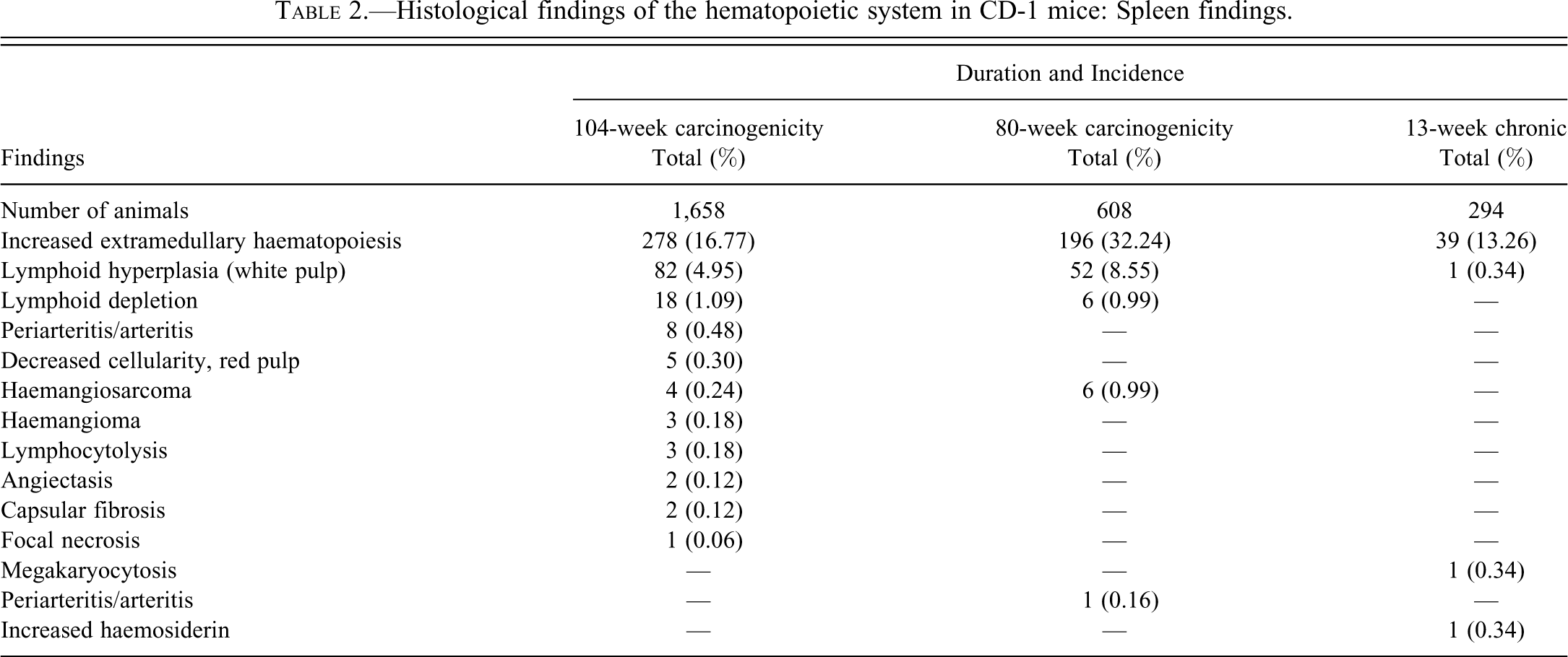
Lymphoid hyperplasia in the spleen may occur in the T-cell zone, the periarteriolar lymphoid sheath (PALS), or in the follicular B-cell zone, and results in compression of adjacent tissue or bulging of the splenic capsule. Affected areas appear as pale foci at necropsy. The finding was recorded mainly in carcinogenicity studies but it was also found in one animal on a 13-week study. Enlarged secondary follicles can be caused by immune stimulation and are common in gavage or intra-peritoneal dosed animals.
Lymph Nodes (Table 3)
Most lymph nodes were unremarkable, with the majority of findings being confined to the mesenteric lymph nodes. The most commonly recorded finding was sinus dilation that typically involved the medullary sinuses. Sinus dilation is often associated with lymphoid atrophy because the medullary sinunses expand as the medullary cords decrease in diameter. Sinus dilation may progress to cystic degeneration. Affected sinuses are dilated with eosinophilic material, presumably lymph, containing lymphocytes, macrophages, and erythrocytes. This lesion is seen as a focal or diffuse aging change.
Histological findings of the hematopoietic system in CD-1 mice: Mesenteric lymph node findings.
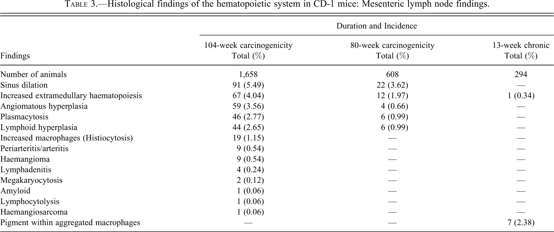
Angiomatous hyperplasia, similar to that seen in Wistar rats, was present in carcinogenicity study animals. This finding is characterized by proliferation of endothelial cells in blood vessels, and there appears to be a continuum of changes from angiomatous hyperplasia to haemangioma, though no haemangiosarcomas have been recorded. Care must be taken when diagnosing this lesion as the hilus region in old animals can look hyperplastic, so the trimming plane must be taken into account.
A few cases of extramedullary hematopoiesis were recorded in mesenteric lymph nodes from mice in both 104- and 80-week-duration carcinogenicity studies and one animal on a 13-week chronic study.
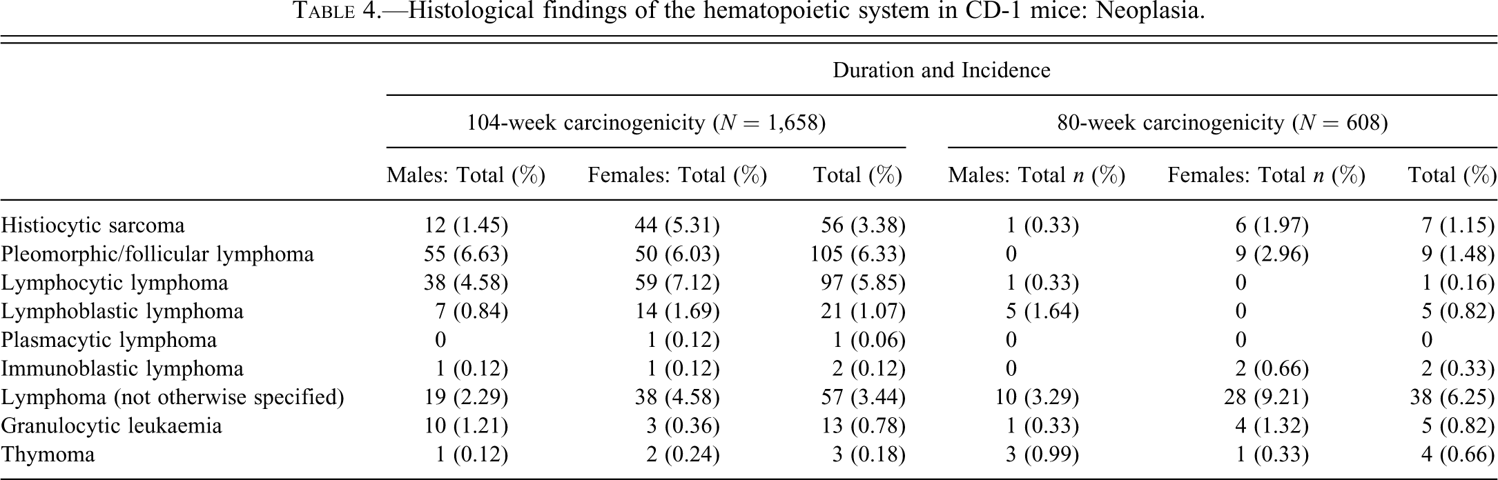
Neoplasia (Table 4)
Lymphoma was the most common neoplastic lesion of the haemopoietic system. Neoplasia of the lymphohaemopoietic system was much more prevalent in 104-week carcinogenicity studies than in 80-week studies, and was not recorded in 13-week studies. In most cases, more than one site was involved. In some animals, however, neoplastic lymphocytes were more prominent in one particular site. Most of the lymphomas were classified as follicular (pleomorphic) cell, followed by lymphocytic, then lymphoblastic, and occasional plasmacytic and immunoblastic. Lymphomas designated “not otherwise specified” were either diagnosed in found dead animals with autolysed tissues or were genuinely too pleomorphic for the study pathologist to give a definitive diagnosis.
Histological findings of the hematopoietic system in CD-1 mice: Neoplasia.
Follicular (pleomorphic) lymphomas tend to arise most commonly in the spleen, causing exaggerated white pulp areas, seen as pale foci at necropsy (Figure 3). Follicular (pleomorphic) lymphomas can have small, medium, or large cell types (or a mixture of all three) that are cohesive with indistinct cell boundaries and moderate pale cytoplasm. Cells of the large type (8–16 µ) have open vesicular nuclei that may be cleaved, and they have a high mitotic index. Cells of the small type (6–10 µ) have condensed nuclei and a low mitotic index. Centrocytes, centroblasts, immunoblasts, macrophages, plasma cells, and T-lymphocytes are also commonly seen admixed amongst the neoplastic lymphocytes (Figure 4). The tumor spreads to involve the Peyer’s patches and/or mesenteric lymph node.
Lymphocytic lymphoma is characterized by small to medium lymphocytes (4–8 µ) with a scant rim of pale cytoplasm and condensed nuclei with stippled chromatin. They form uniform non-cohesive sheets and have a low/rare mitotic index. This tumor tends to arise in the thymus and spreads to involve the spleen. Collision tumours with histiocytic sarcoma have been recorded.
Lymphoblastic lymphoma is characterized by large lymphoblastic cells (7–12 µ) having moderate to scant amounts of pale cytoplasm and nuclei with fine stippled chromatin. The nuclei are round and the cells form non-cohesive sheets so the microscopic field has the appearance of peas boiling in a pan (Figure 5). This is an aggressive tumor with a high mitotic index. Tingible body macrophages are common, giving the classical “starry sky” appearance. The tumor tends to arise in the thymus or bone marrow and spreads to the spleen, lymph nodes, liver, lungs, brain, kidney, gastrointestinal tract, and genitor-urinary tract. “Leukemic overspill” is often a feature of this tumor. The incidence of lymphoblastic lymphoma increases with age; however, it is a common cause of death of unscheduled sacrifice animals under 12 months old.
Plasmacytic lymphoma is very rare and has only been recorded in one animal at this site. This tumor consisted of mature plasma cells (7–16 µ) with abundant pale cytoplasm and clear presence of Russell bodies. The nuclei were round, eccentric with clock face chromatin, and formed non-cohesive sheets. The mitotic index was low.
Immunoblastic lymphomas have been recorded in 4 animals. These consisted of large cells (10–18 µ) with moderate pale eosinophilic cytoplasm and round vesicular nuclei with prominent nucleoli. The neoplastic cells formed non-cohesive sheets with a high mitotic index. These were very aggressive tumors and there was sarcomatous involvement of extra-lymphoid sites—notably in the pancreas.
Histiocytic sarcoma consisted of aggregates/infiltrates of monomorphic or pleomorphic histiocytes, with abundant pale eosinophilic cytoplasm, dark ovoid/elongated nuclei, and indistinct cell borders. Multinucleated giant cells were present in some tumors, although this was inconsistent throughout tumour infiltrates within the same animal and between animals. In some tumor areas, there was pallisading of necrotic cells and cholesterol clefts. Generally these tumors had a high mitotic index, and metastases spread on serosal surfaces as well as via vascular spaces. The tumor most commonly involves spleen, liver, and uterus but also mesenteric lymph nodes and lungs. Hyaline droplets were seen in the proximal convoluted tubule of the kidneys of the majority of mice with histiocytic sarcomas. These droplets have been shown to contain lysozyme degradation products from the tumor cells (Hard and Snowden 1991). In the uterus the neoplastic cells tended to be more spindle-shaped in appearance and resemble those in malignant schwannomas. Histiocytic sarcomas were associated with sudden death in some animals.
Granulocytic leukaemias were reported in several animals. These arise in the splenic red pulp, giving the spleen a greenish tinge at necropsy, and they can involve the liver, kidney, lungs, brain. The tumor is composed of large immature myeloid cells (mainly neutrophilic) with pale staining cytoplasm, notched nuclei, and intracytoplasmic granules. Neoplastic cells are present diffusely in the bone marrow and within vascular spaces.
Benign thymomas were recorded in both 80-week and 104-week carcinogenicity studies. They are composed of neoplastic epithelial components with or without neoplastic lymphocytes that are centrally located within thymic lobules (Figure 6). The epithelial component is generally positive for keratin and appears to be derived from Hassall’s corpuscles or epithelial reticular tissue. The tumor is well encapsulated with a moderate mitotic index even though it is considered benign. Thymomas show an expansive growth pattern and may have slight local invasiveness.
Discussion
The main aim of this study was to determine the incidences of the most common background findings in control Crl: CD-1 (ICR) mice used in chronic preclinical toxicology studies. There are very few reports of this kind in the literature (Maita et al. 1988; Frith et al. 1996, 2001; Son and Gopinath 2004; Baldrick and Reeve 2007; Giknis and Clifford 2010), with the majority of reports being those of C57BL6 and B6C3F1 mice (Tamano et al. 1988; Ward et al. 1999). The findings in this study were generally consistent with the occurrence of similar lesions described in other mouse strains. However, this report presents up-to-date results of incidences and ranges of findings in Charles River Crl: CD-1 (ICR) mice and can serve as a historical control reference for use in correspondence with regulatory authorities.
Lesions of the lymphohaemopoietic system are relatively uncommon in 13-week chronic studies but are seen in higher incidence in carcinogenicity studies of both 80- and 104-week duration. Lymphomas are the most common cause of death of unscheduled sacrifice/found dead CD-1 mice in carcinogenicity studies at this facility, comparable to the findings of Son and Gopinath (2004).
To the best of our knowledge, this is the only report of the incidences and descriptions of background lesions in control Charles River Crl: CD-1 mice used in chronic toxicity studies in recent years. As the incidence of tumor types within the same strain and sex show genetic drift, only studies performed during the previous 3 to 5 years should be used as historical control data. Reference to the incidences reported here should facilitate the differentiation of spontaneous from induced lesions in toxicological safety studies in this strain of mouse. Further representative photomicrographs of the findings described are available on the goRENI website.