
Abstract
The authors performed a retrospective study to determine the incidences and range of spontaneous pathology findings in control cynomolgus monkeys. Data were collected from 570 monkeys (285 animals per sex), aged twelve to thirty-six months, from sixty regulatory studies evaluated at our laboratory between 2003 and 2009. The most common finding overall was lymphoplasmacytic infiltrates observed in the following incidence: liver (60.7%), kidneys (28.8%), heart (25.8%), salivary glands (21.2%), and stomach (12.1%). Inflammation also commonly occurred in the heart, kidneys, lungs, and stomach. The most common degenerative changes were localized fatty change in the liver, myocardial degeneration, and mineralization and pigment deposits in various tissues. Parathyroid, thyroid, and pituitary cysts; ectopic thymus in the parathyroid or thyroid gland; accessory spleen within the pancreas; and adrenohepatic fusion were among the most common congenital findings. Some incidental findings bearing similarities to drug-induced lesions were also encountered in various organs. It is hoped that the results presented here and elsewhere could form the groundwork for the creation of a reliable database of incidental pathology findings in laboratory nonhuman primates.
Introduction
Nonhuman primates are increasingly being used as nonrodent species in preclinical toxicology studies, mainly owing to their close phylogenic relationship to humans and where there is no alternative. Of the available species, the cynomolgus macaque has become the most widely used species, and they are widely available as purpose bred for laboratory use. However, despite their common usage, specific information on the range and incidence of spontaneous background pathology findings in laboratory-raised young adult macaques is not readily available. Because some of these incidental findings may resemble drug-induced findings and thus hinder the interpretation of pathology results, it is important for the pathologist to have available some reference material such as a historical control database or published literature with incidences, range, and histopathological presentation of incidental findings in this species. Both common incidental findings and findings likely to be confused with treatment-related lesions are important in this regard.
Historical background pathology data are particularly important in studies involving nonrodent species because fewer numbers of animals are used, which can greatly increase the chance of some spontaneous finding occurring only in treated animals and can lead to misinterpretation of the finding as treatment effect. When compiling such data, consideration should be paid to some important factors that may affect the composition of the collected data such as the strain and source of the animals used, housing and management and variations in nomenclature and recording levels of the individual pathologists evaluating these studies. Such data also need to be updated regularly to reflect possible changes in these factors over time.
The main aims of this study were to present and discuss summarized results from studies carried out at Charles River, Edinburgh, of the range and incidences of the most common spontaneous lesions of control cynomolgus macaques, and by doing so help provide the basis for further work on organ/system-specific pathology of nonhuman primates. Presentation of some less common or unusual findings, but of toxicological significance, since they can be confused with treatment related changes, was one of the main objectives of this study.
Materials and Methods
Animals
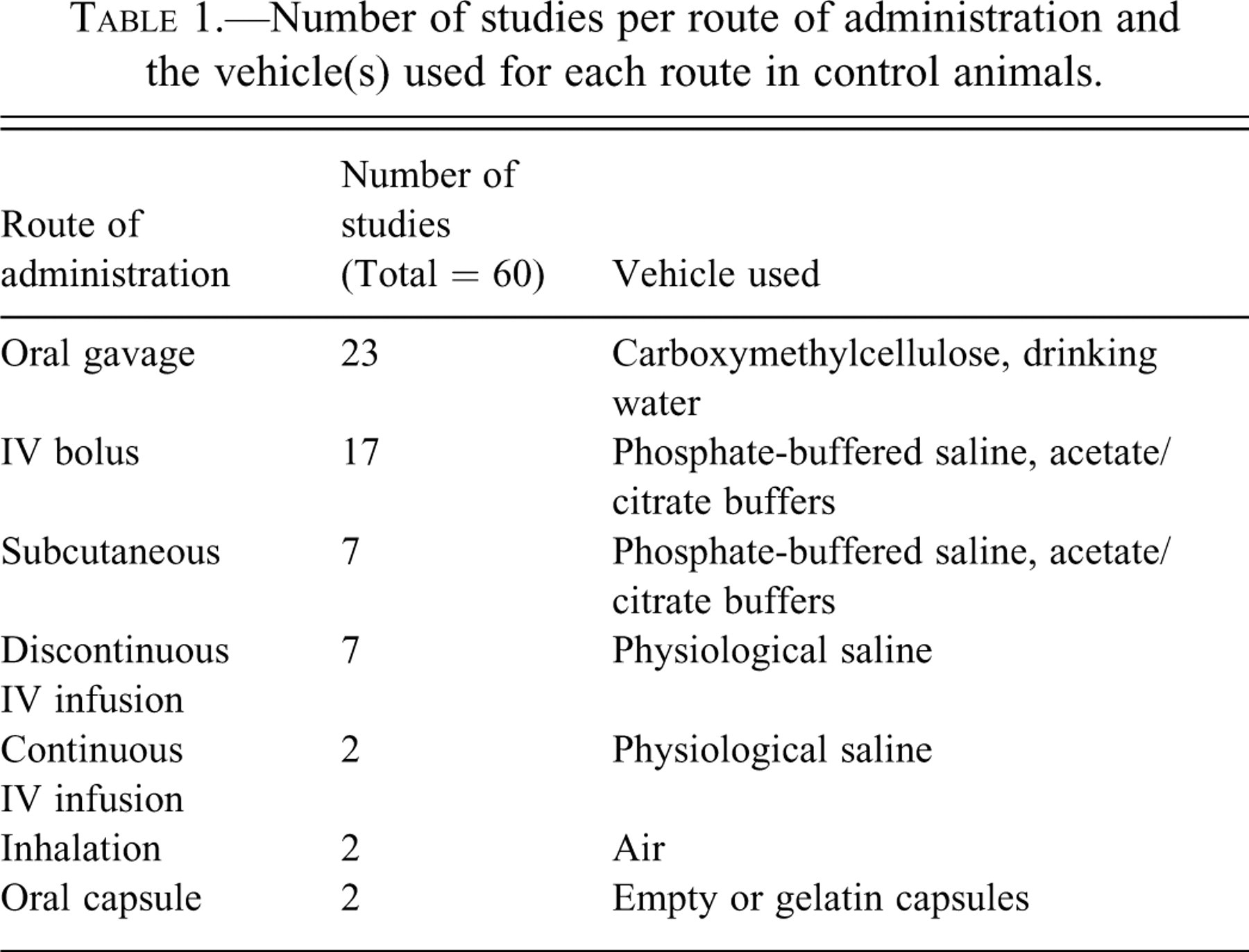
Tissue samples from a total of 570 cynomolgus macaques (285 animals per sex) were obtained from control groups of sixty preclinical toxicological studies evaluated between 2003 and 2009. The animals were purpose bred for laboratory use and came from accredited suppliers (Bioculture, Mauritius; Harlan, UK; Guanxi Grandforest Primate Company, China) from three geographical regions: Mauritius (more than 90% of the animals), China, and Vietnam. All control animals incorporated into the study were obtained from groups of animals that had been sham dosed with an appropriate vehicle. The following vehicles or method of sham dosing were used: empty or gelatin capsules for the oral capsule route, carboxymethylcellulose or drinking water for the oral gavage route, and phosphate- buffered saline, physiologic saline, and acetic or citrate buffers for intravenous or subcutaneous injection routes of administration. For inhalation studies, animals from only the air control groups were used. The total number of studies for each route of administration and the main type of vehicle(s) used for the particular method of administration are shown in Table 1.
Number of studies per route of administration and the vehicle(s) used for each route in control animals.
All animals were between twelve and thirty-six months of age and had body weights ranging between 1.6 and 2.5 kg. They were housed in groups of two or three animals of the same sex and dose group in custom-designed U.K. Home Office–compliant primate cages (Chapter 14, section 21, UK Animals [Scientific Procedures] Act of 1986). The temperature and humidity were automatically controlled at 21°C ± 4°C and 55% ± 10%, respectively, with a minimum of fifteen air changes per hour. An automatic light cycle of 7:00 AM–7:00 PM (twelve-hour cycle) was maintained. Each individual gang pen had drinking water and food hoppers, and animals were fed a commercial primate diet (Mazuri diet, Special Diet Service Ltd, Witham, Essex, England). Twice-weekly fruit supplements were also offered to all animals.
All studies were conducted in accordance with the UK Animals (Scientific Procedures) Act 1986, which conforms to the European Convention for the Protection of Vertebrate Animals Used for Experimental and Other Scientific Purposes (Strasbourg, Council of Europe).
All animals in the study were serologically tested and confirmed free of the following viral diseases during quarantine: simian immunodeficiency virus (SIV), Cercopithecine herpesvirus 1 (B virus), simian retroviruses type D, rabies, simian T-cell leukemia virus (STLV), measles, and filoviruses. Tuberculin tests; other bacteriology screening tests for Shigella, Yersinia, and Salmonella; and parasitological screening (including malaria smears) were carried out on arrival at the test facility.
Pathological Evaluation
Animals were humanely euthanized by intravenous injection with sodium pentobarbitone and exsanguinated via femoral veins. A detailed necropsy was performed under the supervision of a veterinary pathologist who was either present throughout the necropsy sessions or remained on call after observing the commencement of the necropsy. Tissues were preserved in 10% neutral buffered formalin, embedded in paraffin wax, sectioned to a 4–5 μm thickness, and stained with hematoxylin and eosin. They were examined histopathologically, and the findings were entered directly into a computerized database (PLACES 2000 Instem, Apoloco Limited Systems, Plymouth Meeting, PA, USA).
Study Design
Data were collected retrospectively from control groups of cynomolgus monkey studies evaluated over a period of six years (2003-2009). From this pool of information, studies to be incorporated into the present investigation were selected based on the following criteria: At least one control or untreated group (controlled studies) Equal numbers of male and female animals, with a minimum of three animals per sex per group GLP-compliant toxicological studies with evaluation of a full tissue list
Study material including histological incidence tables, individual animal data listings, and a few selected glass slides were retrieved from the archives and analyzed for pathology findings under each body and organ system. Data were available from sixty controlled studies, with three to eight animals per sex per study, giving a total of 570 control animals. A limited number of slides with lesions of interest were re-evaluated by a qualified veterinary pathologist.
Results
Common Histopathology Findings
Tables 2 –7 present the most common histopathological findings by organ system and the prevalence (range) of each finding per control group of three to eight animals. A few uncommon spontaneous findings of toxicological or clinical significance are also included. Findings were graded on a scale of 1 to 5 represented by minimal, mild, moderate, marked, and severe, respectively.
Histological findings in the lung, heart, and aorta of control cynomolgus monkeys (Macaca fascicularis) and their corresponding incidences across sixty studies (285 animals per sex).
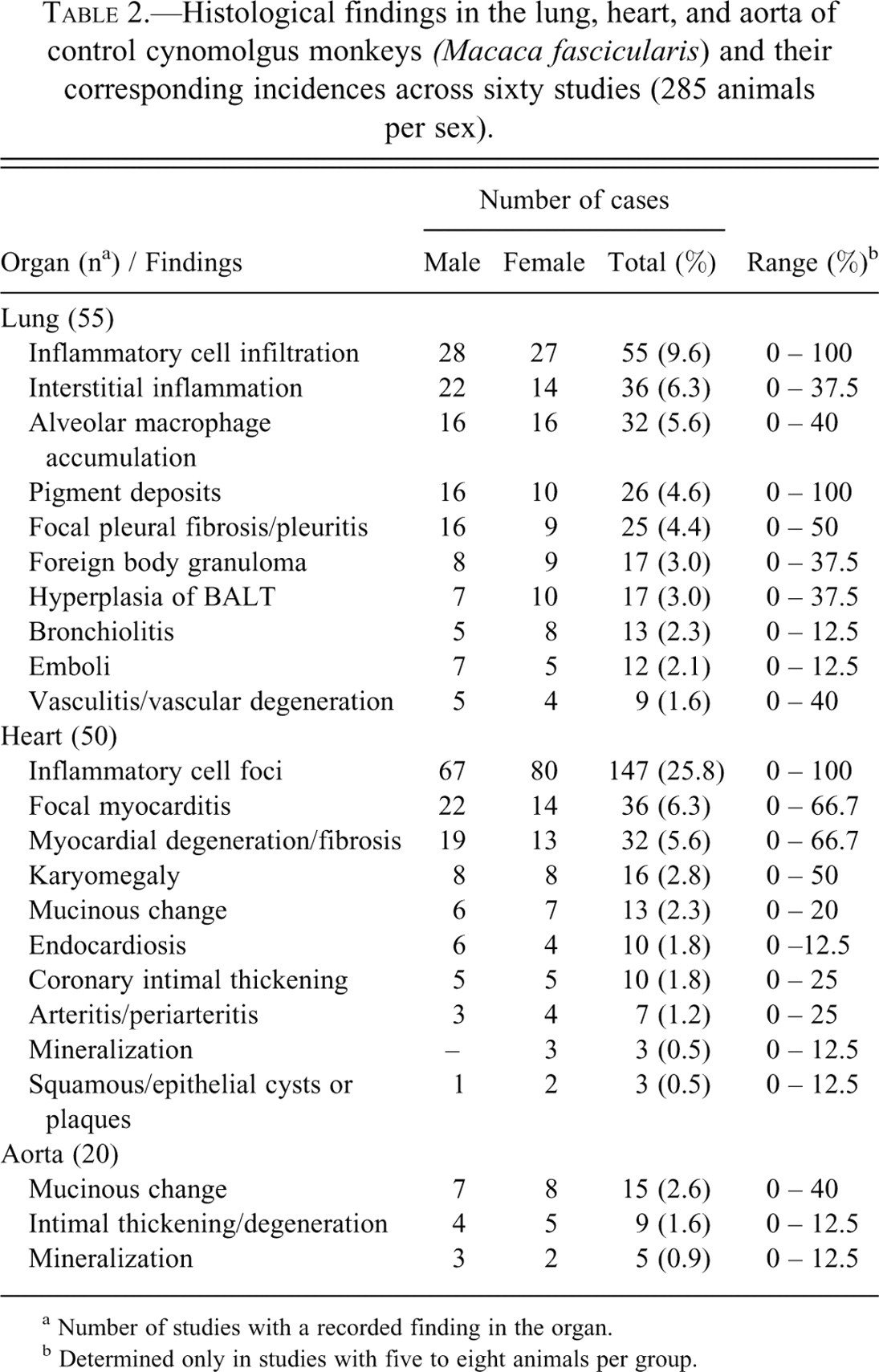
a Number of studies with a recorded finding in the organ.
b Determined only in studies with five to eight animals per group.
Histological findings of the liver, gall bladder, exocrine pancreas, and salivary glands of control cynomolgus monkeys (Macaca fascicularis) and their corresponding incidences across sixty studies (285 animals per sex).
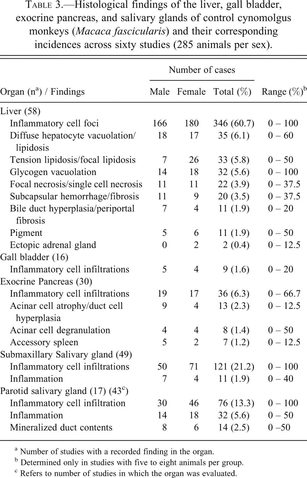
a Number of studies with a recorded finding in the organ.
b Determined only in studies with five to eight animals per group.
c Refers to number of studies in which the organ was evaluated.
Histological findings of the stomach, small intestines, large intestines, esophagus, and tongue of control cynomolgus monkeys (Macaca fascicularis) and their corresponding incidences across sixty studies (285 animals per sex).
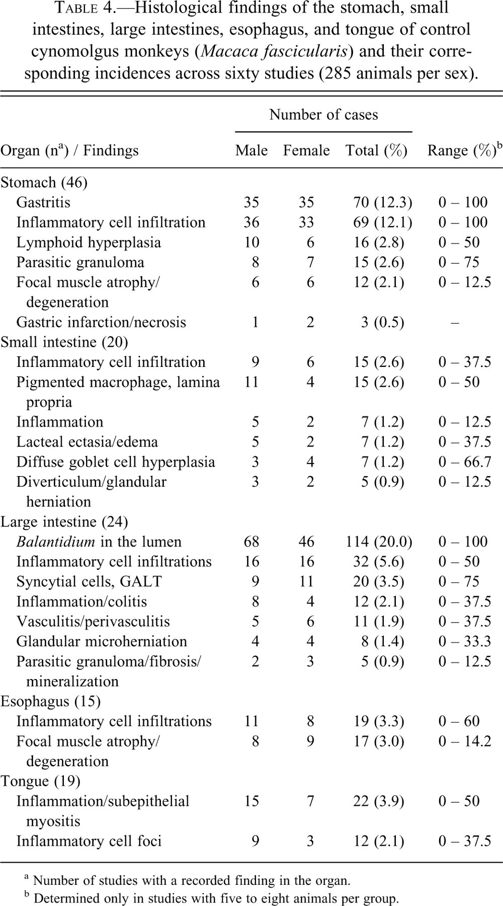
a Number of studies with a recorded finding in the organ.
b Determined only in studies with five to eight animals per group.
Histological findings of the urinary, reproductive, and endocrine systems of control cynomolgus monkeys (Macaca fascicularis) and their corresponding incidences across sixty studies (285 animals per sex).
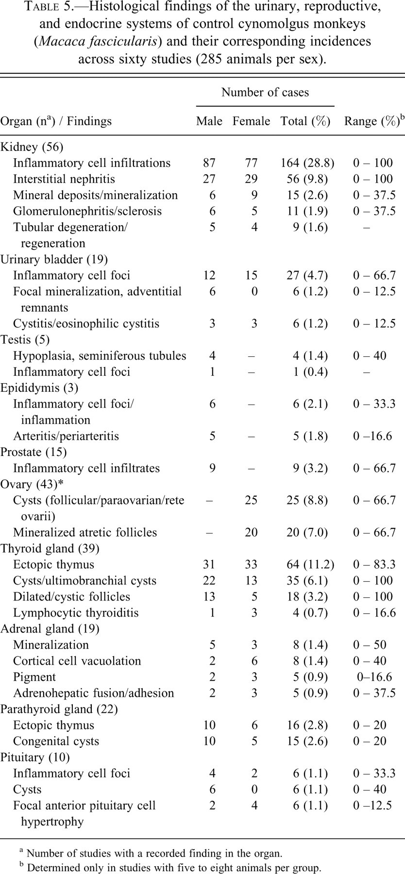
a Number of studies with a recorded finding in the organ.
b Determined only in studies with five to eight animals per group.
Histological findings of the hematopoietic and the lymphoid systems of control cynomolgus monkeys (Macaca fascicularis) and their corresponding incidences across sixty studies (285 animals per sex).
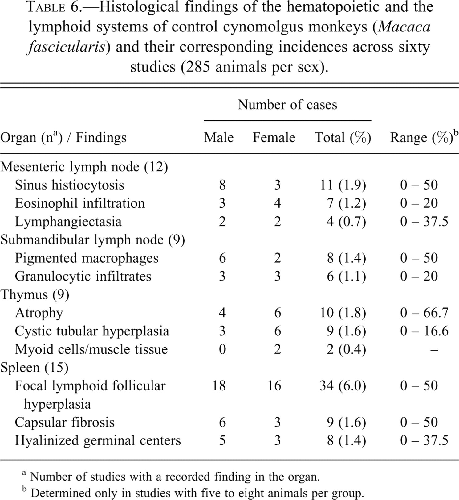
a Number of studies with a recorded finding in the organ.
b Determined only in studies with five to eight animals per group.
Histological findings of the skin and the musculoskeletal and nervous systems of control cynomolgus monkeys (Macaca fascicularis) and their corresponding incidences across sixty studies (285 animals per sex).
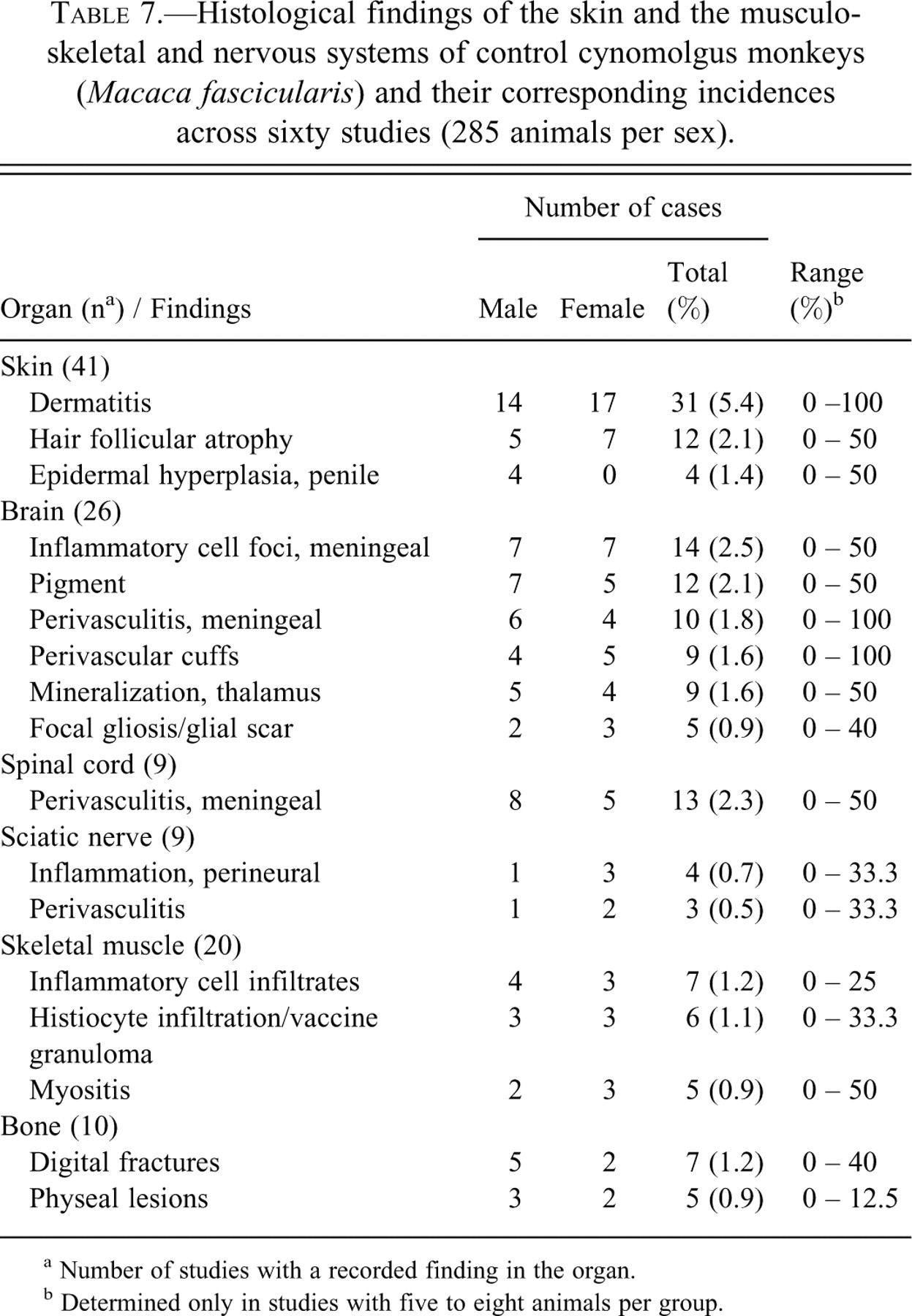
a Number of studies with a recorded finding in the organ.
b Determined only in studies with five to eight animals per group.
Inflammation and mononuclear inflammatory cell infiltrates were the most commonly recorded pathological processes in most tissues and were considered to represent different stages of the same pathological continuum by most pathologists, even though umbrella terms were rarely applied for the two processes. They were most commonly recorded in the liver, kidneys, heart, salivary glands, lungs, and stomach. In general, inflammatory cell infiltrations were characterized by interstitial accumulation of lymphocytes and plasma cells in varying proportions (lymphoplasmacytic), with little or no evidence of damage to parenchymal tissue such as renal tubules, gastric glands, cardiac myocytes, or hepatocytes. Moderate grades of lymphoplasmacytic infiltration were common in organs such as salivary glands, esophagus, stomach, urinary bladder, lungs, and kidneys. They were often associated with lymphoid nodules or follicles around salivary gland ducts, small arteries in the urinary bladder, and bronchi and blood vessels in the lungs. Inflammation was recorded when there was evidence of degeneration and necrosis of parenchymal tissues in association with minimal to moderate inflammatory cell infiltrates, usually of a mixed nature, or when other obvious signs of inflammation such as edema and fibrin depositions were present. Nevertheless, the distinction between the two processes, which often occurred simultaneously in most organs, was not always clear, particularly in the heart.
Findings by Organ System
Cardiovascular and Respiratory Systems
The incidence and range of spontaneous findings of the heart in control cynomolgus macaques are presented in Table 2. As the results indicate, incidental findings occur commonly in the heart. Minimal to mild idiopathic inflammatory cell infiltrates and minimal to moderate focal myocarditis (Figure 1a) were present in more than a quarter of the animals, and occasionally, 100% of control animals in a study were affected. Focal inflammatory cell infiltrates were mainly composed of uniform and bland aggregates of lymphoplasmacytic cells with little or no associated damage to the cardiac myocytes, whereas myocarditis was characterized by some pathologic change to cardiac myocytes such as necrosis, karyomegaly, or fibrin deposition. The inflammatory cell infiltrate associated with focal myocarditis was usually of a mixed nature and included granulocytes and macrophages in addition to lymphoplasmacytic cells.
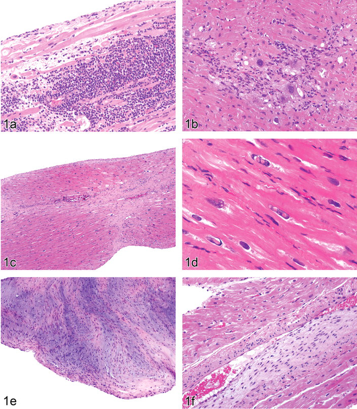
Figure 1(a). Focal myocarditis with a predominantly plasmacytic infiltration (with moart cells and Russell bodies) in the subendocardial region of the left ventricle of a young male cynomolgus monkey (Macaca fascicularis). H & E, × 200.
These inflammatory lesions were rarely observed in deeper parts of the myocardium, as they were mostly confined to subendocardial or subepicardial/epicardial areas, including the epicardial fat, and showed no differences in distribution of lesions between the apex and the base of the heart. Simultaneous occurrence of myocarditis and focal lymphoplasmacytic cell infiltrates in the same heart were common. Focal or multifocal myocarditis was also occasionally observed within the same hearts with other changes such as myocardial fibrosis at the papillary muscle, hemorrhage, or myocardial degeneration with karyomegaly.
Idiopathic myocardial degeneration or cardiomyopathy was recorded in a few studies but with a moderately high prevalence in those studies in which it occurred. The finding was characterized by minimal to moderate localized to extensive degeneration or necrosis of cardiac myocytes with mild to marked hypertrophy, karyomegaly, and vacuolation of cardiac myofibers (Figure 1b), with or without minimal inflammation or fibrosis. In earlier lesions, only vacuolation of cardiac myofibers and karyomegaly with increased nuclear basophilia were present, whereas in more advanced cases, inflammation and fibrosis or mineralization were present (Figure 1c). In some rare and severe cases of cardiomyopathy, the hypertrophic cardiac myofibers could occasionally be observed to contain large intracytoplasmic eosinophilic granules and intranuclear inclusions thought to represent invagination of the cytoplasm and its organelles into the nucleus (Figure 1d). Areas of the heart most commonly affected by myocardial degeneration were, in decreasing order, the apex, the interventricular septum (just below the atrioventricular valves), the papillary muscle, and the subendocardial areas of both ventricles. Acute hemorrhagic necrosis or fibrosis at the papillary muscle, resembling ischemic lesions observed with β-agonists and other cardioactive drugs (Greaves 2000), were also encountered in occasional animals. There were more male animals with myocarditis and myocardial degeneration than females, but the difference was not statistically significant.
Some of the uncommon but important findings seen in the heart of cynomolgus monkeys such as congenital ectopic epithelial and squamous cysts/plaques, arteritis/periarteritis, mineralization, and valvular endocardiosis (Figure 1e) or mucification of the valves and subendocardial areas or the tunica intima of great vessels, are listed in Table 2.
Accumulation of mucopolysaccharides in the intima of the aorta, subendocardial areas of the base of the heart, or coronary arteries without lipid accumulation was a common incidental finding in animals. Another related finding observed uncommonly in the coronary arteries, and to a lesser extent the aorta, was focal thickening of the intima of the blood vessels with fibrosis or mucin accumulation leading to formation of atherosclerotic lesions and near-occlusion of the affected blood vessel (Figure 1f). The atherosclerotic lesions and intimal thickening were formed by infiltration of the intima by smooth muscle cells, mucins, and fibrous tissue with little or no foam cells or extracellular lipids.
Compared with the heart, incidences of spontaneous findings in the lungs of cynomolgus macaques were not just less numerous but also of considerably lower severity and significance. The most common findings were recorded at necropsy, where focal adhesions between lung lobes or adherence to the parietal pleura were common but of little or no clinical significance. Lung adhesions observed at necropsy were not always associated with histopathology findings, but at times focal pleural or subpleural fibrosis and chronic pleuritis were observed. Perivascular or peribronchial inflammatory cell infiltration, peribronchial bronchial-associated lymphoid tissue (BALT) hyperplasia, minimal to mild focal interstitial inflammation or focal subpleural fibrosis, focal foreign body granuloma, emboli, and lung pigment were some of the more common histopathology findings in the lungs. Focal foreign body granulomas were associated with either inhalation of tiny particles of food/plant material or with embolism. Hair emboli were the most commonly encountered form of embolism and were almost exclusively associated with the intravenous form of drug delivery. With intravenous infusion studies, focal to multifocal pulmonary thrombi, diffuse interstitial pneumonia, pulmonary edema and periarteritis (Figure 2a), and localized broncho-pneumonia (associated with thrombi and infarction) were encountered at particularly high incidences, which often corresponded to the incidence and severity of thrombophlebitis at the cannula tip.
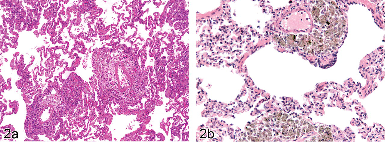
Figure 2(a). Mild interstitial inflammation with periarteritis and edema associated with continuous intravenous infusion in the lung of a control female cynomolgus monkey (Macaca fascicularis). H & E, × 100.
Dark brown to black lung pigment was observed in alveolar macrophages with perivascular or peribronchial distribution (Figure 2b), but it was not associated with lung mites (Pneumonyssus sp.) or other pathological changes of the lung parenchyma. However, in some moderate to marked cases, most of which were noted at necropsy, the aggregates of pigment-laden macrophages tended to form nodules that bulged into the alveolar space. Similar pigment deposits were also present in bronchial lymph nodes in affected animals, and in the absence of mites or any associated damage to the lung parenchyma, a diagnosis of pneumoconiosis was considered for this pigment.
Digestive System
Common lesions encountered in the liver, gall bladder, pancreas, salivary glands, tongue, esophagus, and gastrointestinal tract are shown in Tables 3 and 4. Mononuclear cell infiltrates or inflammatory cell foci in the liver, salivary glands, tongue, and esophagus; localized fatty change or tension lipidosis in the liver; and gastritis were among the most common lesions.
Gastritis was one of the most commonly and consistently recorded findings, and incidences of 100% affected animals were reported in nearly half of the studies. The majority of the lesions were subacute to chronic in nature, with moderate to marked grades being recorded more commonly in the gastric antrum than the fundic/body region. Chronic gastritis was characterized by mild to marked lymphoplasmacytic cell infiltration of the mucosa and submucosa, an increase in lymphoid follicles and mucosal atrophy with attenuation of glandular epithelium, separation and scarcity of glands, and an accompanying increase in the lamina propria. In some severe cases, a few neutrophils or a mixture of cells within gastric glands (glandular micro-abscesses) and mucosal erosion were observed (Figure 3a). Lymphoplasmacytic infiltration of the superficial layers of the mucosa and the formation of prominent lymphoid nodules associated with chronic gastritis were often observed as white nodules at necropsy, but no clinical signs were reported in the majority of the cases. Helicobacter pylori and Helicobacter heilmannii–like organisms were frequently seen both in gastritis cases and in normal-appearing sections, even though investigative staining for bacterial identification was rarely performed.
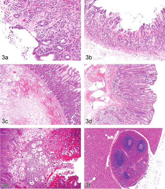
Figure 3(a). Chronic gastritis with erosion and atrophy of the antral mucosa in a young male cynomolgus monkey (Macaca fascicularis). Spiral-shaped bacteria (Helicobacter species) are visible on the luminal surface and within glands. H & E, × 200.
Acute gastritis, which occurred at a much lower incidence than chronic gastritis, was often characterized by hemorrhage, erosions, ulceration, or glandular micro-abscesses and occurred more commonly in the fundus/body of the stomach (Figure 3b).
Other findings recorded at low incidences in the stomach include mucosal herniation and diverticuli, parasitic granulomas (mostly identified as Oesophagostomum species), and gastric infarction. Gastric infarction was reported as a rare incidental finding noted at necropsy as a pale or red focal area affecting any part of the serosal surface of the stomach. On histology, there was evidence of vascular compromise characterized by hemorrhage, necrosis, and fibrin deposition of the submucosa and muscularis, with the mucosa largely unaffected (Figure 3c). As the animals were clinically normal until the scheduled time of kill, the cause and pathogenesis of this finding were not determined.
Although the incidences of histopathology findings observed in the intestine were much lower than those recorded for the stomach, findings in the intestine, especially the large intestine, were more clinically significant, with diarrhea being one of the most common and important clinical signs observed in control animals or untreated, pretrial animals. Idiopathic or nonspecific diarrhea occurred more commonly than diarrhea associated with an etiologic agent. Clinically significant diarrhea was usually associated with chronic colitis, characterized by mild to moderate submucosal lymphocyte and plasma cell infiltration; a few neutrophils within dilated, shortened, or irregular crypts (crypt micro-abscesses); attenuation of enterocytes; glandular micro-herniation; mucosal hemorrhage; and hyperplasia of mucosal epithelium with a reduction in numbers of goblet cells (Figure 3d). Frank ulceration was rarely encountered, but micro-ulcers and erosions caused by the rupture of crypt micro-abscesses were more common. The gross finding of focal reddening of the mucosa, especially at the ileo-cecal junction, was the most commonly recorded necropsy finding associated with chronic colitis. Histology analysis of samples collected from around the ileocecal junction revealed that a considerable proportion of chronic colitis cases also involved parts of the distal ileum.
Other intestinal findings were uncommon, but accumulation of pigmented macrophages in the lamina propria of the small intestines (mostly jejunum and ileum), Balantidium sections within the lumen or in the mucosa, edema, and glandular micro-herniations unassociated with inflammation were variably recorded. Parasitic granulomas with or without sections of nematode parasites in the walls of the intestines, isolated crypt micro-abscesses, cystic dilation of intestinal crypts and lacteal ectasia, and mucosal diverticuli were some of the rarer findings occasionally encountered in this system. Balantidium protozoa were rarely seen invading the mucosa, except in those cases where the animals had been subjected to some form of stress. Frequently, the parasite was seen seemingly gaining access to the intestinal wall via glandular micro-herniation into the gut-associated lymphoid tissue (GALT) (Figure 3e).
Cases of infection with some opportunistic pathogens such as Campylobacter jejuni were very rare and occurred at a frequency of approximately one case per year, whereas Yersinia species were isolated in a single case of ulcerative colitis with multiple abscesses in the liver and spleen. Two confirmed cases of Shigella were also reported in control animals in the six-year period investigated.
Foci of inflammatory cells within the liver parenchyma or in the periportal areas was the most common finding recorded in these studies. The finding was usually of a minimal severity and was occasionally associated with negligible necrosis or apoptosis of a few hepatocytes. The cell type was predominantly lymphoplasmacytic, and a few macrophages or neutrophils were encountered in cases of single-cell necrosis of adjacent hepatocytes or in the periportal or subcapsular areas. No necropsy findings or clinical chemistry changes were associated with this incidental finding. Minimal to moderate grades of focal subcapsular hemorrhage, inflammation, or fibrosis were recorded with some frequency in the liver. The lesions were usually associated with some necropsy findings such as pale or red foci on parietal surfaces of the liver and were thus considered to represent areas of traumatic injury to the liver such as might occur during manipulation of the animals. Tension lipidosis, which was almost always associated with a necropsy finding of pale focus adjacent to attachment sites of ligaments, was more common in females than in males (p < 0.05). Other minor findings such as diffuse vacuolation or lipidosis, glycogen accumulation, pigmentation in Kupffer cells, and periportal fibrosis were variably recorded by the pathologist, with some considering these findings “normal.” Congenital findings in the digestive tract included ectopic pancreatic tissue in the small intestine, accessory splenic tissue in the pancreas (which was almost always located at the tail of the organ or within the adipose tissue surrounding the tail); (Figure 3f), and ectopic adrenal tissue within the liver capsule.
Urogenital and Endocrine Systems
Common lesions encountered in the urogenital and endocrine systems are outlined in Table 5. Minimal to moderate focal interstitial nephritis (Figure 4a) was the second most commonly encountered inflammatory lesion after inflammatory changes in the heart. It was more commonly observed in the outer cortex or subcapsular regions of the renal cortex and invariably involved surrounding tubules or glomeruli. The inflammatory infiltrate associated with interstitial nephritis were predominantly lymphoplasmacytic, with occasional intratubular polymorphonuclear cell infiltrates (tubulointerstitial nephritis). Tubular regeneration, interstitial fibrosis, intra-epithelial pigment, and mineralization were occasionally observed in association with chronic interstitial nephritis.
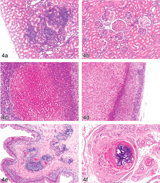
Figure 4(a). Interstitial nephritis with lymphoplasmacytic cell infiltration and associated tubular damage in the outer cortex of the kidney in a young female cynomolgus monkey (Macaca fascicularis). H & E, × 100.
Glomerular lesions were uncommon but not rare and included mild to moderate cases of both focal membranous and proliferative glomerulonephritis and glomerulosclerosis. However, focal and chronic membranous or membranoproliferative forms of glomerulonephritis with sclerosis were more common. The lesions were characterized by glomerular enlargement, thickening of the Bowman’s capsule, hypercellularity of the mesangium with narrowing or obliteration of capillary lumen, and global or segmental deposition of periodic acid–Schiff (PAS)–positive eosinophilic material (Figure 4b). The degree of deposited eosinophilic material varied greatly from minimal segmental deposition to marked global deposition of collagen-like substances resembling collagenofibrotic nephropathy. Periglomerular or interstitial fibrosis in adjacent parenchyma and glomerulocystic changes characterized by atrophy of glomerular tufts and dilation of Bowman’s capsule were other associated findings. There were no gross pathological findings or systemic disease associated with the glomerular changes, and no cases of generalized glomerulonephropathy were encountered.
Intracapsular ectopic adrenal cortical tissue attached to the outer cortex of the kidney, cuboidal metaplasia of the parietal epithelium of the Bowman’s capsule, pigment within the epithelium of the medullary rays, intracytoplasmic pseudo-inclusions of the uroepithelium (including that of the urinary bladder), and multinucleated cells of the renal medulla were inconsistently recorded findings, and most pathologists considered them to be either normal anatomic variations in this species or minor findings of little pathological significance.
Ectopic thymus in the thyroid or parathyroid gland; thyroid, parathyroid or pituitary cysts; ovarian squamous cysts; adreno-hepatic fusion (Figure 4c) or adhesion; and foci of ectopic adrenal tissue in the liver (within the capsule) (Figure 4d), testes, epididymides and prostate were some of the most common congenital findings associated with the endocrine and urogenital systems. There were twice as many congenital cysts of the thyroid gland, including ultimobranchial cysts and dilated/cystic thyroid follicles, in males compared to female animals (p < .05). Minimal to mild grades of lymphocytic thyroiditis were uncommonly recorded in a few animals, mostly females, and were characterized by the diffuse formation of primary or secondary lymphoid follicles within the thyroid, surrounded by a few remaining and distended follicles with hypertrophic thyroid follicular cells. Minimal to moderate grades of focal hypertrophy of the anterior pituitary cells were not uncommon.
Fusion between liver and right adrenal tissue, known as adrenohepatic fusion (AHF), was observed much more commonly than adrenohepatic adhesion and was distinguished by the lack of fibrous tissue between the two parenchymal tissues in the former. Adrenohepatic fusion was not a very common finding, although its prevalence within certain studies was often high, affecting as many as three out of eight control animals in such studies. The lesion was characterized by the presence of hepatic tissue composed of normal-appearing hepatocytes located mostly between the medulla and zona reticularis, but occasionally in the cortex or hilar region of the right adrenal gland. In some cases, the right adrenal gland was observed to be attached to the liver at necropsy, and the histological picture in such cases was suggestive of liver tissue growing into the adrenal gland, whereas in other cases in which there were no associated necropsy findings, the hepatic tissue appeared to be completely enclosed within the adrenal capsule on the routine longitudinal sections of the adrenal gland. The most common finding in the adrenal glands, though, was focal mineralization at the junction of the medulla and cortex (or occasionally within the zona reticularis), which is believed to be caused by dystrophic calcification of remnants of the fetal adrenal cortex (Lowenstine 2003) and was considered to be of no pathological significance.
The urinary bladder was one of the organs with a moderately high number of incidental findings. The most common findings encountered in this organ were lymphocytic foci, usually with a perivascular distribution within the submucosa or the muscularis (Figure 4e). In some cases, these lymphocytic foci were associated with a periarteritis of small- to medium-sized arteries or cystitis. Cystitis was less commonly encountered than inflammatory cell infiltration and was occasionally associated with perivascular infiltration by eosinophils (eosinophilic cystitis). Eosinophilic infiltration of the bladder usually affected the entire wall, from the mucosa to the adventitia, without any evidence of parasitic infestation. Mineralization of functional or vestigial arteries (thought to be remnants of umbilical arteries) was also commonly observed in the adventitia of the bladder (Figure 4f), with early stages of the lesions presenting as focal degeneration, fibrosis, foreign body macrophage accumulation, and pigmentation of arterial walls. The significance of this finding was not established, and no associated necropsy findings were observed.
Owing to the sexual immaturity of the majority of the animals, reproductive tracts of both male and female animals were among the least affected organs, with very little pathology recorded in the respective tissues. Occasionally, mild to moderate arteritis/periarteritis was observed in the epididymis, whereas inflammatory cell foci within the prostate were not uncommon. A single case of ectopic adrenal cortical tissue within the epididymis was also recorded. In the female reproductive tract, the most common incidental findings were mineralization in the ovary, ovarian cysts, squamous cysts within the ovary, and ectopic ovarian follicles in the uterus, mesometrium, or other parts of the broad ligament of the uterus.
Hematopoietic and Lymphoid Systems
Mild to marked lymphoid follicular hyperplasia/prominent lymphoid follicles in the spleen was the most common finding in the hematopoietic and lymphoid systems. Hyperplastic follicles that resulted in compression of adjacent tissue or bulging of the splenic capsule (Figure 5a) were often observed at necropsy as raised pale foci. Hyalinization of germinal centers (antigen-antibody complex material), fibrosis, and histiocytic accumulation in the center of the prominent lymphoid follicles were other findings associated with lymphoid follicular hyperplasia in the spleen and therefore usually not recorded separately. Other findings recorded in the spleen include focal capsular fibrosis, granulocytic infiltration, pigment, and capsular hemorrhage/thrombi. In the thymus, atrophy, mineralization, and cystic tubules were the few incidental findings encountered.
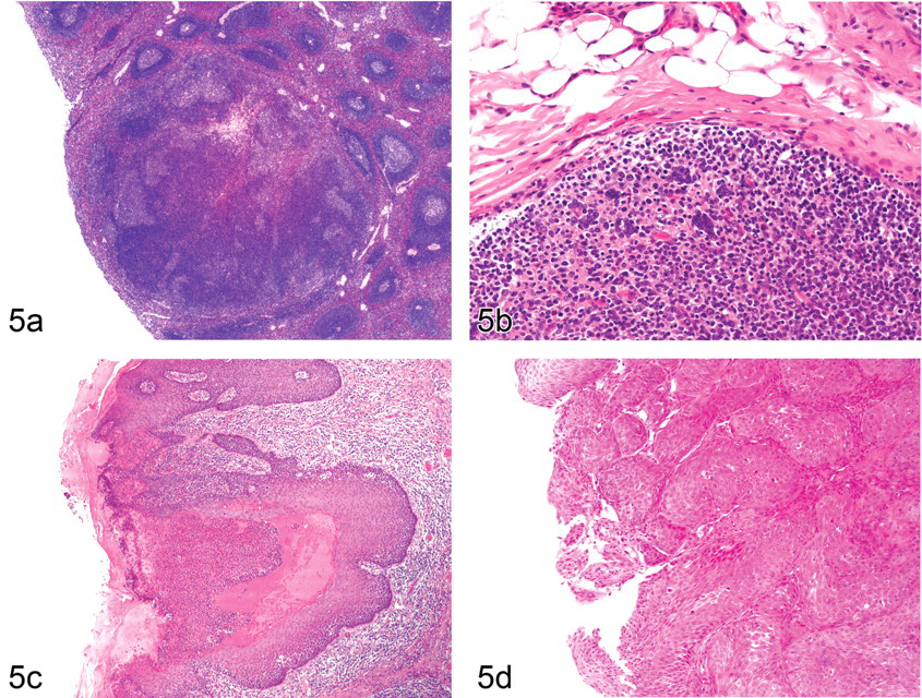
Figure 5(a). Spleen of a young male cynomolgus monkey (Macaca fascicularis) showing marked lymphoid hyperplasia and coalescing of lymphoid follicles resulting in capsular bulging and compression. A small central pale area of lymphocytic necrosis, fibrosis, and macrophage accumulation can be visualized. Necropsy findings of raised pale foci were associated with this finding. H& E, × 50.
Most lymph nodes had unremarkable findings, with the exception of pigment deposits in the bronchial and, rarely, submandibular lymph nodes, granulocytic infiltrates in the submandibular lymph nodes and histiocytosis in mesenteric lymph nodes of animals with inflammatory lesions. A few cases of extramedullary hematopoiesis were recorded in cutaneous-draining lymph nodes (local to injection sites) and other internal nodes such as the lumbar and mediastinal lymph nodes. Lymphoid foci in the bone marrow were frequently observed and were usually associated with prominent lymphoid foci in other sites such as the BALT of the larynx and lung, salivary glands, spleen, and urinary bladder and the GALT of the intestines and stomach. Multinucleate lymphocytic syncytia resembling the Warthin-Finkeldy bodies associated with measles virus were observed with some frequency in the BALT of the larynx and the GALT of the large intestines (Figure 5b). No necrosis of lymphocytes was present within the lymphoid nodules, nor were viral inclusions present within the cells. The cause of these syncytia in animals known to be free of measles and other viral diseases is unknown.
Integument, Mammary Gland, and Musculoskeletal and Nervous Systems
Common lesions of the skin, mammary gland, and the musculoskeletal and nervous systems are listed in Table 7. Most skin findings were traumatic injuries or treatment site findings, and other lesions such as parasitic granulomas were very rare. However, in some male animals, a pseudocarcinomatous hyperplasia of the prepuce and penile mucosa, characterized by marked to severe inflammation of the epidermis and tissues below it, hyperpigmentation and marked epidermal hyperplasia with prominent rete pegs, was encountered with some frequency (Figure 5c). The inflammatory cell infiltrates were composed of a large proportion of eosinophils and lymphoplasmacytic cells, and inflammation of the epidermis of the prepuce and the mucosa of the penis involved ballooning degeneration of cells, vesiculation, and ulceration. Occasionally, similar inflammatory changes were observed on the tongue of affected animals, with similar eosinophilic cellular infiltrates. Other skin and subcutis findings included fat necrosis of subcutaneous fat and fat within umbilical hernias. Reducible umbilical hernias were a common necropsy finding, usually without a histological correlation.
A squamous cell carcinoma of the inner part of the upper lip (Figure 5d) was the only neoplastic finding observed in the entire study.
In the central nervous system, the most commonly encountered lesions were centered around blood vessels. Perivascular inflammatory cell infiltrates and perivasculitis in the meninges of the brain and spinal cord (Figure 6a), perivascular cuffs within the brain (Figure 6b), perivascular melanin and lipofuschin pigment, and mineralized bodies in the basal ganglia were some of the most common findings. Perivascular and meningeal melanin pigment (positive with Masson-Fontana stain) was often noted at necropsy as an intense blackening of large parts of the temporal lobe and was seen on histology to be perivascular in distribution and affecting only the cortical areas. Perivascular cuffs composed of lymphocytes were a common finding observed mostly within the cerebellar peduncle, periventricular areas, and around the vessels of the choroid plexus.
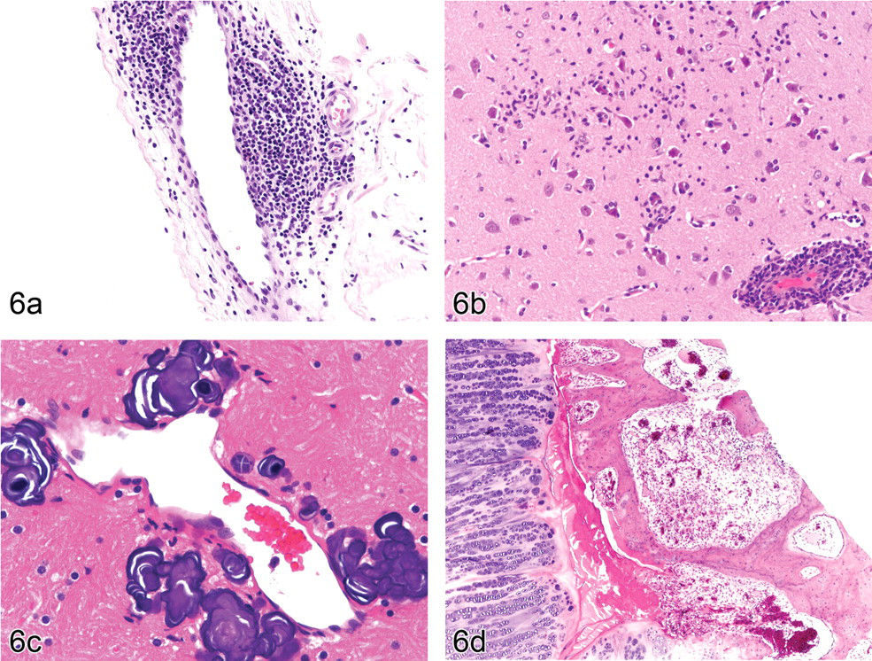
Figure 6(a). Spontaneous perivasculitis or cuffing of a medium-sized vein in the meninges of the spinal cord of a young male cynomolgus monkey (Macaca fascicularis). H & E, × 200.
Deeply basophilic staining or dark purple mineralized bodies were observed with some frequency in the various parts of the basal ganglia such as the globus pallidus and the putamen (Figure 6c). Smaller mineralized bodies were often perivascular in distribution and were associated with other changes in the perivascular space such as pigment accumulation or some small amorphous eosinophilic structures. Larger bodies were arranged in concentric onion ring circles that did not always exhibit a proximity to blood vessels. Coronal sections passing through the mammillary bodies showed most mineralized areas, and the incidence of this finding was a direct function of the number of coronary sections taken, as dictated by the study protocol.
Focal gliosis with neuronal necrosis and neuronophagia, focal glial scars with pigmented microglial cells, and some small spheroid bodies were some of the rare but unexpected and unexplained incidental findings in the brain. Eye findings were extremely rare, and only inflammatory cell infiltration (lymphocytic) of the ciliary body, uvea, or conjunctiva was ever recorded.
Lesions of the skeletal muscle were also rare and limited to focal myositis resulting from trauma; vaccine granulomas characterized by an aggregate of large, foamy histiocytes within the thigh muscle; and sarcocystis parasites found mostly within the tongue muscle and not associated with inflammation. Skeletal lesions were confined mostly to traumatic fractures and developmental or idiopathic bone and cartilage abnormalities such as physeal osteochondrosis or dyschondroplasia and metaphyseal dysplasia consisting of foci of persistent hypertrophic chondrocytes. Some cases of metaphyseal dysplasia resembling rickets were often associated with growth plate (micro) fractures (Figure 6d).
Discussion
The main aims of this study were to investigate and outline the incidences of the most common background findings in control cynomolgus macaques used in preclinical toxicology studies and briefly describe some unusual and significant findings that may be confused with treatment-related findings. There are very few reports of this kind in the literature (Drevon-Gaillot et al. 2006; Ito et al. 1992; Lowenstine 2003; Shimoi et al. 1998). Most other published reports are based on studies in zoo or wild-caught primates or in older animals (Bennett et al. 1998; Scott 1999), which predictably have a spectrum of pathological findings that is slightly different from that of laboratory-raised animals used in toxicological studies.
The findings in this study were generally consistent with the incidence and occurrence of similar lesions described in the literature. These findings include gastritis (Drevon-Gaillot et al. 2006; Scott 1999), interstitial nephritis (Bennet et al. 1998; Drevon-Gaillot et al. 2006), inflammatory lesions of the heart (Chamanza et al. 2006; Drevon-Gaillot et al. 2006; Keenan and Vidal 2006), inflammatory cell infiltrations in the liver (Foster 2005; Ito et al. 1992), tension lipidosis of the liver (Drevon-Gaillot et al. 2006), mineralization in the adrenal gland (Kast et al. 1994; Majeed and Gopinath 1980), pneumoconiosis in the lungs, pigment in the lamina propria of the small intestine (Drevon-Gaillot et al. 2006; Scott 1999), and lung findings associated with continuous intravenous infusion (Lilbert and Burnett 2003). In addition, a well-presented and extensive review of incidental findings and nonlesions in primates and their possible etiologies has also been published (Lowenstine 2003), and most of the findings discussed in this particular paper were evident in our study. However this paper presents up-to-date results of incidences and prevalence of a wider range of spontaneous findings by organ system and from a large population of laboratory-bred animals from a single laboratory with consistent husbandry.
The causes of a few of the more common findings such as gastritis, heart findings, and lymphoid hyperplasia in various organs in laboratory primates remain unclear, but various etiologies have been proposed. Helicobacter species (H. pylori and H. heilmannii–like organisms) are suspected of playing a causative role in gastritis (Reindel et al. 1999). However, the association between these bacteria and clinically significant gastritis in most cases in this study was not conclusive. In fact, most clinically apparent ulcerative gastritis cases with little inflammation present did not have evidence of bacterial organisms, whereas large numbers of bacteria could be seen in normal stomach sections or sections with minimal to mild inflammatory cell infiltrates, leading to the suggestion that stress might have a role in some forms of gastritis in primates.
Another intriguing finding in the stomach was idiopathic gastric infarction. The causes of the few cases of this condition encountered in this study were never established, but gastric infarction is reported in the literature to be associated with an underlying clinical condition such as pancreatitis or cystitis in cynomolgus monkeys (Fikes et al. 1996). However, in man, gastric infarction is also known to occur in young children with no obvious explanation (Wanek et al. 1988).
The specific causes of spontaneous findings in the kidneys of laboratory-bred animals are also not well documented. However, in general, glomerulonephritis in nonhuman primates is associated with infectious agents or conditions that cause chronic antigen–antibody formation (Bennet et al. 1998). In captive-bred animals, such conditions include long-term catheterization or dental disease. Interstitial nephritis, on the other hand, has not been associated with any etiological agent in normal, healthy, laboratory-bred animals. It was also reported to be the most frequently observed lesion at necropsy in zoo primates, but no possible cause was suggested (Bennet et al. 1998).
High incidences of inflammatory lesions of the heart have been reported in cynomolgus monkeys, in which stress and the release of catecholamines were suggested as possible causes (Chamanza et al. 2006). Catecholamine-induced stress was also considered to be the most likely cause of spontaneous cardiomyopathy in cynomolgus monkeys in a recent report (Zabka et al. 2009). Findings such as hypertrophic cardiac myofibers, karyomegaly, and vacuolation have been reproduced in a Rhesus macaque (Macaca mulatta) with active angiomatous pheochromocytoma (Vogel and Fritz 2003) or injected with catecholamines (Khullar et al. 1989). Repeated bouts of endogenous catecholamine release associated with captivity and routine manipulation during toxicology studies is believed to cause focal or multifocal damage to the myocardium (Keenan and Vidal 2006; Lowenstine 2003). Intranuclear (pseudo) inclusion bodies in hypertrophic cardiac myofibers resulting from intranuclear invagination of cytoplasmic organelles, including mitochondria, have been reported in man (Engedal et al. 1977).
One of the most common observations in general was the frequency and extent of lymphoid hyperplasia in various organs in control study animals. Formation of lymphoid follicles in the bone marrow, salivary glands, stomach, lungs, and urinary bladder and the presence of hyperplastic or prominent lymphoid follicles in the spleen are considered to indicate heightened nonspecific immunosurveillance in these animals that have been reared in a relatively disease-free environment and are known to be free from major pathogens. Periodontal disease and tonsillitis are other common causes of lymphoid hyperplasia and granulocytic infiltrates in the submandibular lymph nodes.
The other objective of this study was to report and describe a selection of some unusual or uncommon findings that have been observed in control animals, some of which are of toxicological significance as they may be misinterpreted as treatment-related findings. Some of the findings such as myocardial karyomegaly, myocardial degeneration/fibrosis (Keenan and Vidal 2006; Zabka et al. 2009); mucopolysaccharide accumulation in the subendocardial areas, heart valves, great vessels and aorta (Scott 1999); collagenofibrotic-like lesions of the glomeruli (Adachi et al. 2005); inflammatory cell infiltration of the uvea (Sinha et al. 2006); lymphocytic thyroiditis (Guzman and Radi 2007); pigment deposits in the lung (Drevon-Gaillot et al. 2006; Ito et al. 1992); mineralization in the brain (Wadsworth et al. 1995; Yanai et al. 1994); adreno-hepatic fusion (Mousa and van Esch 2004); accessory spleen in the pancreas (Lau 1973); cuboidal metaplasia of the parietal layer of the Bowman’s capsule (Kaspareit et al. 2004); and subcapsular hemorrhage and fibrosis in the liver (Shimoi et al. 1998) have all been reported previously. However, mineralization of adventitial blood vessels and unidentified vascular remnants suspected to be vestiges of umbilical blood vessels (also seen in the dog) have not been reported to the best of our knowledge. We have also reported on some unusual findings such as focal gliosis and glial scars in the brain, inclusions in hypertrophic cardiac myofibers, and uncommon presentations of ectopic tissue in various organs, which have not previously been reported in cynomolgus macaques.
There were more similarities than differences between this present study and previous studies reporting on incidences of background lesions in control cynomolgus monkeys. Any differences could have resulted from differences in the age, source, and genetic background of the animals, the predominant study types conducted, management and housing, variations in recording levels among study pathologists, and the tissue harvesting methods and trimming planes employed by the laboratory. For instance, the incidence of findings such as accessory spleen, which were found almost exclusively at the tail of the pancreas, depended on the trimming plane for this organ and a careful inspection at necropsy, whereas the incidences of minor and largely insignificant findings such as pigment in the lamina propria of the small intestine, the cytokeratin (tonofilaments)-associated cytoplasmic (pseudo)-inclusions in the uroepithelium, and intraepithelial pigment in renal medullary rays depended on the recording levels and experience of the evaluating pathologist. Because the majority of our animals were from Mauritius, no attempt was made to analyze the results based on the geographical origin of the animals.
To the best of our knowledge this is the most comprehensive study of the incidences, range, and descriptions of background lesions in control cynomolgus monkeys used in toxicity studies reported in recent years. It should facilitate the differentiation of spontaneous from induced lesions in toxicological safety studies in this species. Further work on specific organ systems or in other species of laboratory primates used in preclinical safety testing is encouraged so as to provide more detail and increase the available literature on laboratory nonhuman primates.