
Abstract
The use of the miniature swine as a nonrodent species in research has continued to expand for over a decade, and they are becoming routinely used both in experimental pharmacology and as a therapeutic model for human diseases. Miniature swine models are regularly used for studies designed to assess efficacy and safety of new therapeutic compounds given through different routes of exposure and are used as an alternative model to rodents, canines, or nonhuman primates. Translational preclinical swine study data presented here support the current understanding that miniature swine are the animal model of choice for the assessment of drugs targeting endocrine, dermal, and ocular disorders. Because research investigators need to be familiar with some of the important features of the models developed in the miniature swine in order to place clinical and experimental findings in their proper perspective, relevant references and data from these models will be presented, compared, and partially illustrated.
Keywords
Overview
While human clinical trials work through clearly defined phases to evaluate the various effects of new therapeutic entities (new chemical entities [NCEs] or new biological entities [NBEs]), trials utilizing animal models of disease are less defined and mainly conducted in support of a therapeutic rationale. These animal studies are sometimes referred to as proof-of-concept trials, and their main objective is to be able to translate pharmacology and efficacy data from nonhuman species into humans. Because the selection of the most appropriate animal model is an important component of this process, another research objective is to identify animal models that can improve the prediction of efficacy and safety outcomes and thus contribute to decreasing the number of human clinical trials by eliminating early on any chemical or biological drug entities showing lack of effectiveness or presenting severe adverse effects.
Human Clinical Trials
The major goals of phases I and III human clinical trials for NCEs and NBEs are to evaluate their potency and pharmacokinetic, pharmacodynamic, and therapeutic efficacy. Additionally, bioequivalence human clinical trials aim to demonstrate the similar nature of generic or biosimilar products to chemical or biological medicinal products, respectively. Although there are some variations in the definitions of the early phases of human clinical trials, conducting preclinical trials with animal models consistently fits into the general approach of human drug development.
Animal Models
An animal model is defined as any condition found in an animal that is of value in studying a biological phenomenon; it is a pathological mechanism of an animal disorder useful in studying human disease. Animal models may be spontaneous—monogenetic, multigenetic, environmental—or they may be induced, such as surgically, pharmacologically, or by transgenesis. In contrast to human clinical trials, the major objective of animal models of diseases is to be able to translate their clinical pharmacology and therapeutic efficacy to humans in order to identify candidates for clinical trials in drug development selection or to improve the prediction of study outcomes in clinical trial modeling.
There are three major components that make a particular animal model a valuable model for experimental and translational medicine: drug exposure, modeling of the disease, and relevance to human health. The drug exposure component, or comparative pharmacokinetics, relates to the pharmacokinetics and metabolism of the drug of interest; for example, determining whether the animal species is metabolizing the drug in question in a manner similar to human metabolism of the same drug and whether the level of exposure is identical and/or proportionate and can be used to predict human efficacious or safe starting doses. The disease modeling component, or pathology parallelism, relates to the ability of modeling the disease in the species of interest, specifically determining whether the disease can be experimentally induced. Relevance to human health, or translational value, relates to the disease relevance to the human condition, determining whether the same pathological mechanisms are at play or if it is just the manifestation of a phenotypic similarity.
For abovementioned reasons, the identification and characterization of animal models of diseases having a high translational value have become more and more important at a time when the severe limitations of rodent models, due to their smaller size and difference in dermal structure and mechanism of wound healing compared to humans, have been fully recognized. Swine have historically played a role in experimental medicine but have not always been amenable to be a laboratory species due to their prohibitively large size and housing requirements. The development of miniature swine has effectively created a role for swine in laboratories by providing breeds of a more manageable size; the benefits of this are shown in Table 1.
Miniature Swine versus Domestic Swine Use in Pharmacology and Toxicology.
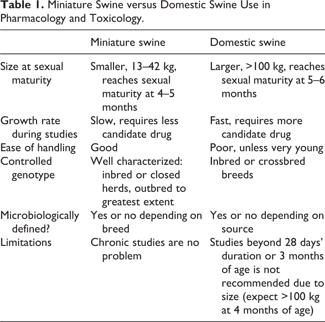
The objective of this publication is to expand on the different ways miniature swine serve as translational models, above and beyond the traditional classical cardiovascular surgical models typically pursued in domestic swine, that are far more valuable than other laboratory animal species for the benefit of human clinical trials and ultimately for the promotion of human health.
Endocrinology Models
Miniature swine are becoming increasingly recognized as models for type 1 diabetes (T1DM) and glucodynamic studies. The comparability of the pharmacodynamic response of these diabetic models with that of humans to known marketed insulin products (both rapid acting and long acting) is the overarching consideration of how to best utilize these models. The pharmacodynamic response to various marketed insulin products in miniature swine should approximate those in humans in order for the model to be both valuable and predictive; apparent differences must be recognized ahead of use. Monitoring of morning fasted blood glucose (FBG) levels in diabetic miniature swine is a necessary element for clinical management and to assess the consistency (through FBG range assessment) for overnight-fasted animals across the pool of available diabetic models.
Induction of Diabetes
Alloxan, or 2,4,5,6-pyrimidinetetrone, is used to induce both T1DM and type 2 diabetes mellitus (T2DM). Alloxan’s molecular shape is similar to glucose, allowing it to be transported into the cytosol of pancreatic beta cells by the glucose transporter 2 located in the plasma membrane. Once in the cytosol, alloxan selectively inhibits glucose-induced insulin secretion with a thiol group that binds to the enzyme glucokinase in the beta cells; alloxan additionally induces redox cycling and generates reactive oxygen species that subsequently induces destruction of the pancreatic beta cells (Rohilla and Ali 2012).
Once induced, multiple types of insulin are used to maintain normoglycemia in the T1DM swine. Although intermediate-acting Humulin® N (Eli Lilly, Indianapolis, IN) is used most commonly, long-acting Lantus® (Sanofi, Bridgewater, NJ) and rapid-acting Humalog® (Eli Lilly) are also used.
Biomarkers of Diabetes
At our present facility, glucose measurements are performed on whole blood with AlphaTRAK® 2 handheld glucometers or on plasma or serum using a Beckman Coulter AU480 clinical chemistry analyzer which uses hexokinase as the reaction enzyme. Circulating insulin and C-peptide levels are measured using a porcine-specific ELISA (Mercodia, Uppsala, Sweden). Typical circulating insulin and C-peptide levels in normal and diabetic Yucatan miniature swine are presented in Table 2. In stabilized diabetic Yucatan miniature swine that were induced through alloxan-induced destruction of pancreatic beta cells, the blood glucose levels increased 7-fold, from a mean of 58.7 mg/dl in normal miniature swine to a mean of 429 mg/dl in diabetic miniature swine, compared to normal miniature swine that were fasted overnight for approximately 18 hr followed by feeding.
Circulating Levels of Insulin and C-peptide in Type 1 Diabetes Mellitus (T1DM) Yucatan Miniature Swine.
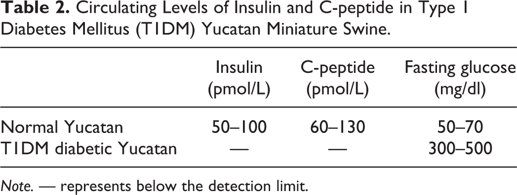
Note. — represents below the detection limit.
Insulin Pharmacodynamics Experimental Design
In order to compare the pharmacodynamic responses, rapid-acting and long-acting insulin products were tested in T1DM for effect onset, peak, and duration times as reflected by changes in FBG, and these data were compared with the reported human parameters. In the miniature swine, typical insulin pharmacokinetic and pharmacodynamic studies designed to collect this information consisted of two different study durations—short term and long term. In short-term studies, animals are generally fasted, and insulin is withheld for approximately 18 hr prior to dosing to allow any systemic maintenance insulin to be eliminated and the blood glucose levels to stabilize. Animals are fed before or at the time of dosing. Blood glucose level is measured using whole blood samples with a handheld glucometer to screen for hypoglycemia, and the remainder of each sample is placed in an appropriate blood collection tube to be processed for plasma or serum collection. In long-term studies, the animals are fed and given their regular maintenance insulin approximately 12 hr before dosing for the study and are not generally fed at the time of dosing. The remainder of the study is similar to a daylong study; the animals are fed and administered regular maintenance insulin immediately upon completion of the last time point of glucose testing for the study.
Type 1 Diabetes
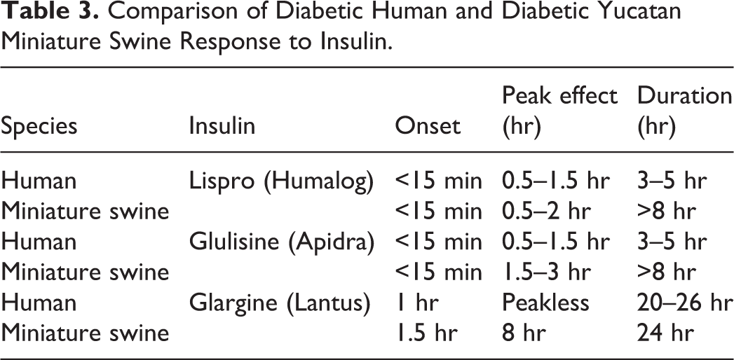
Pharmacodynamic effects from various insulin products with known properties of onset, peak, and duration times in humans were studied in the alloxan-induced type 1 diabetic Yucatan miniature swine, for comparative purposes. For this study, the well-known prototypical marketed insulin products Humalog®, Apidra®, and Lantus® were administered subcutaneously at mealtime to separate groups of animals, then the blood glucose profiles were recorded over the next 8 hr using handheld glucometers for rapid-acting insulin (Humalog®, Apidra®) or over 24 hr for long-acting insulin (Lantus®). Onset, peak, and duration were determined by reviewing the collective group profile figures. Blood glucose profile data in the diabetic Yucatan generally compared well with the published human glucodynamic data for time to onset of effect and peak effect for the rapid-acting insulins tested (Table 3). These data suggest the Yucatan diabetic model, under these conditions, has similar pharmacodynamic responses to humans for onset and peak effects but not necessarily for duration for the rapid-acting insulin. In humans, the long-acting insulin showed similar onset and duration, but it showed a peak effect in miniature swine, but no peak was normally reported at all in humans. These differences in pharmacodynamic response could be due to the duration of fasting, the high doses of insulin administered to the miniature swine, the use of a small number of animals in these trials, or a partial biological difference in the glucodynamic profiles of the various insulins between miniature swine and humans.
Comparison of Diabetic Human and Diabetic Yucatan Miniature Swine Response to Insulin.
Type 2 Diabetes and Metabolic Syndrome
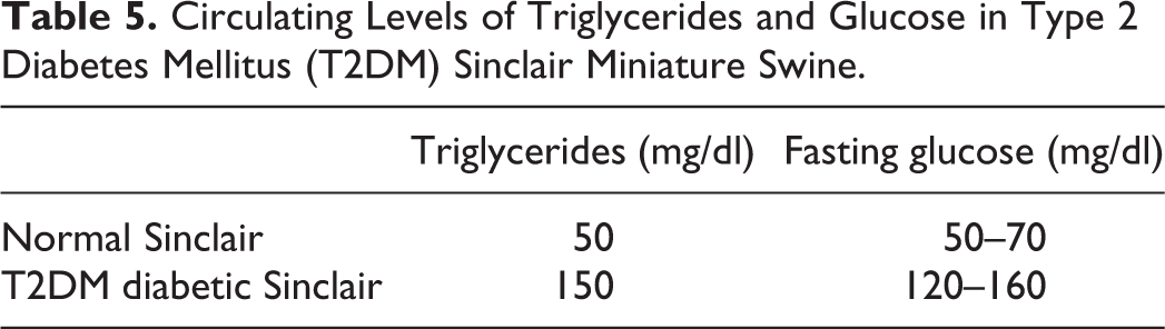
Miniature swine are also an ideal model for T2DM, which has a multifactorial genetic cause in humans, because they are genetically close to humans, susceptible to both spontaneous and diet-induced obesity, have a dyslipidemia profile similar to that of humans and can exhibit all aspects of metabolic syndrome. Type 2 diabetic miniature swine require no extra maintenance and are used for intravenous glucose and meal tolerance testing, incretin studies, and also metabolic studies. Dyslipidemia may be induced in many breeds of miniature swine by feeding a high-fat atherogenic diet as illustrated in Yucatan miniature swine in Table 4. The Yucatan miniature swine have been considered a superior breed for atherosclerosis studies (M. M. Swindle 1992). The Ossabaw pig is also sensitive to diet-induced dyslipidemia (Lee et al. 2009). Sinclair miniature swine, with a origin similar to the Göttingen and Ossabaw miniature swine, readily develop dyslipidemia as well. Adult castrated male Sinclair miniature swine maintained on a high-fat diet for 3 months demonstrate an increase in fasting triglyceride levels and a moderate increase in fasting blood glucose, when low-dose alloxan is administered to partially ablate the population of pancreatic beta cells (Table 5). Type 2 diabetic miniature swine can be maintained at either obese or normal body condition and thus provide many options for studying the various facets of metabolic syndrome from insufficient insulin secretion to reduced insulin sensitivity.
Mean Lipid Profiles of Yucatan Miniature Swine in Response to Being Fed a High-fat Diet.
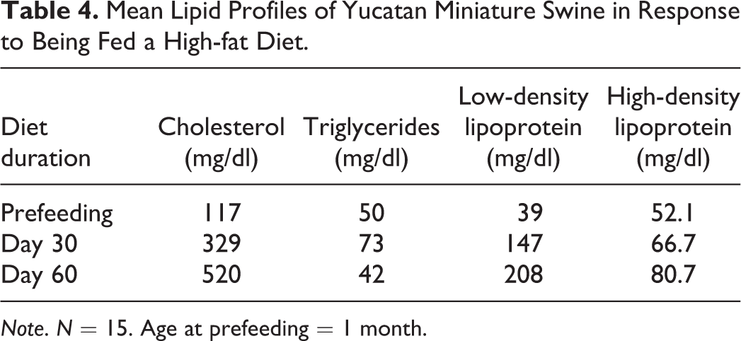
Note. N = 15. Age at prefeeding = 1 month.
Circulating Levels of Triglycerides and Glucose in Type 2 Diabetes Mellitus (T2DM) Sinclair Miniature Swine.
Dermal Models
Swine have been used extensively in dermal research because of the comparability of their integument to that of humans. During the past half century, they have been used in preclinical dermal toxicology, dermal pharmacokinetics, dermal phototoxicity, dermal wound healing studies, and a broad array of other biomedical research applications (Brown, Stricker-Krongrad, and Bouchard 2013; Gad, Stricker-Krongrad, and Skaanild 2015). Reviews of the use of swine in such studies have been previously published (Fujii et al. 1997; Gad, Stricker-Krongrad, and Skaanild 2015; Monteiro-Riviere and Riviere 1996; M. M. Swindle 2007). In the field of toxicology, swine skin has been used for acute and repeat-dose dermal toxicology, dermal absorption, allergic contact dermatitis, phototoxicity, and photosensitization studies. Models have been created both in vivo and in vitro with skin membranes and grafts. Both miniature and domestic breeds have been used for these types of studies; however, miniature breeds such as Sinclair, Yucatan, Hanford, and Göttingen may be more advantageous due to their smaller size at sexual maturity. Using these miniature breeds allows investigators to conduct experiments in mature (rather than pediatric) animals with a consistent size and health status. Each breed may be utilized in some aspect of dermal toxicology (Brown, Stricker-Krongrad, and Bouchard 2013; Gad, Stricker-Krongrad, and Skaanild 2015; Svendsen 2006; M. M. Swindle et al. 2012). The value of miniature swine dermal models for preclinical safety is confirmed by Ganderup (2012) who reviewed miniature swine safety and efficacy data on 43 marketed drugs with previously reported adverse responses. Approximately 50% of the reviewed drugs had a dermal indication, and 27 drugs had both human and miniature swine data to enable a comparison. Overall, the predictive value of miniature swine safety and efficacy studies to human outcomes of all reviewed drugs were 89% and 100%, respectively. Select models of the expanding landscape of dermal studies and applications discussed below include transdermal absorption, skin stripping, evaluating topical reactions, and various aspects of wound healing.
Comparative Anatomy and Function
Dermal anatomic and physiologic similarities between miniature swine and humans include a sparse hair coat, a relatively thick epidermis, epidermal turnover kinetics, lipid composition, lipid biophysical properties, and arrangement of dermal collagen and elastic fibers. The differences are the interfollicular muscle, the distribution and function of apocrine versus eccrine sweat glands, thickness of the stratum corneum (SC), the basement membrane epitopes, and cytochrome P-450 biotransformation isoenzymes (Svendsen 2006; M. M. Swindle et al. 2012).
Transdermal Absorption
In general, the miniature swine is accepted as an appropriate model for topical agent testing, and skin penetrance is second only to macaques in its similarity to humans for both lipophilic and hydrophilic drugs. There are other factors that make the miniature swine ultimately superior to macaques as an animal model, including the adhered dermal structure, much less hairy surface area compared to macaques, and their ease of handling. Human permeability is higher than pigs for most compounds tested (Panchagnula, Stemmer, and Ritschel 1997), but miniature swine are still a recognized predictive model for human drug candidate dermatopharmacology studies (Simon and Maibach 2000).
Skin Stripping for Dermal Penetration
Tape stripping is a simple and effective method for removing the SC (Figure 1) and is commonly employed during in vivo studies investigating the percutaneous penetration and disposition of topically applied candidate drugs as well as in investigations of drugs intended to restore damaged epithelial barriers (Escobar-Chavez et al. 2008). One study was performed with the objective to assess the remaining thickness of the SC following 0, 10, 20, 30, 40, and 50 repetitions of tape stripping of skin on 3 young adult male Yucatan miniature swine weighing 33–36 kg each. Following the clipping of the pelage over the dorsal lumbar and thoracic areas, six 5 cm × 5 cm sites were demarcated, and the skin was stripped using 1.8-mm clear acrylic adhesive tape applied with uniform, firm pressure. The analysis of results by light microscopy showed an inverse pattern of SC thickness to the number of tape stripping repetitions. After 20 strippings, the number of remaining SC layers was reduced from 11–15 to 2–6, and 50 passes were required to remove nearly all the SC in each animal. No immediately detectable underlying changes were observed in the epidermis or dermis. These data demonstrate that miniature swine skin can be stripped of SC in a linear fashion based upon repetition of the technique and suggest that this is an acceptable model in miniature swine.
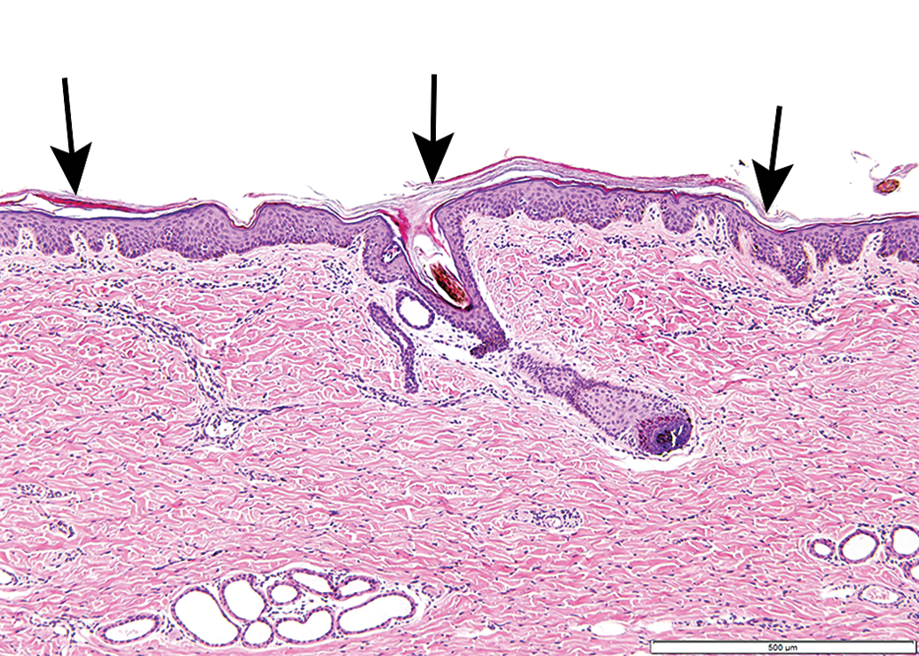
Hematoxylin and eosin staining of normal Yucatan miniature swine skin. Arrows point to the stratum corneum, the layer removed with tape stripping.
Clinical Evaluation of Topical Reactions
Draize scoring, developed by John Draize (1951), provides a method of grossly assessing the degree of inflammation based on quantifying the values that are at risk of interpretation bias or being considered insignificant. Originally developed for use in rabbits, it has been modified for use in both swine and humans and thus is often referred to as the modified Draize score. In dermal studies, erythema and edema need quantified analysis. Erythema and edema are each graded on a scale ranging from 0 to 4, with a maximum total possible score of 8 for irritation encompassing both erythema and edema. For edema, this ranges from 0 = “no edema” to 4 = “severe edema raised >1 mm and extending beyond the area of exposure”; and for erythema, this ranges from “no erythema” to “severe erythema or slight eschar formation.” Slight, well defined, and moderate to severe are graded as 1, 2, and 3, respectively.
Dermal Inflammation
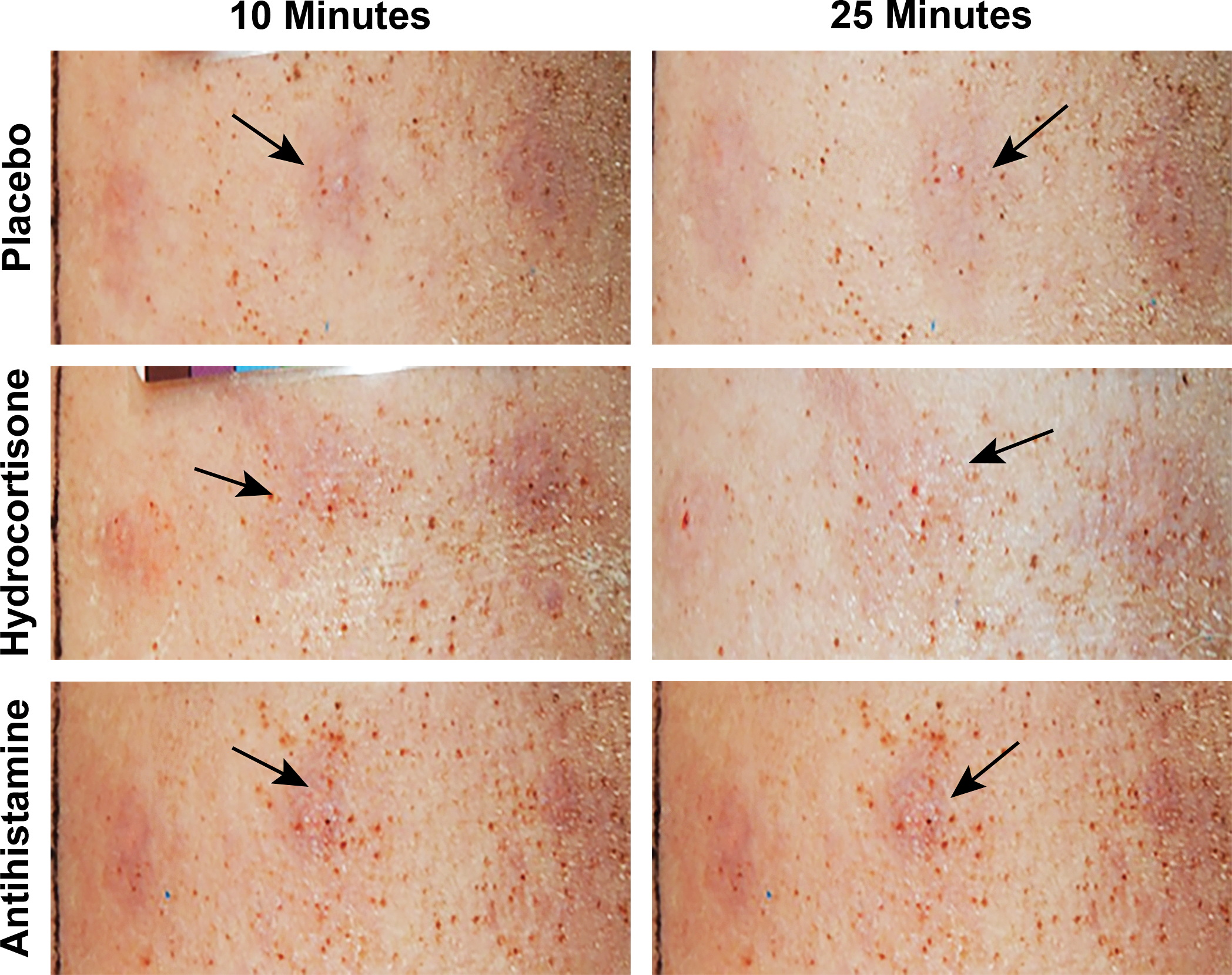
Urticaria is dermal edema that results from vascular dilation and leakage of fluid into the skin in response to molecules released from mast cells. Human studies assessing efficacy of topical and oral therapies for the treatment of urticarial reactions frequently involve inducing wheal and flare reactions with intradermal injections of histamine (Clough, Boutsiouki, and Church 2001; Cole, Clough, and Church 2001; Reddy and Singh 1976). To develop a comparable and reproducible dermal urticaria model in miniature swine, Hanford miniature swine, ages 3–18 months, were pricked on their backs with a skin test device (Lincoln Diagnostics Inc, Decatur, IN) that was loaded with a vehicle or varying concentrations of histamine. The irritation and wheal and flare responses of the dermis were visually evaluated with Draize scoring, as described above, and wheal size measurement. The reactions of the skin were assessed at 10, 20 or 25, and 45 min after test formulation application. Histamine dose-dependently induced skin irritation at both 10 and 20 min after treatment, and responses began diminishing at 20 min after treatment (Table 6). The most prominent erythema and edema responses were observed at 10 min after treatment, which slightly diminished 20 min after treatment. Histamine also caused skin wheals that ranged between 4 and 7 mm in diameter. After establishment of urticarial responses, the miniature swine were pricked on their backs again with 25 or 50 mg/ml histamine, and the test sites were then topically treated with placebo, hydrocortisone cream, or antihistamine cream after 15 min in order to determine whether the histamine-induced wheals would then respond to commonly available over-the-counter products. Both hydrocortisone and antihistamine reproducibly inhibited urticaria that was induced by 25 mg/ml histamine, and both had an even more robust effect on the more intensive urticaria induced by 50 mg/ml histamine (Figures 2 and 3). Noting that the human studies evaluated the preventive effects of therapeutic agents by treating before implementing histamine-induced wheal and flare (Clough, Boutsiouki, and Church 2001; Cole, Clough, and Church 2001; Reddy and Singh 1976), it is expected that the response to treatment in the present model is reduced compared to what it would be if the same preventive process was studied. Therefore, this urticaria model can currently be used for the testing topical treatments for dermal irritation and inflammation; it also has the potential to expand and be further developed as a model.
Effects of Different Concentrations of Histamine on Dermal Irritation in a Hanford Miniature Pig Measured with Draize Scoring and Wheal and Flare.
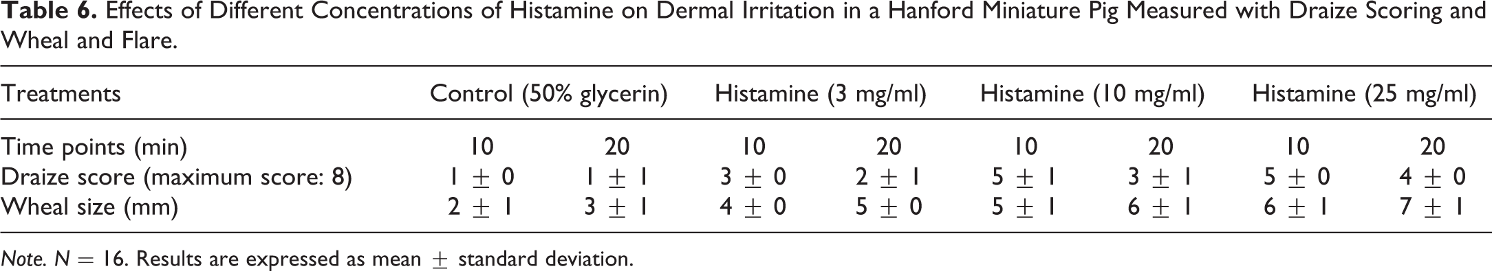
Note. N = 16. Results are expressed as mean ± standard deviation.
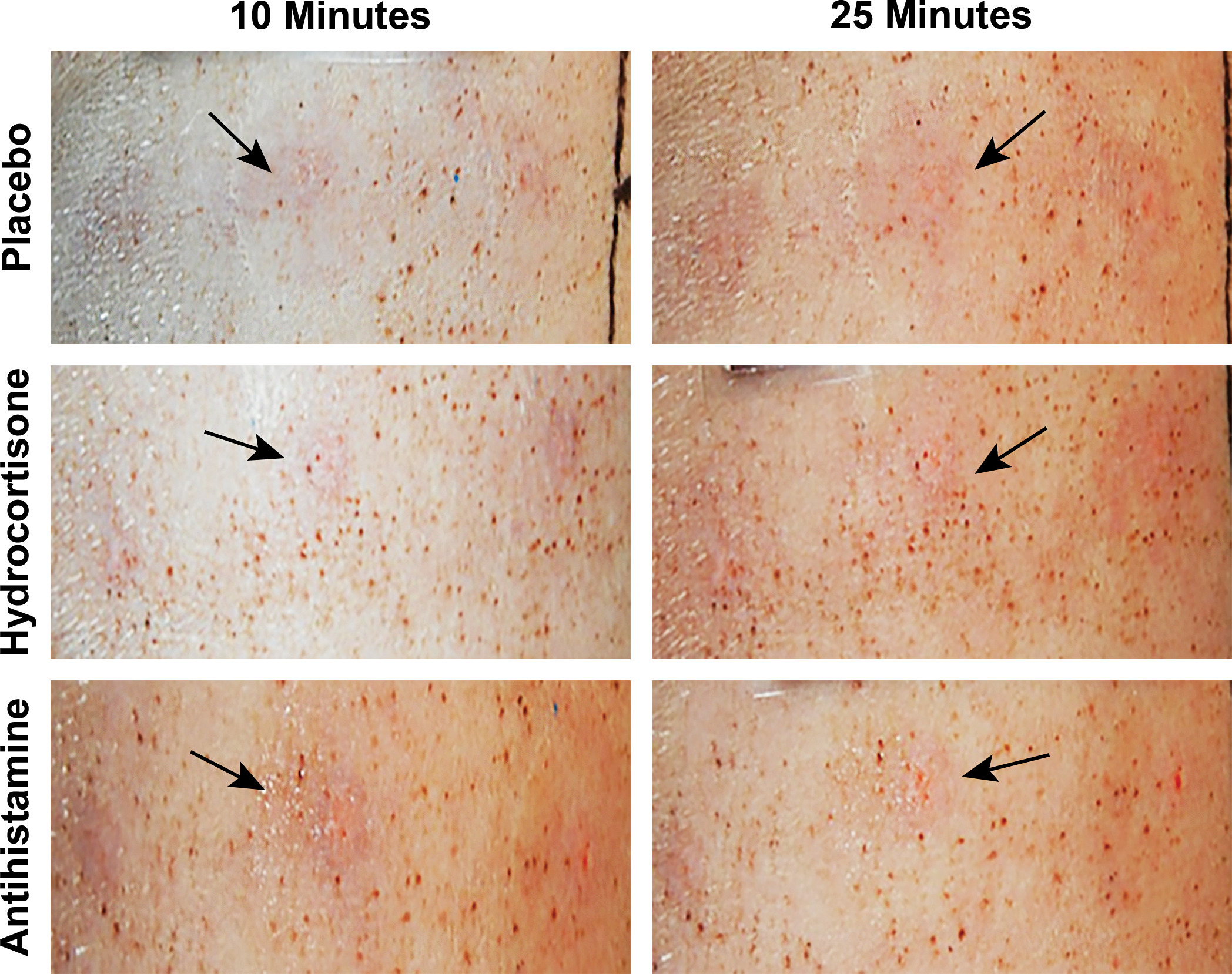
Observation of urticaria resolution 10 and 25 min after treatment with placebo, hydrocortisone, or antihistamine, beginning 15 min after pricking with 25 mg/ml histamine. Arrows point to areas of urticaria. Responses 45 min after treatment are not shown; urticaria was almost completely resolved in both hydrocortisone and antihistamine treatment groups.
Observation of urticaria resolution 10 and 25 min after treatment with placebo, hydrocortisone, or antihistamine, beginning 15 min after pricking with 50 mg/ml histamine. Arrows point to areas of urticaria. Responses 45 min after treatment are not shown; urticaria was almost completely resolved in both hydrocortisone and antihistamine treatment groups.
Psoriasis is a long-lasting autoimmune disease characterized by patches of abnormal skin. These skin patches are typically red, itchy, and scaly. They may vary in severity from small and localized to diffuse. In the miniature swine, daily intradermal challenge with chemokine (interleukin [IL]-23) induces a psoriasis-like skin phenotype, with erythema, epidermal hyperplasia, hyperkeratosis, parakeratosis, spongiosis, and dermal leukocyte infiltration. Eight 3 cm × 3 cm sections are delimited within the dorsal scapular to dorsal lumbar region on each animal, 4 sections on each lateral side. Each animal receives daily intradermal administration of IL-28 within each section for 7 consecutive days. Each section is scored for plaque characteristics such as erythema, scaling, and induration using a modified psoriasis area and severity index introduced by Fredriksson and Pettersson (1978). Skin histology can be performed to assess epidermal thickness, parakeratosis, and marker of angiogenesis and proliferation by immunohistochemistry (e.g., CD31 and Ki67, respectively). This model can subsequently be utilized to evaluate the response to treatment using products that are being developed.
Wound Healing
For many decades, swine have been a standard model for wound healing. Rabbit and rodent models are used in wound healing mainly due to lower expense and can be used for screening tests; however, they have significant differences from humans including a dense pelage and a thin epidermis and dermis, and they heal predominately through wound contraction. Since pigs have fixed skin, their gross healing characteristics including having similar elastic properties quantify them as being similar to humans. Swine and humans have a comparable dermal–epidermal ratio of 10–13:1, and the actual skin thickness varies between regions of the body (Sullivan et al. 2001). Reepithelization is an important component of wound healing in swine as in humans. Farm pigs may have exaggerated wound healing because of their rapid growth characteristics; miniature swine have been used for chronic wound healing models because they more accurately simulate adult human healing rates (Chvapil and Chvapil 1992).
Excisional and Incisional Wound Healing
Partial thickness cutaneous wounds in swine undergo significant reepithelization as a primary component of the healing process. Partial thickness wounds are created with a dermatome that can be set at varying depths. The size of the lesions created varies between studies, but generally they are square lesions within the range of 3.0 cm on each side (M. M. Swindle 2007; Bolton, Pines, and Rovee 1988; Chvapil 1992; Mertz, Hebda, and Eaglstein 1986; Ordman and Gillman 1966; Kerrigan et al. 1986; Sullivan et al. 2001; Wang et al. 2001; Singer and McClain 2003; Middelkoop et al. 2004). Full-thickness excisional wounds are created to the depth of the fascia using either scalpel incisions or biopsy punches. These deep wounds have wound contraction and granulation as the initial predominate healing processes. Excision wounds at this depth made with a scalpel are generally of 3 to 5 cm size on each side. Circular lesions of 1-2 cm diameter may be created with biopsy punches. These wounds tend to heal by development of scar tissue. The depth of the wounds varies from 0.8 to 1.5 cm depending upon location on the body. The most common area is bilaterally on the flank. Wounds of this size usually have a volume of 10 to 15 ml. Total number of full-thickness wounds are generally limited to 8 per animal. Wounds of this size would be expected to achieve 40% reduction with granulation in 7 to 8 days (M. M. Swindle 2007; Chvapil 1992; Middelkoop et al. 2004). Incisional wounds are made using a scalpel and are also studied for testing new closure devices or suture materials. The depth and location may vary depending upon the goals of the study. Incisional wound healing is quicker than the other types of wounds, and each animal should be used as its own control.
Geometric Determinants of Wound Healing
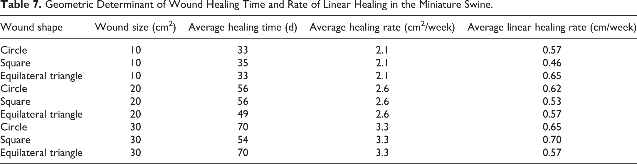
To investigate the relationships between wound geometry and wound healing rates, wounds of varying size and shape were created in adult Yucatan miniature swine. The primary outcomes of interest were time to complete healing (defined as full epithelialization), healing rate (defined as absolute change in area from baseline over time), and linear healing rate (defined as change in wound “radius” over time). Although larger wounds took longer time to heal than the smaller ones, there was no appreciable difference in healing rate associated with the wound shape, as shown in Table 7. For example, the average healing time for 20 cm wounds was 56, 56, and 49 days for circles, squares, and triangles, respectively. Mean healing rates were 2.1, 2.6, and 3.3 cm2 per week for 10, 20, and 30 cm2 wounds, respectively. Results for the outcome of absolute wound healing rates were calculated using planimetry data. There was a strong correlation between healing rate and initial wound area. It was determined that linear healing rates were largely unaffected by either initial wound area or wound shape.
Geometric Determinant of Wound Healing Time and Rate of Linear Healing in the Miniature Swine.
Ischemic Wound Healing
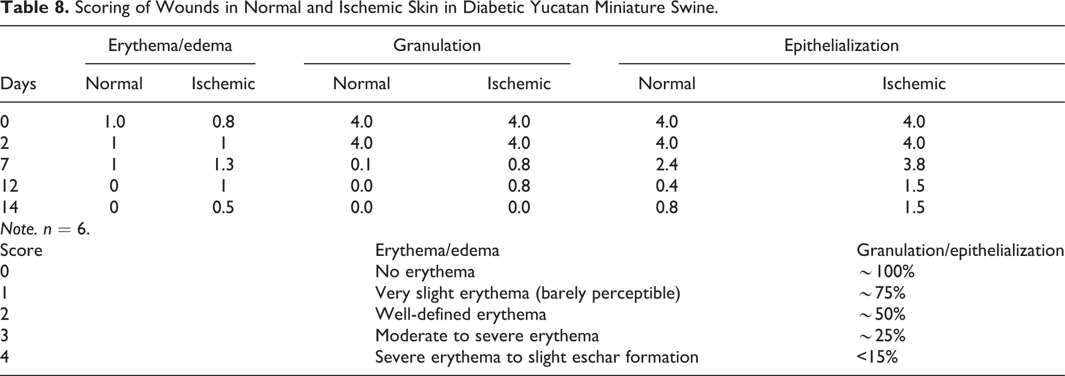
Swine have long been utilized as models to study the effects of treatments on skin flaps and grafts due to their physiologic similarities to humans, as discussed above (M. M. Swindle 2007). Kerrigan et al. (1986) published a detailed summary of the various types of flaps and grafts studied in plastic surgery. Skin flaps are generally made on the dorsal flank, the buttocks, or the limbs, and they are generally full-thickness flaps. Tissue ischemia of wounds in diabetics is a consequence of developing vascular complications, which limit the supply of blood and blood-borne products to the wound site and severely impairs the wound healing process. In order to establish a model of chronic ischemic wound healing in diabetic miniature swine, the wound healing process was investigated in bipedicle ischemic flaps in chemically induced diabetic miniature swine. Bipedicle flaps (5 cm × 15 cm) were created, a silicone film was placed underneath some of the bipedicle flaps to create ischemic conditions, then 0.8-cm diameter center punches were created and scored during healing for erythema, edema, granulation, and epithelialization (Table 8). Erythema and edema were more prominent in ischemic skin wounds. Additionally, granulation and epithelialization were delayed in ischemic skin wounds, with the delay in epithelialization being the most noticeable difference between the two wound types.
Scoring of Wounds in Normal and Ischemic Skin in Diabetic Yucatan Miniature Swine.
Ophthalmology Models
As with the endocrine and dermal models, the use of miniature swine for ophthalmology models is varied and expanding. The anatomic similarities to human eyes, along with comparable physiologic processes, enable miniature swine to be good candidates for surgical procedures and reliable for model development and subsequent testing of potential therapeutic agents (Kyhn et al. 2008; Rosolen, Rigaudiere, and Le Gargasson 2003; Sachs et al. 2005).
Comparative Anatomy and Function
The orbit of swine is open at the lateral aspect, as compared to the closed orbit of humans in which the eye is completely enclosed by bone. There are 7 extraocular muscles attached to the orbital wall in deep fossae, while humans have 6. In addition, humans have an annular ligament, the annulus of Zinn, surrounding the optic nerve that is absent in swine (M. M. Swindle 2007; Adams 1988; Curtis, Edwards, and Gonyou 2001). Pigs, unlike humans, have a translucent nictitating membrane that crosses the eye horizontally from the medial canthus; this membrane serves as a protective “third eyelid” and contains a nictitans gland for lubrication. The globe dimensions range from 19.6 to 25.0 mm in height, 21.9 to 25.1 mm in width, and 19.4 to 22.4 mm in depth. The cornea is horizontally oval in shape. The sclera and iris are usually pigmented. The retina is very similar to humans (M. M. Swindle 2007; K. E. Swindle and Ravi 2007; Reilly et al. 2008, Ruiz-Ederra et al. 2005; Shafiee et al. 2008; Kyhn et al. 2008; Iandiev et al. 2006; Czajka et al. 2004; Sachs et al. 2005; Petters et al. 1997). The pig does not have a tapetum lucidum, a true macula, or a fovea; they do have a cone-rich visual streak (Fernandez de Castro et al. 2014). Pigs have binocular vision and some degree of color vision.
Uveitis
A miniature swine model of uveitis is induced by an acute intravitreal injection of 200 ng bacterial lipopolysaccharide in saline under anesthesia. All miniature swine are examined with slit-lamp microscopy and indirect or direct ophthalmoscopy throughout days 1 to 6 postinjection. Corneal neovascularization, vitreous opacity, and anterior chamber cells and flare are evaluated, and clinical scoring is performed using Hogan’s classification (Hogan, Kimura, and Thygeson 1959; Kimura, Thygeson, and Hogan 1959). Aqueous and vitreous are collected at necropsy; the presence of leukocytes in the vitreous is evaluated by a total cell count using a hematocytometer, and a differential cell count is performed in the aqueous solution under a microscope after staining. In addition, inflammatory cells in sections of the anterior and posterior segment structures are quantified by a grading scale. Drug treatments, such as methotrexate, mercaptopurine, corticosteroids, or triamcinolone can be evaluated in this model (Gilger et al. 2013).
Retinal Detachment
The predominant number of porcine ophthalmic models in the literature involves the retina because of the anatomic and physiologic characteristics described above. Retinal detachment in humans can develop for a variety of reasons including trauma and metabolic disorders. The condition can be created surgically in swine (Iandiev et al. 2006). A lateral canthotomy is created, and a circumscript vitrectomy is performed in the region of the detachment. The vitreous is replaced with physiologic saline. A subretinal injection of saline followed by 0.25% sodium hyaluronate is administered using thin glass pipettes. This results in a rhegmatogenous detachment of the retina in the selected area. Clinical ophthalmic examinations are performed postoperatively, and the vitreoretinopathy is graded on a scale of 0 to 5 using the Silicone Study Classification System (Machemer et al. 1991). Briefly, grade 1 indicates an inner retinal wrinkling, while grades 3–5 indicate the number of quadrants with retinal detachment. Using this model, drug therapies and experimental surgical corrective treatments can be tested (Boone et al. 1996).
Cataracts
Diabetic miniature swine are routinely screened for clinical ocular abnormalities including visible “mature” cataracts (Table 9, Figure 4). Over the course of a 6-month period, the prevalence was 30% (80 positive animals of 266 animals). The most recent incidence (past 2.5 months) was 20.4% (38 positive animals with 60 affected eyes from a pool of 186 previously negative animals). Eighteen animals had bilateral and 20 animals had unilateral cataracts (oculus dexter: 31 and oculus sinister: 29). Cataract onset ranged from 2 to 19 months postinduction of diabetes with an average onset of 11 months. Cataracts were detected earlier in animals when euglycemia was intentionally less controlled, which supports the current predominant theory of glycation-induced cataract development. Interestingly, swine, unlike humans, are not capable of glycating their hemoglobin due to the lack of penetration of glucose into the red cells. Miniature swine with cataracts appear to function acceptably well despite the assumed visual handicap.
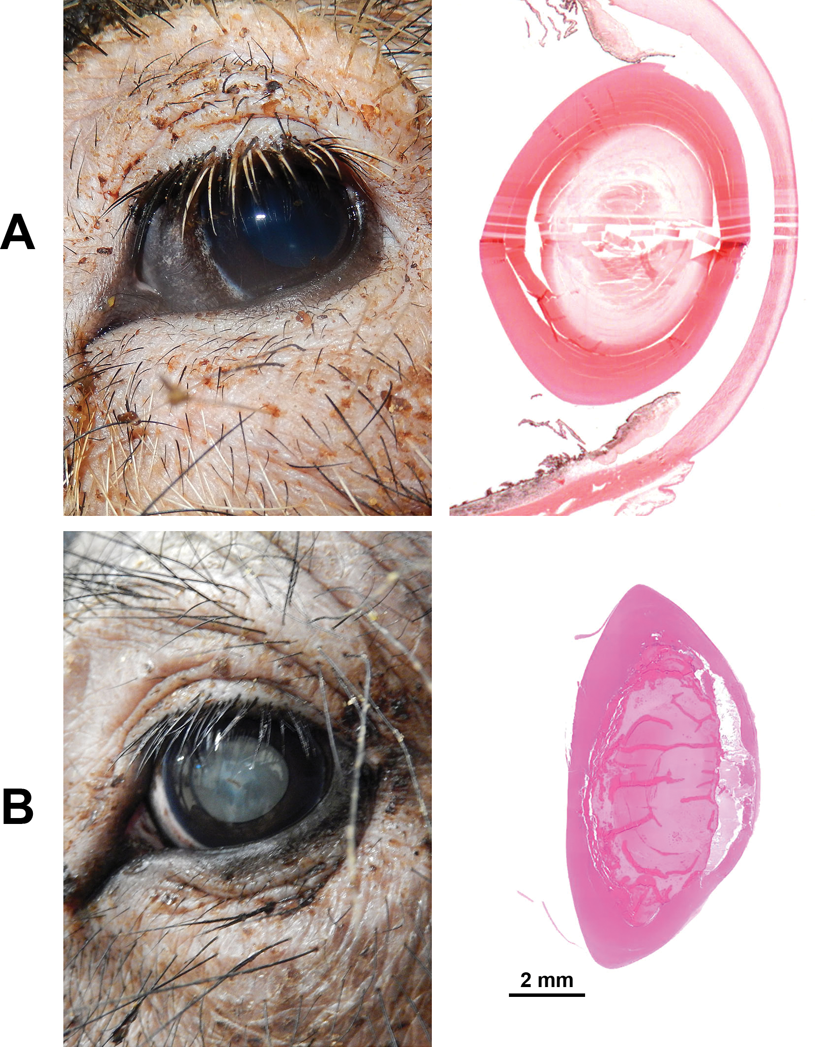
Gross and histologic comparison of normal (A, top) lens and lens with cataracts (B, bottom) in Yucatan miniature swine. Hematoxylin and eosin stain.
Prevalence of Cataracts over a 2.5-month Period in Diabetic Yucatan Miniature Swine.a
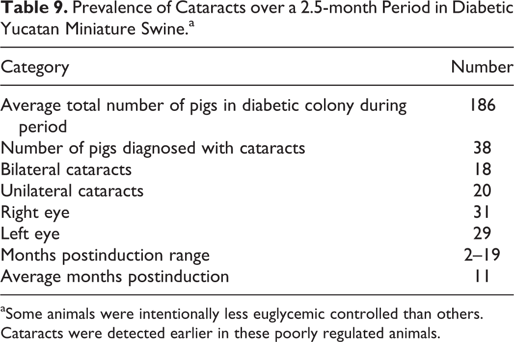
aSome animals were intentionally less euglycemic controlled than others. Cataracts were detected earlier in these poorly regulated animals.
Glaucoma
A model of glaucoma via surgical induction of increased intraocular pressure (IOP) has been created in Yucatan miniature swine. In this study, animals had bilateral IOP measurements performed prior to surgical intervention to establish a baseline IOP. IOP measurements were taken with a Tono-pen VET™ veterinary tonometer. In order to reduce venous drainage from the eyes, episcleral veins were scarified by cauterization in each eye. IOPs were periodically measured for several weeks postcauterization surgery. Pharmacologic intervention was then begun with a commercially available synthetic prostamide analog with ocular hypotensive activity. Drops were applied once daily, and IOPs continued to be measured. After 7 weeks of daily treatment, eye drops were discontinued, and IOP measurements were obtained continuously.
All animals presented with significant increases in IOP during postsurgical intervention (p ≤ .005) and significant decreases in IOP with pharmacological therapy (p ≤ .0006). Pretreatment mean IOP was 19 ± 4 mmHg, postsurgery mean IOP was 24 ± 5 mmHg, posttreatment mean IOP was 18 ± 4 mmHg, and recovery mean IOP was 20 ± 4 mmHg. Therefore, Yucatan miniature swine could be considered a viable model for surgically induced glaucoma. Also, it has been shown that the miniature swine eye is responsive to pharmacological therapy to reduce IOP and as such could be a potential model for future pharmacological research.
Diabetic Retinopathy
A miniature swine model was also developed and characterized for long-term effects of diabetes mellitus on retinal function and structure. Nine animals were used: 3 were nondiabetic, 3 had been diabetic for 5 to 8 months, and 3 had been diabetic for 13 to 17 months. Electroretinograms were performed under isoflurane anesthesia using a portable, full-field, flash system. A-wave, b-wave, and oscillatory potential (OP) amplitudes were evaluated. At maximum scotopic intensity, some animals had increased a-wave amplitudes when compared with the normal animals. B-wave amplitudes appeared normal or reduced. At standard scotopic intensity, OP amplitudes appeared increased or normal. Animals were euthanized, eyes enucleated, and fixed in 2.5% glutaraldehyde in sodium cacodylate buffer for transmission electron microscopy. Images of capillaries from inner nuclear (INL) and inner plexiform/ganglion cell (IPL/GCL) layers were obtained from each diabetic and each normal miniature swine. Basement membrane thickness was significantly increased (p < .001) in the diabetic miniature swine, both in the INL and in the IPL/GCL. ERG results varied across all animals; the strongest correlation was between basement membrane thickness and OP amplitude. It has previously been found that diabetes is responsible for basement membrane thickening in miniature swine, making them a useful model for studying mechanisms and treatments at various stages of diabetic retinopathy (Hainsworth et al. 2002).
Translational Research
The identification and characterization of animal models and diseases having a high translational value have become more and more important at a time when the limitations of rodent models have been fully recognized. As has been described, the miniature swine can play a role beyond its classical use as a model for surgical studies. The miniature swine is a relevant model for the evaluation of therapeutics aimed at treating endocrine diseases such as T1DM and T2DM, inflammatory diseases, wound healing, dermal diseases other than wound healing, and multiple ophthalmic diseases. The use of miniature swine in biomedical research is continuing to grow. Exciting new advances in model development and the relevance of miniature swine in the study of human disease will continue to increase in prominence in the future.
Footnotes
Authors’ Contribution
Authors contributed to conception or design (AS, CS); data acquisition, analysis, or interpretation (AS, GB); drafting the manuscript (AS); and critically revising the manuscript (AS, CS, GB). All authors gave final approval and agreed to be accountable for all aspects of work in ensuring that questions relating to the accuracy or integrity of any part of the work are appropriately investigated and resolved.
Declaration of Conflicting Interests
The author(s) declared no potential conflicts of interest with respect to the research, authorship, and/or publication of this article.
Funding
The author(s) received no financial support for the research, authorship, and/or publication of this article.