
Abstract

Significance Statement
In this study, we emphasize a giant pituitary adenoma associated with nasal involvement. This highlights that the diagnosis of extended giant pituitary adenoma is essential to avoid accidental injury during surgery.
Introduction
Pituitary adenomas are benign tumors, but can rarely extend into nasal cavity, and may reach giant dimensions before diagnosis. Prolactinomas with a diameter of more than 40 mm are defined as giant adenomas. 1 Magnetic resonance (MR) imaging is a frequently used imaging method in the diagnosis and preoperative workup of these tumors and provides relevant information for surgical planning.1,2 We here present MR imaging findings of a 72-year-old patient with giant prolactinoma extending into the nasal cavity.
Case Presentation
A 72-year-old woman presented with headache and endocrine dysfunction without other history of neurological or systemic complaints. On physical examination, the arterial blood pressure was 165/95 mm Hg in both arms. Nasal examination revealed an exophytic mass sitting on the posterior-superior wall of the nasal cavity. Biochemical evaluation revealed hyperprolactinemia (68.9 ng/mL, range 1.3-47 ng/mL) and low-luteinizing hormone and follicle-stimulating hormone levels (4.6 IU/L, range 5-20 IU/L; 4.01 IU/L, range 5-20 IU/L, respectively). Magnetic resonance (MR) imaging demonstrated a herniation of giant pituitary mass (47 × 52 × 64 mm) into the sphenoid sinus and nasal cavity (Figure 1(A) and (C)). Intense contrast enhancement was seen in the lesion on postcontrast T1-weighted images (Figure 1(B) and (D)). MR image also showed compression to the nasal cavity air column by the originating tumor from the pituitary gland. Concordantly, the diagnosis of the mass was confirmed as giant prolactinoma via clinical, laboratory, and MR imaging findings. Sagittal (A) and coronal (C) T2-weighted MR images reveal a herniation (arrows) of giant pituitary mass (asterisk) into the sphenoid sinus and nasal cavity. Sagittal (B) and coronal (D) postcontrast T1-weighted MR images show a solid mass with intense contrast enhancement.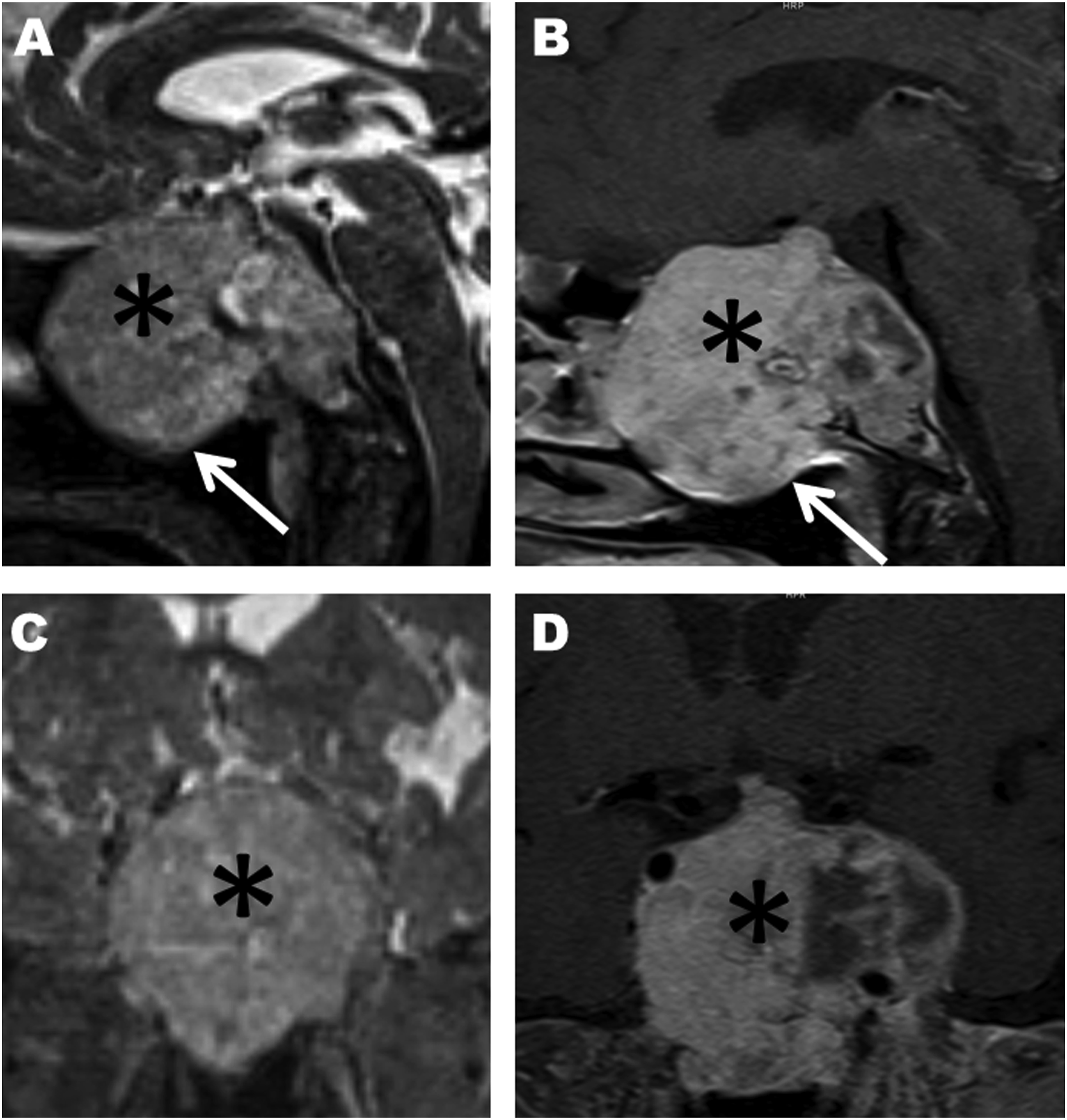
Conclusion
The demonstration with computed tomography (CT) of pituitary adenoma is frequently difficult. However, it can easily visualize the bone defect in the skull base. MR imaging is essential for the evaluation of giant pituitary adenoma to confirm the extent of tumor and the possible infiltrative processes and to plan for the safest surgical approach.1-3 Radiologists must avoid misdiagnosis because giant pituitary adenoma may erroneously mimic sinonasal or nasopharyngeal tumors.
Giant pituitary adenomas are rare benign tumors but can extend into nasal cavity. The ultimate extension and relationship with the adjacent anatomic structures of the tumor can be detected by noninvasive diagnostic methods such as MR imaging. Thus, surgical complications can be significantly reduced.
Footnotes
Declaration of Conflicting Interests
The author(s) declared no potential conflicts of interest with respect to the research, authorship, and/or publication of this article.
Funding
The author(s) received no financial support for the research, authorship, and/or publication of this article.