
Abstract
To describe a rare case of spindle cell tumor in the nasal cavity of a young female, along with its successful surgical management using a low-temperature plasma knife. A 38-year-old female patient presented with a 6-month history of nasal blockage, hyposmia, and local tenderness. The patient denied a history of definite diagnosis or special treatment. A preoperative magnetic resonance imaging scan revealed the size and extent of the tumor in her nasal cavity. She underwent endoscopic surgery for the sinus using a low-temperature plasma knife. The procedure was performed without complications, and her symptoms resolved within 1 month. The postoperative pathology report suggested a spindle cell tumor in her nasal cavity. Nasal spindle cell tumor is rare and usually presents with no specific clinical symptoms, which should be taken into consideration during diagnosis. In this case, we demonstrate that the effectiveness and safety of sinus surgery can be improved and aided by a low-temperature plasma knife and nasal endoscopy.
Keywords
Introduction
Spindle cell tumors are spindle-shaped or short spindle-shaped with varying lengths. 1 Tumors may be derived from either epithelial or mesenchymal tissues, and mostly occur in the breast, calcaneus, skin, and subcutaneous soft tissues.2,3 Spindle cell-predominant tumor types are divided into benign and malignant forms, and their diagnosis relies on pathological examination. 4
A case of a benign spindle cell tumor, referred to as a spindle cell lipoma (SCL), was first described by Enzinger in 1975. 5 Nearly 90% of all SCLs occur in male patients, predominantly in the neck and torso. The SCLs are often mistaken for liposarcomas and excision is the recommended treatment. Malignant tumors, including nasal spindle cell sarcomas, are considered high-grade lesions pathologically. Nasal spindle cell sarcoma is found in the nasopharynx, oropharynx, and thyroid gland in most cases, with rare occurrences in the nasal cavity and maxilla. 6 Management generally includes surgical resection and radiotherapy (RT).
Here, we report a rare case of a benign spindle cell tumor in the nasal cavity, resulting in a progressive loss of smell and stuffy nose, which was completely and successfully excised using a low-temperature plasma knife.
Case Report
A 38-year-old Chinese female patient presented with a mass in her right nasal cavity without obvious inducement, accompanied by local tenderness, sneezing, runny nose, and occasional nasal congestion for 6 months. The patient had no particular history of headache, blood in the nasal mucus, hyposmia, or loss of hearing or vision. The patient denied a history of a definite diagnosis or special treatment.
Nasal endoscopic examination revealed a pink neoplasm in the right nasal cavity, with a smooth surface and a clear boundary close to the inferior turbinate and nasal septum. Due to occlusion of the neoplasm, its root and structures including the middle meatus, middle turbinate, and olfactory cleft could not be observed (Figure 1). Nasal endoscopic observation preoperatively (A), and intraoperatively (after complete removal of the tumor) (B). The triangle represents the neoplasm, while the round dot indicates the nasal septum.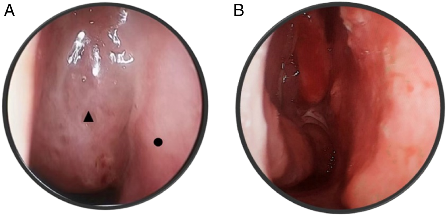
Enhanced magnetic resonance imaging (MRI) revealed an irregular mass in the right nasal cavity measuring 15 × 40 × 50 mm3, with normal and low signal intensities on T1W1 and T2W1, respectively. The enhanced MRI sequence revealed an unclear boundary between the lesion and the right superior and middle turbinate with moderate to significant enhancement of the lesion (Figure 2). Expression of the inflammatory factor interleukin-6 (IL-6) was 5.06 pg/mL, slightly higher than the normal (reference range, 0–3.4 pg/mL). Coronal images in the enhanced magnetic resonance imaging (MRI) scan show the soft tissue in the right nasal cavity (A–-F from different orientations).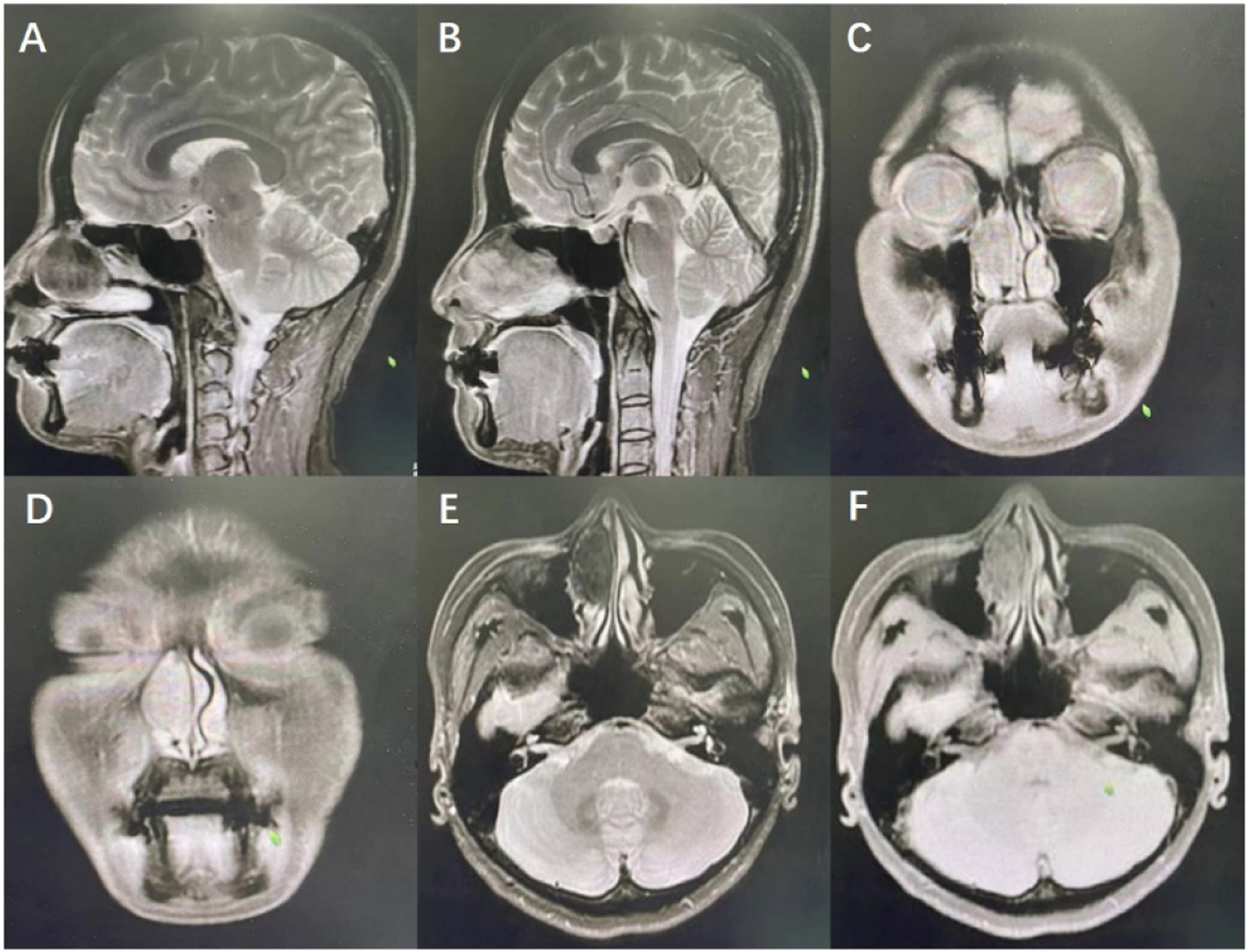
Endoscopic excision was performed under general anesthesia after excluding surgical contraindications. Intraoperative adrenaline (4 mg in 40 mL saline) was administered to thoroughly reduce mucosal secretion. The nasal forceps excised portions of the neoplasm in stages until the root of the neoplasm was located in the middle and upper parts of the nasal septum. A low-temperature plasma knife (MC401, MECHAN Co.Ltd. Chengdu, CHINA) was then used to cut the mucosa .5 cm away from the root of the neoplasm visualized under the nasal endoscope. Subsequently, the mucosa along the root was stripped and cut to completely remove the neoplasm without damaging the septal cartilage and bone.
Histopathological examination revealed a spindle cell tumor with mild cell morphology, interstitial collagenization and blood vessels, and a slit-like tendency toward benign lesions (Figure 3). . The gross specimen of the neoplasm after excision (A) and paraffin sections of the neoplasm (B). Hematoxylin & Eosin staining (40 x).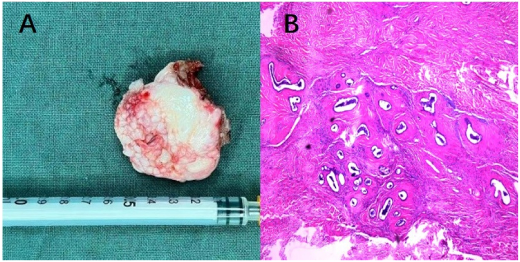
In the first and second months after surgery, endoscopic observation confirmed that the wounded mucosa in the nasal cavity had healed and smoothened, being completely covered by epithelium. The patient recovered from nasal congestion, and no bleeding was reported. The patient was free of neoplasms in the nasal cavity and sinus. The tumor was located in the nasal cavity without the involvement of the main blood vessels and nerves, and was completely removed by surgery; as such, the patient’s course was good after surgery. Radiotherapy, which is generally not considered for benign spindle cell tumors, was not performed. We plan to continue following-up with the patient and pay close attention to any changes in condition.
Discussion
Although the etiology of spindle cell tumors remains unclear, genetic and environmental factors may play a role in their formation. Spindle cell tumors predominantly affect the dermis and subcutis of the distal extremities, and occur only infrequently in the nasal cavity. Symptoms are mostly determined by the location of the tumor. Nasal spindle cell tumors present with symptoms according to the involved site when they reach a certain size, and it is difficult to define the nature of the tumor prior to surgery. MRI can help detect the location and boundaries of the tumors. A surgical approach may be planned preoperatively according to the imaging findings. Histopathology is important for differential diagnosis and treatment strategies. At present, treatment options for spindle cell tumors in the nasal cavity are scarce, and no relevant diagnosis or treatment guidelines exist. Studies on benign nasal spindle cell tumors of the respiratory tract are limited. Tastemel et al reported a case of a 4-month-old baby with nasal obstruction due to a spindle cell hemangioma in the ethmoid sinus obliterating the nasal passage. 7
The preoperative physical examination and imaging findings of our patient were consistent with the diagnosis of a benign tumor. Intraoperatively, the tumor was found to compress the nasal septum and middle turbinate; however, lack of an ulcer or erosion was consistent with the appearance of a benign tumor.
A low-temperature plasma knife includes a conductive medium (salt), resulting in a high concentration of plasma in the periphery of the electrode, comprising highly ionized particles. 8 These particles guarantee that the current does not generate a large amount of heat through the tissue, thereby preventing visible damage. 9 Scientists have developed low-temperature plasma coagulation instruments with minimal risk of burn injury by reducing the plasma current that flows through the tissue. 10 The application of nasal endoscopy combined with a low-temperature plasma knife for nasal surgery can reduce mucosal injury and consequently enhance postoperative mucosal recovery and epithelialization. Complete resection of the tumor with a low-temperature plasma knife viewed under a nasal endoscope can not only provide a clear surgical field, define the scope of the tumor, allow for its complete remove removal, but also reduce intraoperative bleeding. The advantages of low-temperature plasma, accurate localization of the tumor, and surgical expertise, together ensured minor local damage. The final diagnosis was made according to the postoperative pathology.
The main manifestations of spindle cell tumors are progressive unilateral nasal congestion and bulging, local pain, and olfactory disorders, which are nonspecific and determined by the location and size of the neoplasm. The large size of the tumors and their tendency to compress the surrounding tissue may result in confusion with the signs of malignancy.
Therefore, clinical symptoms, endoscopic observation, and enhanced MRI scans are insufficient for the diagnosis of spindle cell tumors, and pathological examination is indispensable to distinguish benign from malignant nasal tumors.
Owing to its rarity, more cases of nasal spindle cell tumors need to be reported to determine its prognosis. Endoscopic nasal resection of spindle cell tumors using a low-temperature plasma knife is safe and effective. This case report details the occurrence of spindle cell tumors in the nasal cavity, and our findings serve as a reminder that, although these neoplasms are rare, it is prudent to consider them in the differential diagnosis of a nasal mass.
Footnotes
Declaration of Conflicting Interests
The author(s) declared no potential conflicts of interest with respect to the research, authorship, and/or publication of this article.
Funding
The author(s) disclosed receipt of the following financial support for the research, authorship, and/or publication of this article: This research was supported by the National Natural Science Foundation of China (Grant No. 12172082).
Ethics Approval
Ethical approval to report this case was obtained from the Ethics Review Committee of the Second Affiliated Hospital of Dalian Medical University
Informed Consent
Written informed consent was obtained from the patients for the anonymized information to be published in this article.
Data Availability Statement
All data and materials generated or used during the study appear in the submitted article.