
Abstract
Cyanide is an environmental neurotoxin which has been reported to arrest the normal functioning of the brain. This study investigated the protective properties of methanol and flavonoid-rich extracts of the leaves of Spondias mombin on redox status, cholinergic dysfunction and electrolyte disturbance in cyanide-induced neurotoxicity in rats. Male Wistar rats were orally pre-treated with Spondias mombin methanol leaf extract (SMC) (50, 100 and 150 mg/kg), flavonoid-rich extract (SMF) (25, 50 and 75 mg/kg) or quercetin (20 mg/kg), followed by intraperitoneal administration of 2 mg/kg potassium cyanide. Cyanide intoxication caused brain damage in rats as echoed in the deleterious alterations to activities/levels of endogenous antioxidants and biomarkers/enzymes linked with electrolyte imbalance and neurotoxicity. Pre-treatment with SMC and SMF significantly attenuated these KCN-induced imbalances (p < 0.05). The results suggested that the protection conferred by SMC and SMF probably involves attenuation of oxidative stress and regulation of ionic homeostasis. SMF displayed a better apparent ameliorative activity than SMC and 75 mg/kg SMF offered the best protection suggesting that flavonoids probably contributed to the protective effect of Spondias mombin leaf.
Introduction
Cyanide is an environmental agent found in various forms which could be produced by plants containing cyanogenic glycoside and from industrial waste during the decomposition of compounds by heat in the absence of oxygen (e.g. electroplating). 1 Inhalation or ingestion of cyanide affects the body systems such as vascular, respiratory, sensory, cardiac, visual and metabolic systems. 2 Cyanide is an inhibitor of complex IV which induces mitochondria dysfunction and alters ATP generation. It primarily exerts its toxic effects by binding and inhibiting cytochrome C oxidase, a metalloenzyme, thereby impairing enzyme and cell function. An assault of cyanide will result in the reduction of oxygen (histotoxic hypoxia), that is sufficient to cause tissue damage. Cyanide has been found to contribute to toxicity by inhibiting other important enzyme systems such as peroxidase, phosphatase, tyrosinase and dehydrogenase activities. 3 Cyanide toxicity induces changes in the brain due to the impairment of the mitochondrial system. 4 There is accumulating evidence that during synaptic transmission, the brain is sensitive to toxicity from many environmental agents, resulting in neurological injury which could lead to neurodegenerative diseases. 5 Cyanogenic compounds prevent the mitochondria from maintaining ATP synthesis 6 and also cause oxidative stress. 7 Thus, a defect in mitochondria integrity has a deleterious effect on cellular functions of the brain. 3 These properties of cyanide have made it a good chemical to model neurotoxicity.
Medicinal plants contain inherent active ingredients and are employed in the treatment of diseases or to relieve pain. 8 Medicinal plants are used as therapies for the prevention and treatment of disease conditions including neurodegenerative diseases, owing to the presence of mixtures of different biologically active phytochemicals. Spondias mombin is a plant that belongs to the Anacardiacae family. It is found in the tropical rainforests of Nigeria and other countries of the world. 9 S. mombin is traditionally used for the treatment and management of numerous conditions that include neurological diseases such as epilepsy and psychosis.10,11 Almost every part of the plant is useful ranging from its thick corky bark to the leaves, flower and fruits. It has been reported that the plant extract possesses purgative, antioxidative, antihelmintic, analgesic, haemostatic, antibacterial, abortifacient, 12 anti-anaemic, anti-diabetic 13 and hypoglycaemic 14 properties. Other reports have shown that the leaf extract of the plant elicited anxiolytic, sedative, antiepileptic and antipsychotic effects in mice and rats.9,15–17 This study was designed to evaluate the protective effects of leaf extracts of Spondias mombin against cyanide-induced neurotoxicity.
Materials and methods
Chemicals
Thiobarbituric acid (TBA), trichloroacetic acid (TCA), Ellman’s reagent (DTNB), sulphosalicyclic acid (SSA), glutathione (GSH), naphthyl ethylenediamine dihydrochloride (NED), acetylthiocholine iodide, epinephrine, catechin, kaempferol, quercetin, isoquercetin, epicatechin and rutin were obtained from Sigma-Aldrich, USA. All other reagents and chemicals used were of analytical grade. Randox diagnostic kits for total protein assay and Teco diagnostic kits for electrolyte assays were bought from Randox Laboratories Limited, Crumlin County, Antrim, UK and Teco Diagnostics, California, USA, respectively. Methanol, acetic, caffeic, ellagic and chlorgenic acids were obtained from Merck (Darmstadt, Germany).
Plant collection, preparation of crude extract and flavonoid-rich extract of S. mombin
Fresh Spondias mombin leaves were collected from a host tree on the campus of the Federal University of Technology, Akure, Nigeria (Latitude: 07°30′43″N and longitude: 05°13′70″E). The leaf was authenticated at the herbarium of Botany Department, University of Ibadan, Ibadan, Nigeria (Voucher number 16747). The leaves were air dried. The dried leaves were pulverised and macerated in 80% (v/v) methanol for 72 h. Thereafter, the mixture was filtered, the filtrate was subjected to rotary evaporation to remove the solvent and the aqueous extract was freeze-dried to obtain the crude extract of Spondias mombin (SMC).
Five gram of the crude extract was dissolved in 100 ml of distilled water and 100 ml of 10% H2SO4 was subsequently added. The aliquot was boiled at 100°C to evaporate to 100 ml. The solution was filtered and the residue was dissolved with warm 95% ethanol (50°C). It was then filtered again and the filtrate was evaporated to dryness to give the flavonoid-rich extract (SMF). 18
Qualitative phytochemical screening
The extracts (SMF and SMC) were phytochemically screened for the purpose of detecting the presence of different phytochemicals. The tests (saponins, tannins, alkaloids, flavonoids and cyanogenic glycosides) were carried out according to methods previously described19–21 with slight modifications in the quantity of extracts used.
Test for alkaloids
Crude extract (0.1 g) was dissolved in 5 ml of 1% (v/v) hydrochloric acid in a steam bath and filtered. About 1 ml of Dragendorff’s reagent was added to 1 ml of the filtrate. Formation of a black colouration indicated the presence of alkaloids. 19
Test for saponins
The tendency of saponins to produce frothing in an aqueous solution was observed. About 1 g of extract was dissolved in 10 ml of water inside a test-tube and the mixture was shaken. Frothing indicated the presence of saponins. 19
Test for tannins
The extract (1 g) was dissolved in 5 ml of distilled water and filtered. Thereafter, 1 ml of ferric chloride was added to the filtrate. A blue-black or blue-green precipitate indicated the presence of tannins. 19
Test for phlobatannins
One gram of extract was boiled with 5 ml of 1% (v/v) aqueous HCl. A red precipitate indicated the presence of phlobatannins. 19
Test for anthraquinones
The extract (5 g) was dissolved in 10 ml of benzene and filtered. About 10% (v/v) ammonia solution (5 ml) was added to the filtrate and the mixture shaken to observe for the presence of pink, red or violet colour in the ammoniacal lower phase indicating the presence of anthraquinones. 19
Test for steroids
Extract (0.2 g) was dissolved in 2 ml of acetic acid and 2 ml of concentrated H2SO4 was added. A violet to blue-green colour indicated the presence of steroids. 20
Test for flavonoids
Extract (0.5 g) was dissolved in 5 ml of ethanol and the mixture was shaken and filtered. To 1 ml of the filtrate, few drops of 80% (w/v) alcoholic KOH were added. A yellow colouration confirmed the presence of flavonoids. 21
Test for terpenoids
Extract (0.2 g) was mixed with 2 ml of chloroform (CHCl3). Then, 3 ml of concentrated H2SO4 was carefully added to form a layer. A reddish brown colour at the interface indicated the presence of terpenoids. 20
Test for cardiac glycosides (Legal’s test)
Extract (0.5 g) was dissolved in 2 ml of pyridine and five drops of 2% (w/v) nitroprusside and 20% (w/v) sodium hydroxide were added. A yellow colour at the interface confirms the presence of cardenolides. 19
Quantification of compounds by High Performance Liquid Chromatography-Diode Array Detector (HPLC-DAD) Analysis
Chromatographic analysis was carried out by the method described by Sabir et al. 22 SMC was analysed at a concentration of 12 mg/ml. The presence of nine antioxidant compounds were investigated namely; caffeic acid, chlorogenic acid, ellagic acid, catechin, epicathechin, quercetin, isoquercetin, rutin and kaempferol. The flow rate and injection volume were set at 0.8 ml/min and 40 μl, respectively and the wavelength for catechin and epicatechin were set at 280 nm, caffeine, ellagic acid and chlorogenic acids at 327 nm, and quercetin, isoquercetin, rutin and kaempferol at 365 nm. The samples and mobile phase were filtered through 0.45 µm membrane filter (Millipore). Stock solutions of standard references were prepared in the mobile phase at a concentration range of 0.020–0.200 mg/ml for quercetin, isoquercitrin, rutin, kaempferol, catechin and epicatechin, and 0.050–0.250 mg/ml for chlorogenic, caffeic and ellagic acids. All chromatographic operations were carried out at ambient temperature and in triplicate.
Experimental animals and grouping
Fifty-four male albino rats of Wistar strain weighing 210–240 g were used for the experiment. Animals were kept in a room under natural light and dark cycle and had free access to rat chow (Vital Feeds, Lagos, Nigeria) and water ad libitum at the animal house of Department of Animal Production and Health, The Federal University of Technology, Akure, Nigeria. Animals were used in accordance with the International Guide for the care and use of laboratory animals published by National Institute of Health, 1985. We divided the 54 animals into nine groups with six animals per group as follows;
This study was conducted using a single dose of KCN (2 mg/kg, i.p.).23,24 Quercetin was used as the reference standard because of its well-established biological and neuroprotective properties in various pharmacological studies.25,26
Following the administration of cyanide, animals were left for 1 h in their cages. After 1 h, the animals were sacrificed by overdose of anesthetic. The brain was excised, rinsed in 1.15% ice-cold potassium chloride solution and then homogenised in ice-cold phosphate buffer (pH 7.4) using Teflon homogenise (1:9 (w/v) ratio of tissue to buffer). The brain homogenate was centrifuged at 7000 rpm for 20 min at 4°C to obtain the supernatant which was used for biochemical analyses.
Biochemical estimation
Estimation of markers of oxidative stress
Reduced glutathione (GSH) Level
The level of GSH was estimated according to the method of Beutler et al. 27 A GSH standard curve was prepared and GSH content of the sample was estimated from the curve.
Glutathione peroxidase (GPx) activity
GPx activity was evaluated according to the method of Haque et al. 28
Determination of lipid peroxidation
Lipid peroxidation was evaluated by measuring the level of malondialdehyde (MDA), thiobarbituric acid reactive substances (TBARS), according to the method described by Varshney and Kale. 29 The MDA level was calculated according to the method of Ádám-Vizi and Seregi. 30 Lipid peroxidation was calculated with a molar extinction coefficient of 1.56 × 10⁵ M−1cm−1 and was expressed as units/mg protein.
Evaluation of superoxide dismutase (SOD) activity
SOD activity in the brain homogenate was determined by the method of Kakkar et al. 31
Estimation of concentrations of electrolytes
Determination of Ca2+ level
Calcium concentration was determined spectrophotometrically using an assay kit as described by Akinmoladun et al. 26 A purple coloured complex was formed when calcium reacts with cresolphthalein complexone. The absorbance was measured against the reagent blank at 578 nm and the intensity of the coloured complex is proportional to the concentration of calcium. Level of Ca2+ was expressed in mmol/mg protein.
Determination of Mg2+ level
Magnesium ion concentration was determined spectrophotometrically using an assay kit as described by Akinmoladun et al. 26 A coloured complex of magnesium in alkaline medium is formed with calmagite, producing a red complex measured at 530 nm. The coloured complex formed is proportional to the concentration of magnesium. Level of Mg2+ was expressed in mmol/mg protein.
Determination of Na+ level
Sodium ion concentration was determined spectrophotometrically using assay kit as described by Akinmoladun et al. 26 The method for determining Na+ level is based on the reaction of sodium with a selective chromogen to produce a chromophore measured at 550 nm and varies inversely as sodium ion concentration in the brain homogenate. Level of Na+ was expressed in mmol/mg protein.
Determination of K+ level
Potassium ion concentration was evaluated spectrophotometrically using an assay kit as described by Akinmoladun et al. 26 The level of K+was determined using a prepared mixture of Na tetraphenylboron to produce a turbid suspension of potassium tetraphenylboron to which the absorbance was taken at 500 nm and is proportional to concentration of potassium in the sample. Levels of K+ was expressed in mmol/mg protein.
Enzyme activities assay
Evaluation of acetylcholinesterase (AChE) activity
Activity of acetylcholinesterase (AChE) was evaluated according to the method described by Ellman et al. 32 A reaction mixture of 0.1 ml 5,5′-Dithiobis-(2-nitrobenzoic acid) (0.01 M DTNB) and 2.6 ml of 0.1 M phosphate buffer (pH 8.0) was prepared. 0.04 ml of the brain homogenate was added to the total volume of the reaction mixture and was incubated for 5 min at room temperature. The substrate, 0.075 M acetylthiocholine iodide was freshly prepared and 0.04 ml was added to the mixture. The absorbance was read for 3 min at 30 s intervals at 420 nm (UV-1100 Macy Instruments Inc., China). AChE activity was calculated using a molar extinction coefficient of 1.36 × 10⁻⁴ M/cm and the result was expressed in µmol/min/mg protein.
Estimation of total protein concentration
Concentration of the total protein was estimated using Biuret method with Randox assay kit. One millilitre of Biuret reagent was added to 0.02 ml of the brain homogenate and the mixture was incubated at 25°C. The absorbance was measured on a spectrophotometer at a wavelength of 546 nm.
Statistical analysis
Results were expressed as mean ± standard deviation (SD). Data were analysed using one-way analysis of variance (ANOVA) followed by Duncan’s multiple range comparison test using Statistical Package for Social Sciences (SPSS) software 16.0 (SPSS Inc., Chicago, Illinois, USA). Values having p < 0.05 were considered statistically significant.
Result
Phytochemical constituents in SMC and SMF
The summary of phytoconstituents present in SMC and SMF is shown in Table 1. Tannins, anthraquinones, flavonoids, terpenoids and cardiac glycosides were detected in SMC and only flavonoids were detected in SMF. The proportion of the flavonoids present in SMF was more than in SMC as signified by multiple positive sign.
Phytochemical constituents of Spondias mombin flavonoid-rich extract and Spondias mombin crude extract.

−: not detected; +: detected; SMF: Spondias mombin flavonoid-rich extract; SMC: Spondias mombin crude extract.
Phenolic constituents in SMC
HPLC analysis of SMC showed the presence of the following flavonoids: catechin, chlorogenic acid, caffeic acid, ellagic acid, epicatechin, rutin, isoquercitrin, quercetin and kaempferol denoted by peak 1 to 9, respectively (Figure 1), with their corresponding concentration showed in Table 2.

Representative high performance liquid chromatography profile of Spondias mombin crude extract (SMC). Catechin (peak 1), chlorogenic acid (peak 2), caffeic acid (peak 3), ellagic acid (peak 4), epicatechin (peak 5), rutin (peak 6), isoquercitrin (peak 7), quercetin (peak 8) and kaempferol (peak 9).
HPLC phenol composition of the extract of Spondias mombin crude extract.

SMC: Spondias mombin crude extract; LOD: limit of detection; LOQ: limit of quantification.
Effect of SMC and SMF on the level of reduced glutathione (GSH)
The GSH level in all experimental groups is presented in Table 3. The result showed that the level of GSH in the brain of the KCN-intoxicated rats decreased significantly compared to the control group (p < 0.05). However, rats pretreated with SMC and SMF showed significant increase in the level of GSH (p < 0.05).
Level of GSH and lipid peroxidation in the brain of rats.

Each value represents mean ± SD (n = 6). Values with same superscripts down the column are not statistically different at p < 0.05.
Effect of SMC and SMF on the level of lipid peroxidation
The cyanide-intoxicated rats showed a significant high level of lipid peroxidation (p < 0.05) compared with the control. However, pretreatment with varying dosages of SMF and SMC showed significant decrease in lipid peroxidation compared with KCN-intoxication group (p < 0.05). The dose that offered the best protection against lipid peroxidation was 150 mg/kg SMC (Table 3).
Effect of SMC and SMF on the glutathione peroxidase (GPx) activity
It was observed in Figure 2 that there was decrease in GPx activity in the brain of KCN-intoxicated rats compared with the control (p < 0.05). The decrease was ameliorated in the brain of SMC and SMF treated rats. Pretreatment with 75 mg/kg SMF gave the highest increase in GPx activity (p < 0.05) after KCN-intoxication.

GPx activity in the brain of rats in all experimental groups.
Effect of SMC and SMF on the activity of superoxide dismutase (SOD)
The Activity of SOD in the brain of KCN-intoxicated rats reduced significantly (p < 0.05) compared with the brain of control animals (Figure 3). Treatment with SMF and SMC significantly increased the activity of the SOD compared with KCN-intoxicated rat in a dose-dependent manner (p < 0.05). Pretreatment with 150 mg/kg SMC showed the highest increase in SOD activity (p < 0.05) after KCN-intoxication.
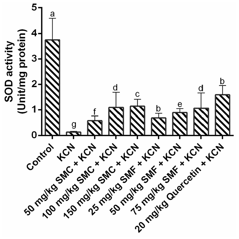
SOD activity in the brain of rats in all experimental groups.
Effect of SMC and SMF in the activity of acetylcholinesterase (AChE)
There was 69.60% increase in AChE activity of KCN-intoxicated rats compared with control (p < 0.05) (Figure 4). The increase in AChE activity was ameliorated in SMC and SMF treated group. The abnormal AChE activity shown by KCN intoxicated rats was reduced by 59.89% in SMC treated rats and 57.68% in SMF treated rats, both at their highest doses.

Acetylcholinesterase (AChE) activity in the brain of rats in all experimental groups.
Effect of SMC and SMF on the concentration of electrolytes
Calcium ion (Ca2+)
As shown in Table 4, there was 89.17% increase in Ca2+ level in the KCN-induced group compared with the control (p < 0.05). The increase in Ca2+ level was significantly mitigated in both SMF and SMC treated rats. SMF pretreatment reduced the level of Ca2+ by 55.0% and SMC by 35.8% at their highest dosages (p < 0.05).
Concentration of electrolytes in the brain of rats.

Each value represents mean ± SD (n = 6). Values with same superscripts down the column are not statistically different at p < 0.05.
Magnesium ion (Mg2+)
Table 4 revealed that the Mg2+ level in the brain of the KCN-induced neurotoxicity group decreased significantly by 75.49% compared to the control group (p < 0.05). Rats pretreated with SMC and SMF showed increase in Mg2+ level in a dosage-dependent manner, with the highest dosages of 150 mg/kg SMC and 75 mg/kg SMF eliciting increases of 57.2% and 69.04% (p < 0.05), respectively.
Sodium (Na+)
In Table 4, there was 65.18% increase in of Na+ level in the KCN-toxified rats when compared to control rats (p < 0.05). The increase in Na+ level was significantly decreased by SMF and SMC pretreatment in a dosage-dependent manner (p < 0.05). Pretreatment with 150 mg/kg SMC and 75 mg/kg SMF significantly caused 40% and 32.02% decrease in Na+ level respectively, compared to KCN-intoxicated rats.
Potassium ion (K+)
In Table 4, KCN-intoxicated rats have a 94.72% reduction in K+ level compared to control rats (p < 0.05). Pretreatment with SMC and SMF caused a significant dose-dependent increase in K+ level (p < 0.05). The highest doses of 150 mg/kg SMF and 75 mg/kg SMC showed the most pronounced effects by increasing K+ level by 87.1% and 78.8%, respectively.
Discussion
The application of herbal medicines in the treatment of various diseases is growing at a fast rate globally, the low toxicity and easy accessibility being major contributors to its global acceptance. Therapeutic actions of medicinal plants have been linked to the presence of biologically active phytochemicals such as saponins, terpenoids, flavonoids and phenolic acids, which possess powerful free radical scavenging and antioxidant activities. 33 In the present investigation, Spondias mombin remarkably ameliorated the oxidative and neurotoxic effects induced by potassium cyanide. Phytochemical screening of the extracts revealed the presence of polyphenolic compounds which was confirmed by the HPLC analysis of SMC. The protective potentials of flavonoids present in flavonoid-rich plants that are ethnomedicinally used for treating neurological dysfunctions have been reported to be through interaction with the cellular and molecular pathphysiologies implicated during synaptic transmission, potentiation and plasticity. 34
Cyanide is a hazardous chemical that is poisonous to the brain, leading to loss of muscular function as observed in experimental neurological disorders. 35 The toxic effect of cyanide is attributed predominantly to anoxia 36 and hypoxia 37 following inhibition of cytochrome c oxidase, a terminal mitochondrial respiratory chain enzyme. 36 Anoxia in the brain, causes free radical generation and a number of antioxidant enzymes are inhibited, leading to oxidative stress.38,39 Therefore, it is clear that oxidative stress is linked with cyanide neurotoxicity. The antioxidant defence and lipid peroxidation (LPO) inhibitory capacity demonstrated by SMC and SMF in the brain of the KCN intoxicated rats revealed that these extracts have ameliorative ability to reduce oxidative stress in a stress-mediated pathology. This corroborates the findings of Ozkul et al. 40 Ogundele et al. 41 revealed that cyanide toxicity involves production of ROS via cyanide binding to the iron and copper in the active site of cytochrome c oxidase. This anomaly was revealed from the result of cyanide intoxicated animals without polyphenol treatment. Decreased activity of SOD indicated participation of superoxide radical which could be hydrolysed to hydrogen peroxide, which can produce a highly toxic hydroxyl radical. 2 GPx, an endogenous antioxidant enzyme, catalyses the reduction of hydrogen peroxide to water and their corresponding stable product using cellular glutathione as the reducing agent. 37 Also, reactive oxygen species (ROS) can cause the production of malondialdehyde (MDA), an end product of lipid peroxidation. 33 The reduction in the level of GSH in the brain of the rats can be linked to the oxidation of GSH by reactive oxygen species generated due to cyanide-induced assault. Pre-treatment with SMF and SMC ameliorated the effect of cyanide intoxication by significantly increasing the level of GSH, reducing the level of lipid peroxidation and boosting the activities of GPx and SOD to combat the oxidative effect generated by KCN. These results revealed that S. mombin has antioxidative capacity to mitigate the oxidative stress generated by cyanide-induced neurological damage.
The principal mechanism of detoxicating cyanide in the body is through enzymatic conversion by the mitochondrial enzyme rhodanese (through the thiosulphate-cyanide sulphur transferase) to thiocyanate which requires a source of sulphur36,37 that is usually offered by thiosulphate or other biological compounds such as those found in flavonoid-rich plant extracts. It is therefore possible that the ameliorative effect of S. mombin is due to the presence phytochemicals such as flavonoids in the SMF or it may be that the attenuating effects of SMC could be effected by other phytoconstituents present, which may involve a different detoxification pathway from that of thiocyanate formation.
As revealed in our findings, cyanide intoxication caused Ca2+ overload resulting from uncontrollable influx of Ca2+ that can lead to uncontrollable depolarisation of neuronal membrane. This could result in increased level of Na+ and decrease in the level of K+ simultaneously, and also, reduction in the level of Mg2+ in the brain. This anomaly could arise as a result of uncoupling of the electron transport chain by cyanide. Mg2+ is a cofactor for enzymes in many metabolic pathways such as the glycolytic process to aid adequate breakdown of glucose to the precursor for TCA cycle and also blocks N-methyl-D-aspartate (NMDA) receptors 41 and Ca2+ channels. 27 Cyanide activates the NMDA receptor causing influx of calcium ion into the cell, which could also result in ionic imbalances when Na+/K+ pump is blocked,38,41 and initiate accumulation of fluid. 42 SMF and SMC ameliorated the imbalances in these electrolytes which could be by interfering with channels, pump and receptors of these electrolytes or by counteracting the effect of the cyanide on mitochondria functioning.
A previous report has shown that cyanide toxicity significantly caused decreased levels of neurotransmitters in brain of rats, 43 prompting us to evaluate the activity of AChE. AChE, a serine hydrolase enzyme hydrolyses acetylcholine (ACh) into choline and acetic acid. The ameliorative capacity of SMF and SMC to mitigate alteration to the activity of AChE revealed that S. mombin can influence neuronal signal transmission through its effect on cholinergic system dysfunction as a result of cyanide-induced neurotoxicity. As shown by our findings, S. mombin extracts facilitated the inhibition of AChE activity and removed the restriction imposed on the cholinergic system. Spondias mombin extracts might be demonstrating its anticholinergic inhibitory property through modulation of neuronal signalling pathway.
Generally, the pattern of result observed for SMF and SMC on their ability to mitigate KCN-induced neurotoxicity revealed that both extracts possess neuroprotective activity which could be due to the constituent flavonoids for SMF as revealed from the qualitative phytochemical screening and the presence of polyphenolic compounds as shown from the HPLC fingerprinting of SMC. Therefore, the ability of the Spondias mombin extracts to assuage the neurotoxicity caused by potassium cyanide in rats may be linked to the presence of phenolics and flavonoids such as catechin, chlorogenic, caffeic acid, ellagic acid acid, epicatechin, rutin, isoquercitrin, quercetin and kaempferol which could be acting synergisticallyy in the extracts.
Conclusion
Spondias mombin extract is a potential neuroprotective agent for reversing the deleterious effects of KCN exhibited through its antioxidative capacity, restoration of imbalances in electrolyte flux and its acetylcholinesterase inhibitory property. SMF offered the best protective effect at 75 mg/kg. The detailed mechanism by which it reverses the effects of KCN toxicity on the brain is not well known, this however, requires further investigation.
Footnotes
Declaration of conflicting interests
The author(s) declared no potential conflicts of interest with respect to the research, authorship, and/or publication of this article.
Funding
The author(s) received no financial support for the research, authorship, and/or publication of this article.