
Abstract
Somatosensory evoked potentials (SEPs) are widely used to study the functional integrity of ascending sensory pathways. For animal studies, SEPs provide a convenient method to quantitatively assess the functionality of the nervous system with low invasiveness. Even though they are frequently used in animal models, little attention is paid to the fact that SEPs are vulnerable to contamination from experimental factors such as anaesthetic delivery. In this study, the effect of isoflurane on SEP measurement was investigated in a rat model. The aim was to find out the adjustments for anaesthetic delivery optimizing the quality of the recordings. Two aspects were studied: the effect of isoflurane dosage on the SEP parameters and on the repeatability of the measurements. The SEP quality was found to be best when 1.5% isoflurane concentration was used. This dosage resulted in the best signal-to-noise ratio and equal repeatability of the measurements compared with the others. Our findings can help in refining the anaesthetic protocols related to SEP recordings in a rat model and, by improving the quality of the measurements, potentially reducing the number of subjects needed to carry out studies.
Somatosensory evoked potentials (SEPs) are widely used to study the functional integrity of ascending sensory pathways. In current clinical practice, they are used for various diagnostic purposes such as intraoperative monitoring during high-risk surgery and the detection of hypoxic ischaemic encephalopathy after cardiac arrest.1,2 For animal studies, SEPs provide a convenient method to quantitatively assess the functionality of various portions of the somatosensory pathways with low invasiveness. Even though several sophisticated measures for the quantification of SEPs have been proposed,3,4 the analysis still mainly relies on the determination of simple parameters such as amplitudes and latencies from the averaged signal waveform. 5 Whereas these parameters are rather easy to derive from the recordings, they are also vulnerable to contamination from experimental factors present during data collection. This artefact, increasing the variability of the data, may lead to the use of an unnecessary high number of subjects in the study when, for example, statistical significance is of interest.
Anaesthetics are known to influence neural electrophysiology.6–9 In animal studies, maintaining a steady-state anaesthesia with inhalational agents such as isoflurane is more suitable compared with drugs administered by, for example, injection. However, these gases are known to affect SEP features such as the amplitude of the signal, and thus anaesthesia has a critical influence on the recordings. To this day, no standardized protocol or justifiable recommendation exists for the anaesthetic delivery which guarantees the quality of SEP recordings in the rat model.
In this study, the effect of isoflurane on SEP measurement was investigated in rats. The aim was to find out the appropriate adjustments for anaesthetic delivery to optimize the quality of the recordings. Two aspects were studied: the effects of isoflurane dosage on the SEP parameters and on the repeatability of the measurements. The study was approved by the Institutional Animal Care and Use Committee (IACUC) at the National University of Singapore, Singapore.
To carry out SEP measurements, four screw electrodes were implanted on the cranium of 10 adult female Sprague–Dawley (200–220 g) rats. Rats were ordered through ACUC office at the National University of Singapore and were supplied by the “InVivo Inc.” in Singapore. The electrodes were located above the somatosensory cortex of different limbs according to Figure 1. A fifth electrode was positioned in the right parietal area close to the lambda to serve as a reference. Carboxylate dental cement (3M, St Paul, MN, USA) was applied in order to fix the electrodes to the cranium. During the electrode placement, the animals were anaesthetized with a ketamine (75 mg/kg) and xylazine (10 mg/kg) cocktail.
Block diagram of the experimental system and electrode locations for somatosensory-evoked potential (SEP) measurement. The forelimb recording sites (stars) are located 0.2 mm posterior and 3.8 mm lateral to the bregma. The hindlimb recording sites (circles) are located 2.5 mm posterior and 2.8 mm lateral to the bregma. Reference electrode (square) is located 3 mm lateral to lambda.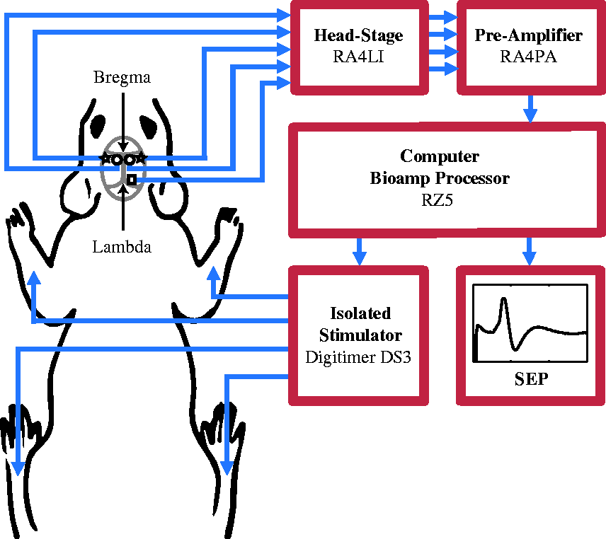
The SEP recordings were carried out as illustrated in Figure 1 at least seven days after the electrode implantation (see Supplement 1 online at http://lan.sagepub.com for details). During the measurement, the rats were anaesthetized with a mixture of isoflurane and 100% oxygen delivered at a rate of 1.3 L/min. The anaesthesia was maintained using a rodent-size anaesthesia mask connected to a diaphragm with a C-Pram circuit (Smiths Medical, Dublin, Ohio, USA) designed to deliver and evacuate the gas. The recordings were made while the isoflurane dosage was increased in a step-like manner from 1.0% to 2.5% using 0.5% increments. At each step, the dosage was kept fixed for 5 min to guarantee the equilibrium before recording the SEPs. To study the effect of the anaesthetic dosage on the repeatability of the measurements, another SEP recording was carried out for each rat at least one day after the first one following the same protocol.
From the SEP waveforms, two parameters were defined: amplitude representing the total peak-to-peak value of the signal within 5–40 ms after the stimulation and N1 latency representing the time from the stimulation to the first negative peak in the signal. The repeatability of the measurement was assessed by calculating the difference in the parameter values between the first and second recordings for each rat. For the analysis, only the hindlimb recordings were used.
The effect of the isoflurane dosage on the SEP waveform, derived parameters, and repeatability of the measurement is illustrated in Figure 2. Increasing the concentration from 1.5% to 2.0% and finally to 2.5% resulted in a statistically significant decrease in the SEP amplitude. Furthermore, a slight increase in the N1 latency was observed which was, however, not statistically significant. Furthermore, the anaesthetic concentration did not have a statistically significant effect on the repeatability of the measurement, evaluated by the difference in the parameter values between the first and second recordings from the rats, suggesting comparable repeatability at all anaesthetic levels. The recordings could not be carried out at 1.0% isoflurane as the rats tended to have some unintentional limb movement at that concentration.
The effect of isoflurane on somatosensory-evoked potential measurement. In the upper row, an example of the averaged signal waveform during 1.5%, 2.0%, and 2.5% isoflurane anaesthesia is given for the same rat. The amplitude (dashed line) and N1 component (circle) are illustrated. Below, the amplitude, amplitude difference, N1 latency, and N1 latency difference values are given during 1.5%, 2.0%, and 2.5% isoflurane anaesthesia for the 10 rats. The values are given separately for the left and right hindlimb recordings. The bars indicate the means and standard deviations. Statistically significant differences between groups are shown above the bars (two-sample t-test; *P < 0.05, **P < 0.01, ***P < 0.001).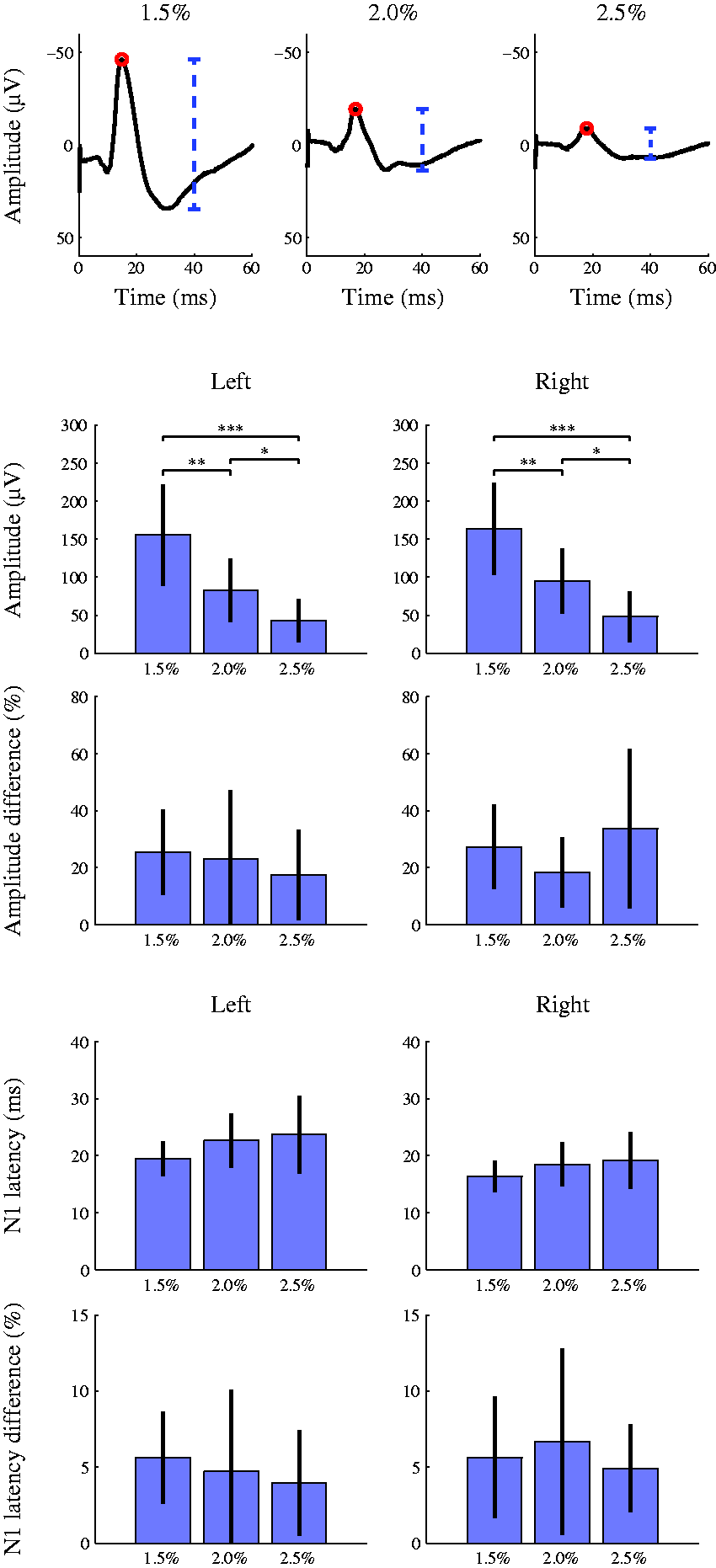
Even though SEPs are often used for quantitative analysis of neural function in animal models in the literature,10,11 little attention is paid to the fact that the measures are vulnerable to contamination from experimental factors such as anaesthetic delivery. Neglecting this source of artefact may lead to increased variability in the data and consequently, for example, raise the number of subjects included in the study unnecessarily. The current study showed the crucial role of a correct anaesthetic dosage in carrying out accurate SEP measurements using rats. In line with previous findings, 12 increasing the anaesthetic concentration significantly suppressed the SEP amplitude, emphasizing the importance of carefully controlled fixed anaesthetic delivery during the measurement and between consecutive recordings. Based on the results of the current study, 1.5% isoflurane concentration is suggested to be used when SEPs are being recorded in a rat model. This dosage resulted in the highest SEP amplitude and thus the best signal-to-noise ratio which is of interest especially when dealing with a model which includes possible neural injury. Lighter concentrations led to inadequate anaesthesia and unintentional limb movement which contaminated the recordings. A 1.5% concentration achieved the best signal-to-noise ratio; and equally, or even more, important to this was the fact that the repeatability of the measurements was comparable with that of the other concentrations. This indicates the stability of the recordings at this lowest feasible concentration which, in combination with this optimal signal-to-noise ratio, guarantees the best conditions for SEP measurements. Even though in this study only the effects of isoflurane were being studied, similar results can be expected when using other anaesthetics with GABAergic pharmacodynamics. With these drugs, a similar approach is suggested when optimizing the anaesthetic delivery to achieve a high quality in SEP recordings.
Footnotes
Declaration of conflicting interests
None declared.
Funding
This work was supported by the Maryland Stem Cell Research Fund (grant number 2013-MSCRFII-0109-00); and the National University of Singapore (grant numbers R-175-000-121-133, R-175-000-121-733, R-175-000-122-112 and R-711-201-026-133).
References
Supplementary Material
Please find the following supplemental material available below.
For Open Access articles published under a Creative Commons License, all supplemental material carries the same license as the article it is associated with.
For non-Open Access articles published, all supplemental material carries a non-exclusive license, and permission requests for re-use of supplemental material or any part of supplemental material shall be sent directly to the copyright owner as specified in the copyright notice associated with the article.