
Abstract
Despite the fact that sheep are a widely used animal model in cardiovascular research, reference values for transthoracic echocardiography in normal growing animals are not available. Eight healthy female lambs underwent two-dimensional, M-mode and pulsed wave Doppler echocardiographic examination at 100 days of age and every three months thereafter over a 12-month period. The study was conducted under sedation with midazolam, butorphanol and constant rate infusion of intravenous propofol. Their growth phase was completed at about one year of age. All the echocardiographic parameters considered were significantly correlated with body weight and age class except for the left ventricular systolic and diastolic diameters. Functional indices were not correlated to body weight or age except for the E-point to septal separation distance (EPSS). Doppler-derived parameters were not influenced by independent variables. Transthoracic echocardiography can be considered an applicable method for cardiovascular research using a growing lamb animal model after appropriate adjustments for age and body size.
The development of animal models of cardiovascular diseases has provided researchers with important insights into pathophysiological processes, and they are essential tools for the development of new therapies. 1 Among large animal models, sheep are considered suitable because of their docility, slow growth and easy housing for long-term follow up. Furthermore, the ovine heart is similar to the human heart since its dimensional and functional parameters are within human reference values and it possesses, similarly to humans, a limited capacity for developing collateral circulation after myocardial ischaemic diseases. 2 Sheep models have been developed to evaluate many aspects of cardiovascular diseases affecting millions of people in the Western world. Among these, Gorman and colleagues investigated the pathogenesis of mitral regurgitation after acute myocardial infarction 3,4 and an ovine model of chronic heart failure has recently been proposed. 5
Recently, researchers have been directing their attention to the development of new prosthetic valves, percutaneous devices 6,7 or graft conduits, 8 using sheep as a preclinical model.
Moreover, new strategies for the correction of congenital heart defects, which require surgical reconstruction of the right ventricle to pulmonary artery continuity using tissue-engineered vascular grafts, have been tested in lambs because the growth of the animal was considered to be of primary importance. 9,10 Infants in whom prosthetic conduits are implanted, in fact, require more than one intervention, in order to adapt the prosthetic valve or conduit size to the heart’s dimensions which increase with age. 9
In all the above-mentioned studies, as well as in the course of routine clinical activity, echocardiography is used as a non-invasive tool for the evaluation of cardiac morphology and function, being a sensitive and accurate method for measuring changes in chamber dimensions and thickness of the cardiac walls in response to a pathological process. 11
Despite the increasing interest in sheep as laboratory animals in cardiovascular research, only a few studies have provided normal echocardiographic reference values for this species. M-mode echocardiographic values in non-sedated adult sheep of various breeds were investigated first by Moses and Ross in 1987, 12 while pulsed wave (PW) Doppler flow patterns and Doppler-derived functional parameters obtained in adult non-sedated Merino ewes were published by Kirberger and van den Berg in 1993. 13 More recently, reference values were obtained for adult male Corriedale sheep and compared with human references for investigational purposes. 2
To the authors’ knowledge, echocardiographic values in healthy growing sheep have not been published so far. The aim of the present study, therefore, is to provide 2-dimensional, M-mode and PW Doppler-derived echocardiographic reference values for healthy growing sheep.
Animals
A total of eight lambs were included in the study. The animals were purchased from a sheep farm (Magonara Maurizio, Villa Estense, Padua, Italy), with ages varying from 80 to 150 days. Upon arrival at the Veterinary Faculty animal facility, all the lambs underwent a complete physical examination in order to assess their health status. The experimental protocol included a complete blood count (CBC) and a biochemical panel (total proteins, albumin, globulins, urea, glucose, total bilirubin, creatinine, AST, γ-GT, LDH, CK, ALT, ALP, calcium, phosphorus, magnesium, sodium, potassium, chloride). Only healthy animals were considered for the study. Prior to the beginning of the study, the animals were housed in two multiple pens (1.4 m2 for each animal) for at least two weeks to acclimatize them to the new housing environment, and they received an oral administration of 7.5 mg/kg of netobimin (Hapadex® 5%, Schering–Plough, Milan, Italy). After 20 days a faecal flotation was performed and, in case of a positive response, the anthelminthic treatment was repeated.
The period of experimental observation lasted about one year, during which the animals were fed a commercial pellet (crude protein 16.0%, crude fibre 10%, crude oils and fats 2.7%, crude ash 10.0%) (Compli Sheep, Tecnozoo, Piombino Dese, Padua, Italy) and hay ad libitum. Fresh water was always available.
The lambs’ health status was assessed daily by a technician and the veterinary surgeon in charge was called in cases of a suspected medical problem.
Materials and methods
The Faculty of Veterinary Medicine of Padua University was approved for the use of sheep as laboratory animals 14 and the project was communicated to the Italian Ministry of Health according to the Italian law for the use of laboratory animals 14 under project registration number 90/08 C21.
Preparation and positioning
Each animal underwent serial echocardiographic evaluations after about 90, 180, 270 and 360 days from the first examination. The animals were fasted 24 h before the procedure in order to reduce the repletion status and the gas content of the rumen, which could be a source of artefacts during echocardiographic examination.
The day of the procedure every animal was weighted and the body surface area (BSA) was calculated from the body weight (BW) using the formula:

Each animal was sedated with midazolam (Midazolam cloridrato, 0.2 mg/kg; Hospira Italia, Naples, Italy) and butorphanol (Dolorex, 0.1 mg/kg; Intervet Italia, Milan, Italy) intravenously and, to maintain the animal in lateral recumbency with minimal physical restraint, a constant rate infusion (CRI) of intravenous propofol (Propovet; Esteve, Bologna, Italy[AQ3]) was started (0.1 mg/kg/min) after a bolus of 0.5 mg/kg intravenously.
The precordial area was clipped from the 3rd to 7th intercostal space (ICS) bilaterally from the sternum to the costochondral junction and the skin was degreased with a denatured ethyl alcohol solution. The dorsal aspect of both forelimbs and the left rear limb were clipped and cleaned in the same way, and 43 × 45 mm disposable patch electrodes (FIAB, Vicchio, Italy) were placed and fixed with tape for electrocardiogram (EKG) recording.
The animals were positioned on an echocardiographic table in right lateral recumbency and then turned onto the left side. The limb beneath was extended forwards. Coupling gel (Acquasonic gel; Parker Laboratories, Fairfield, NY, USA) was liberally applied on the shaved skin. All the examinations were conducted from below the animal by the same experienced operator.
Equipment
The echocardiographic examination was performed using a GE Logic® P5 ultrasonographic device (General Electric Healthcare, distributed by Micra Lab Srl, Cassano D’Adda, Milan, Italy) equipped with a multifrequency micro-convex 6–10 MHz probe (C6–10) or a 1–3 MHz sectorial phased array probe pre-set for echocardiographic examination, with the frequency selected on the basis of the patient’s size. The EKG trace was recorded simultaneously.
Imaging technique
For echocardiographic examination the right parasternal, left caudal and left cranial acoustic windows were used, according to the recommendations for canine and feline animals. 16
From the right parasternal window, the standard long axis, four chamber (inflow view), five chamber (outflow view) and short axis views at the level of the chordae tendineae, mitral valve and aorta were considered.
M-mode examination of the left ventricle was performed from the right parasternal short axis view in order to obtain standard projections according to the recommendations for human 17 and veterinary medicine. 18
Echocardiographic images and cine loops were stored on the hard disk of the ultrasonographic machine and post-processed using the software provided by the supplier.
M-mode measurements of left ventricle diastolic dimension (LVIDd), left ventricle systolic dimension (LVIDs), interventricular septum in diastole (IVSd), interventricular septum in systole (IVSs), left ventricular posterior wall diastolic and systolic thickness (LVPWd and LVPWs), respectively were obtained according to the recommendations of the American Society of Echocardiography,
19
and the mitral valve E-point to septal separation distance (EPSS) was measured as previously described in dogs.
20
Fractional shortening (FS) was calculated as follows:

The sample volume for PW Doppler interrogation was set at 3 mm in order to minimize artefacts related to the movement of the cardiac structures. Flow through the pulmonary artery was evaluated from the right parasternal window short axis view, at the level of the heart base. After a correct alignment between the interrogation line and the vessel was obtained, the sample volume was positioned in the supravalvular position. Pulmonary artery peak flow velocity (PA Vmax) was measured at the midpoint of the Doppler flow spectrum at the point of maximum blood flow velocity.
The left ventricular diastolic flow was recorded from the left caudal window apical view, optimized for the mitral inflow. After the mitral leaflets were correctly imaged and a correct alignment was obtained, the sample gate was positioned inside the ventricular chamber, just distal to the tip of the mitral leaflets. Early diastolic (MV E) and late diastolic (MV A) peak velocities and their ratio (MV E/A) were measured.
The aortic flow was recorded from the left caudal window, positioning the gate at the level of the aortic valve, in the supravalvular position. Aortic peak velocity (AO Vmax) was measured from the baseline to the outer edge of the systolic Doppler flow spectrum; pre-ejection period (PEP) was measured from the beginning of the Q wave on the EKG trace to the beginning of the systolic flow; ejection time (ET) was measured from the beginning to the end of the systolic flow, and PEP/ET was calculated.
Heart rate (HR) was calculated from the R–R distance on the synchronized EKG trace in the same image where the aortic flow was recorded.
Each parameter was measured three times and the average value was used for subsequent statistical analysis.
Statistical analysis
Prior to data processing, all echocardiographic parameters were tested for normality by the Shapiro-Wilk test (PROC UNIVARIATE SAS ©). Correlations (r) were calculated to detect associations between predictive variables (BW, age, BSA, HR) and echocardiographic measurements. Where a significant correlation was found, linear regression analysis was performed. P values < 0.05 were considered significant. Age, BW, BSA, average daily gain (ADG), HR, M-mode and Doppler parameters were analysed using a linear mixed model which included the repeated effect of age class (</ = 100 days; 101–200 days; 201–300 days; 301–400 days and >400 days) and random effects of individual animals to obtain estimated mean values at different times (PROC MIXED, SAS).
Statistical analysis was performed using SAS 9.2 and StatSoft Statistica 10.0 (2011).
Results
All the lambs were in good health status after physical examination, and all haematological and biochemical parameters were within the reference ranges. Despite the first microscopic stool examination showing evidence of positivity for Trichuris spp., Coccidia spp., gastrointestinal strongyles, nematodes and cestodes, all the animals were negative at the microscopic stool recheck, two weeks after anthelminthic treatment.
The mean BW was 30 ± 2 kg and 58 ± 5.03 kg at the beginning and end of the study, respectively. The ADG progressively and significantly increased until the 4th age class (362 ± 33 days) and then decreased, indicating that the growth period was completed at the end of the experiment (Figure 1).
Average daily gain (ADG) in sheep in different age classes. Different letters indicate significantly different values (P < 0,05).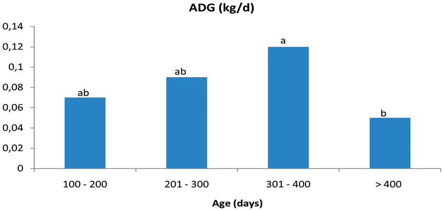
Physiological parameter (HR, systolic and diastolic pressures, body temperature) monitoring was performed 15 min after venous and arterial access: baseline values were within the normal range for this species. The standing position was lost 2–4 min after midazolam–butorphanol injection and administration of the propofol bolus, followed by continuous infusion, started 15 min after sedative administration. All the physiological parameters were recorded every 5 min commencing from sedation. During the procedure no significant changes from baseline values were observed in any animal. Every subject appeared to be well sedated (no reaction to the procedure, spontaneous breathing, blinking and swallowing). Following echocardiographic examination and after discontinuation of propofol infusion, each animal was positioned on the floor and regained the standing position within 15 min.
From the right parasternal window good quality long axis four chamber and five chamber views and short axis views of the left ventricle papillary muscles, mitral valve and heart base were easily obtained from every animal. Even where good quality left cranial and caudal projections were difficult to obtain, good alignments with mitral valve inflow and aortic flow were possible and PW Doppler traces were correctly recorded.
The normality test revealed a normal distribution for all the variables.
Least squares means and pooled standard errors (SE) and P values for age, body weight, body surface area, heart rate and echocardiographic parameters according to age class.
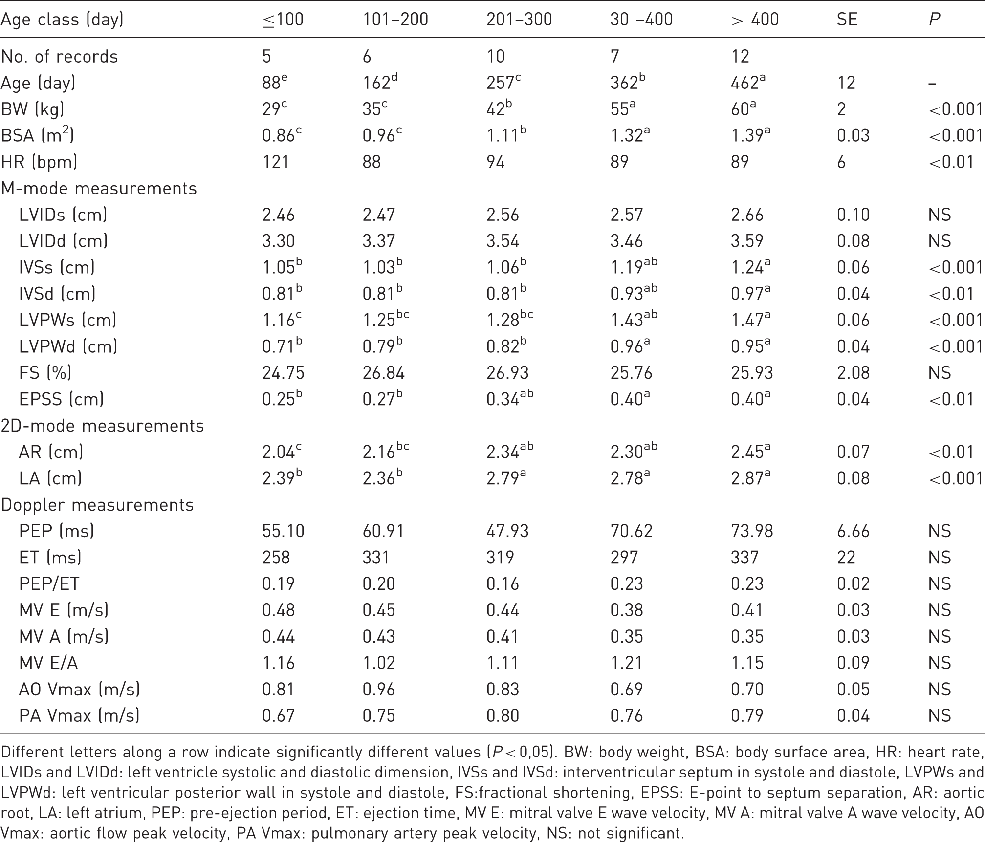
Different letters along a row indicate significantly different values (P < 0,05). BW: body weight, BSA: body surface area, HR: heart rate, LVIDs and LVIDd: left ventricle systolic and diastolic dimension, IVSs and IVSd: interventricular septum in systole and diastole, LVPWs and LVPWd: left ventricular posterior wall in systole and diastole, FS:fractional shortening, EPSS: E-point to septum separation, AR: aortic root, LA: left atrium, PEP: pre-ejection period, ET: ejection time, MV E: mitral valve E wave velocity, MV A: mitral valve A wave velocity, AO Vmax: aortic flow peak velocity, PA Vmax: pulmonary artery peak velocity, NS: not significant.
The statistical analysis revealed that age class was a significant factor for almost all the 2D and M-mode echocardiographic parameters, except for LVIDd, LVIDs and FS. A significant increment in M-mode parameters according to age was generally found up to the 4th age class (mean age 360 days) when the definitive dimensions were reached. On the contrary, EPSS and 2D measurements (LA and AR) increased their dimension until the 3rd age class (mean age 256 days).
No significant age effect was found for Doppler-derived parameters.
Correlation coefficients (r) for echocardiographic parameters versus body weight and heart rate.
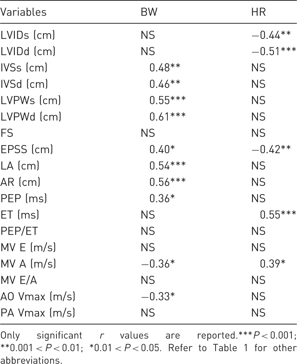
Only significant r values are reported.***P < 0.001; **0.001 < P < 0.01; *0.01 < P < 0.05. Refer to Table 1 for other abbreviations.
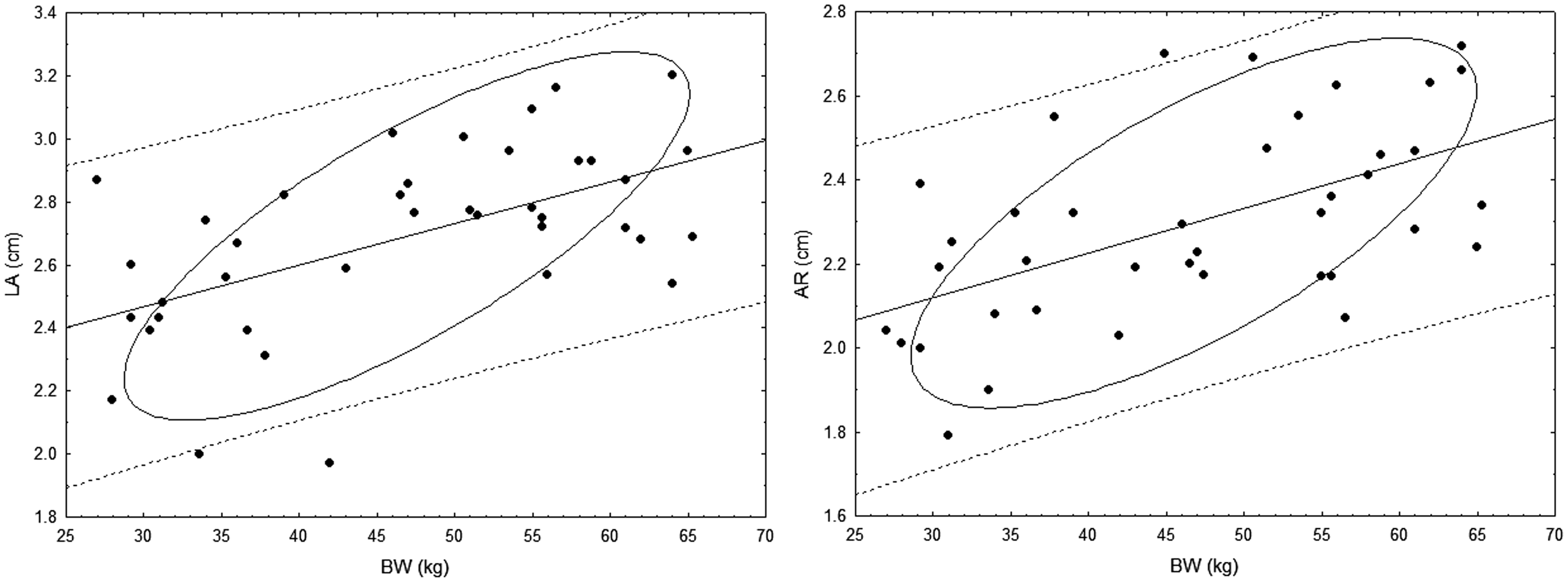
Regression equations calculated for echocardiographic parameters significantly correlated to BW are shown in Table 3 and illustrated in Figures 2 and 3, which show the individual values, the estimated line equation and the 95% predicted and 95% ellipsis confidence.
Graphic representation of the individual values, estimated line equation and the 95% predicted and 95% ellipsis confidence intervals for M-mode-derived echocardiographic parameters significantly correlated with body weight (BW). IVSs and IVSd: interventricular septum in systole and diastole, LVPWs and LVPWd: left ventricular posterior wall in systole and diastole. Graphic representation of the individual values, estimated line equation and the 95% predicted and 95% ellipsis confidence intervals for left atrium (LA) and aorta root (AR) dimensions. BW: body weight. Regression model for echocardiographic parameters and body weight. Refer to Table 1 for other abbreviations.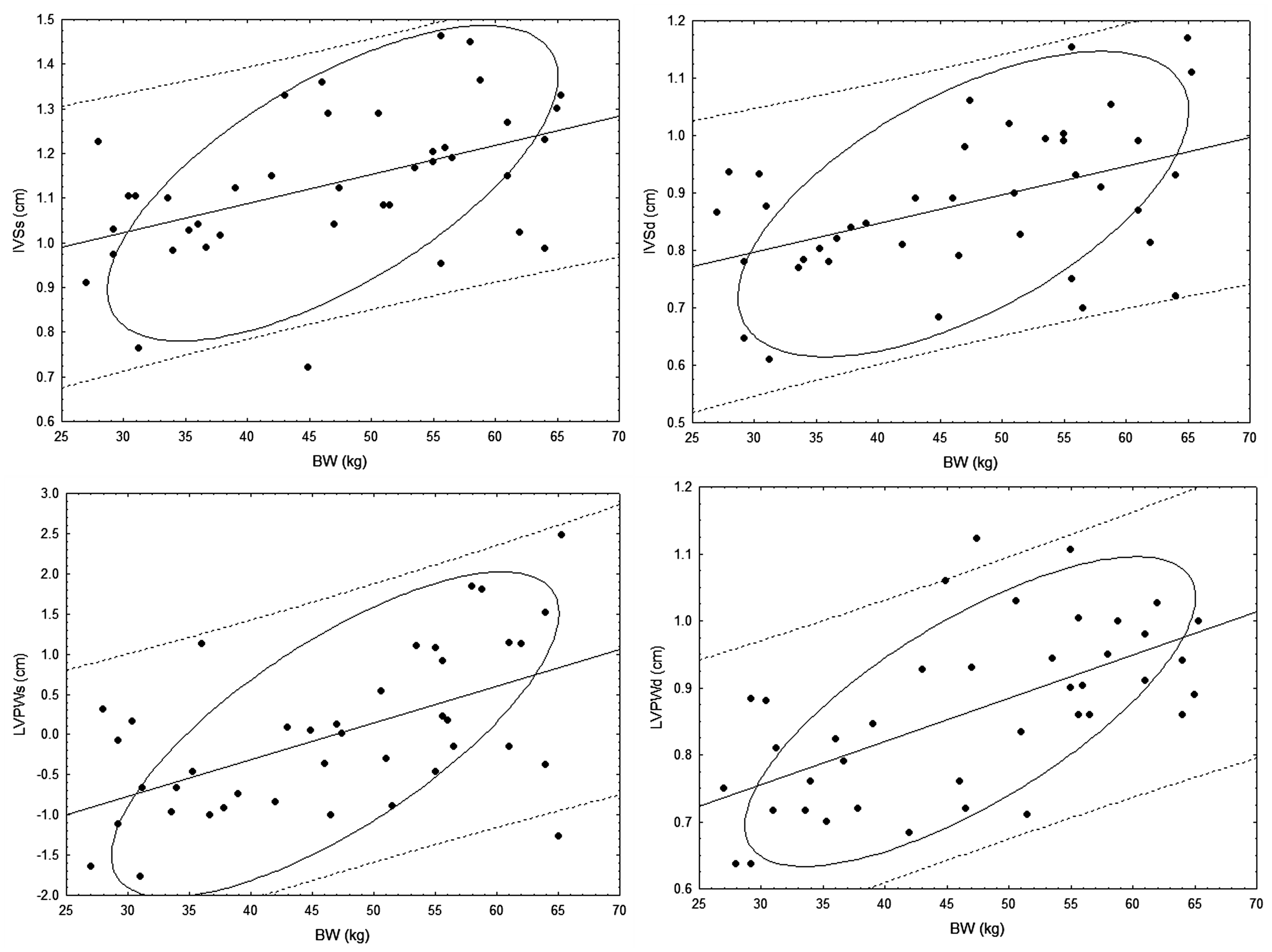
Discussion
Sheep are often used as a laboratory animal model due to their ease of handling. Although commonly used in cardiovascular research, sheep have been considered a difficult species for echocardiographic examination. The acoustic windows are very small 12 and this limits the opportunity for good quality 2D images, irrespective of the position of the animal. 13 This is probably due to the presence of an accessory pulmonary right lobe that envelops the right heart and part of the left ventricle, and due to the compression of the air-filled rumen against the diaphragm. The strategies adopted in the present study to reduce artefacts due to these anatomical aspects (lateral recumbency, 12 h fast) allowed sufficient acoustic window quality, especially in the smallest and thinnest subjects. The presence of a large and thick sternum and the vertical axis of the heart also represent obstacles to obtaining optimal apical views. 22 Despite these limits, a good alignment with the transmitral and aortic flows could be obtained in all the animals, as has also been observed by other investigators. 2
Other acoustic windows have been proposed, but even with these the cardiac apex was not adequately imaged in most animals. 23 In some studies, researchers have utilized a subdiaphragmatic access to image the left ventricle through an upper midline laparotomy. 4 More recently, transesophageal 8 and epicardial echocardiography 24 have been employed in order to better visualize the cardiac structures or to study the haemodynamic performance of native or prosthetic valves. These approaches have allowed an accurate echocardiographic examination but are more invasive and expensive than the transthoracic echocardiography used in the present study.
During the study period, the animals doubled their BW and the ADG progressively increased until the 4th age class. Many of the variables considered in this study (IVSd, IVSs, LVPWd and LVPWs, LA and AR) showed a growth pattern in agreement with the ADG. Interestingly, IVSd was thicker than LVPWd at the beginning of the study. This difference progressively decreased until the two cardiac walls had the same thickness in the adult animal. This finding was previously also observed in dogs 25 and can be considered a normal developmental pattern, possibly a remnant of the right ventricular dominance and hypertrophy during fetal life.
An unforeseen result was the lack of significant correlation of LVIDd and LVIDs with BW. For this reason, regression equations were not calculated for these parameters. In other studies in cats 26 and calves, 27 the left ventricle diameters have been significantly correlated with BW or BSA. In dogs 24 –28 the left ventricle internal diameter doubles its dimension during the first two months of age and then slowly grows until the 7th month. 29 In Spanish foals the left ventricular chamber increases its diameter until 91–180 days in females, and only after 270 days in males. 30 The animals considered in the above cited studies increased their BW many times during the experiment. The lambs in the present study received their first echocardiographic examination at about 100 days of age; thus it was not possible to rule out that an earlier rapid development of the left ventricle may have occurred during the first three months of life and that after that period it continued at a rate too slow to be statistically significant.
To our knowledge, no other echocardiographic studies in growing sheep are available for comparison with our results. The only previously published data were obtained from adult non-sedated sheep of various breeds 12 and these showed a positive correlation between BW and ventricular dimension. For these reasons the growth pattern of the left ventricle chamber in sheep deserves further investigation, in particular in younger lambs from birth to four months of age.
The EPSS was significantly correlated with age, BW and HR. This is considered a functional systolic parameter which increases in case of myocardial failure; and in the dog this is not influenced by breed, age, sex, mass or HR. 20 However, its increase with growth during the first seven months of age 24 was evidenced in a giant breed but not in the beagle, a medium breed. 29 Moreover, an increase in EPSS with BW has also been demonstrated in adult animals of different breeds, and values of less than 6 mm are usually considered normal. 31 The same pattern was observed in growing foals. 30 In our sample this EPSS increased significantly, but the variation was very slight (from 2.5 to 4 mm). Unfortunately, this parameter was not measured in the previously cited studies on sheep or goats 32 and a comparison with our values is not possible.
Among functional parameters, the FS was not significantly correlated with BW or age, nor with HR. This confirms the usefulness of this parameter as a global function index. The absolute values were lower than those obtained in adult Corriedale sheep under light sedation with diazepam 2 and in non-sedated sheep of various breeds. 12
The Doppler-derived parameter results were also in agreement with the range obtained by another author, 20 except for AO Vmax and PV Vmax, which were lower than those previously reported. This could be the consequence of the anaesthetic protocol employed, as discussed afterwards. A less than optimal beam orientation should also be considered as a possible cause of velocity underestimation, but care was taken to avoid this kind of artefact during image recording. No significant correlation was evidenced with age or BW.
The only variable strongly correlated with HR was ET. This effect was completely eliminated in the PEP/ET ratio, which confirms this value as a systolic functional index, independent of other variables, as has been demonstrated over many years 33 .
Particular attention was paid to the effects of the anaesthetic drugs and, in particular, to the propofol infused by CRI during the entire experiment at low velocity. Even though most animals may tolerate restraint in a lateral recumbent position, maintaining this position for the duration of the echocardiographic examination may be stressful. Moreover, Italian law concerning the protection of laboratory animals 14 establishes that every experimental procedure that may cause any kind of stress or pain must be performed under tranquillization or sedation and, in cases of pain, under general anaesthesia, which may influence cardiovascular function. Therefore, we used drugs known to have minimal effects on echocardiographic parameters and at low doses. Anaesthetic drugs may exert dose-related cardiovascular effects; for this reason, we used a drug combination that allowed the desired sedation with minimal doses. In human medicine, balanced propofol sedation, a combination of propofol with small doses of an opioid and/or a benzodiazepine, is routinely used: by combining small doses of several drugs that interact synergistically, each drug’s therapeutic action is potentiated while the side-effects of each are minimized due to the small doses used. The synergism between propofol, opioids and benzodiazepine is profound for both analgesia and sedation endpoints. 34
Many studies have investigated the cardiovascular effects of this drug on haemodynamic and echocardiographic parameters. Propofol significantly reduces FS in humans, 35 producing a dose-dependent reduction in both preload and afterload. In dogs, reductions in cardiac output and myocardial contractility have been reported after injection of an anaesthetic dose. This cardiosuppressive effect is thought to be related to the Ca2+ channel blocking properties of this drug as demonstrated in an in vitro study, 36,37 but these effects were reported at doses higher than those used in the present study. In human patients, sedative doses of propofol and midazolam affect diastolic function to a non-significant degree. 38 However, even if the effects of the sedatives used in this study are probably not significant, it is not possible to rule out that some functional parameters may be affected, in particular those associated with global systolic function like FS, or those associated with load or contractility conditions like LVIDd, LVIDs and Doppler-derived parameters and in particular AO Vmax. Moreover, investigations on the cardiovascular effects of the drug combination used in the present study are not available.
Some limitations have to be recognized in the present study. A larger sample also including males should have been used to assess the gender effect in growing sheep. However we must also consider that females are usually employed as experimental animals due to their meekness; therefore knowledge of the normal values in females are essential when testing the effects of a drug or a procedure on the cardiovascular system.
In conclusion, we have presented the normal echocardiographic parameters for growing sheep. Even if transthoracic echocardiography is a challenging technique due to a poor acoustic window, it represents the simplest and easiest tool for assessing cardiovascular function for research purposes in growing sheep.
Footnotes
Acknowledgements
The study was supported by grants from the University of Padua, the Italian Society of Hypertension, the Association ‘Un Cuore Un Mondo-Padova’, and Fondazione Cariparo to AS.