
Abstract
We measured the endocytic uptake of low-density lipoproteins (LDLs) conjugated to colloidal gold in cultured cells, either by counting gold particles on electron micrographs or by inductively coupled plasma (ICP) mass spectrometry (MS). Both procedures are comparable but the latter requires a considerably shorter time and allows analysis of a much larger sample. In addition, ICP MS, compared to alternative radioactive or fluorescent procedures, offers the major advantage of using the same probe to quantify the endocytic uptake and to follow it by electron microscopy. Therefore, ICP MS analysis provides an easy, rapid, and sensitive quantification of endocytosis that complements the electron microscopic studies.
Keywords
B
Atomic absorption or mass spectrometric techniques can provide an accurate and sensitive determination of gold concentrations, and we have recently developed a procedure to quantify gold associated to cells by ICP MS (Martín de Llano et al. 1996). This technique allows the quantitation of the gold present in a sample atomized at high temperature in a plasma of argon, using a mass spectrometer detector and Rh as an internal standard (Jarvis and Gray 1992; Martín de Llano et al. 1996). To assess its suitability in endocytic studies, here we compared measurements of the uptake of colloidal gold-LDL conjugates via receptor-mediated endocytosis in cultured cells, carried out either by ICP MS or by counting of gold particles on electron micrographs.
LDL was labeled with colloidal gold and the quality of the conjugates was checked (Martín de Llano et al. 1996). We used Chinese hamster ovary (CHO) ldlA7 mutant cells that do not express their functional LDL receptor (Kingsley and Krieger 1984). These cells were either stably transfected with a mammalian transfection vector in which the human LDL receptor cDNA was cloned to derive CHO hLDLR cells or, as controls, mock-transfected with the same vector but without the LDL receptor cDNA (Martín de Llano et al. 1996). CHO hLDLR cells and mock-transfected CHO ldlA7 cells were cultured as described (Martín de Llano et al. 1996) and incubated with colloidal gold-LDL conjugates (final A520 = 0.425) for 0, 15, 30, 60, 120, and 180 min. Control experiments also included incubation of cells either at 4C or in the presence of an excess (50-fold) of unlabeled LDL.
The localization of gold in the cells was as expected (Figure 1). The number of gold particles associated with cells was counted on electron micrographs (Knecht et al. 1986; Aniento et al. 1993) and the gold concentration was also analyzed by ICP MS (Martín de Llano et al. 1996) at various times (Table 1). It is clear that both procedures gave essentially comparable results. In all control incubations there was always a good agreement of ICP MS measurements with the electron microscopic results. Therefore, we conclude that ICP MS measurements of gold associated with the cells is a valid assay to quantitate endocytic uptake. However, compared to the manual counting of gold particles on electron micrographs, ICP MS is simple, sensitive, and time-saving procedure. Each measurement is carried out in 2 min, whereas the time required by an experienced worker to count the number of gold particles on 30 micrographs (which in addition represent only an insignificant part of the whole sample measured by ICP MS) was, on average, 1-2 hr for each time point. In addition, it is possible to distinguish, using different temperatures (e.g., 4C and 37C), between LDL-gold bound to the plasma membranes and that internalized into cells. The assay described has several major advantages over radioactive and fluorescent procedures. These include the possibility of storing the samples for prolonged periods, which allows assay of large numbers of samples simultaneously. In addition, the high specificity and sensitivity of the ICP MS measurements offers a less dangerous alternative to the use of radio-active probes. However, perhaps the most notable advantage of the ICP MS procedure is that the same probe (i.e., colloidal gold-labeled ligand) can be used to localize the label at the electron microscopic level.
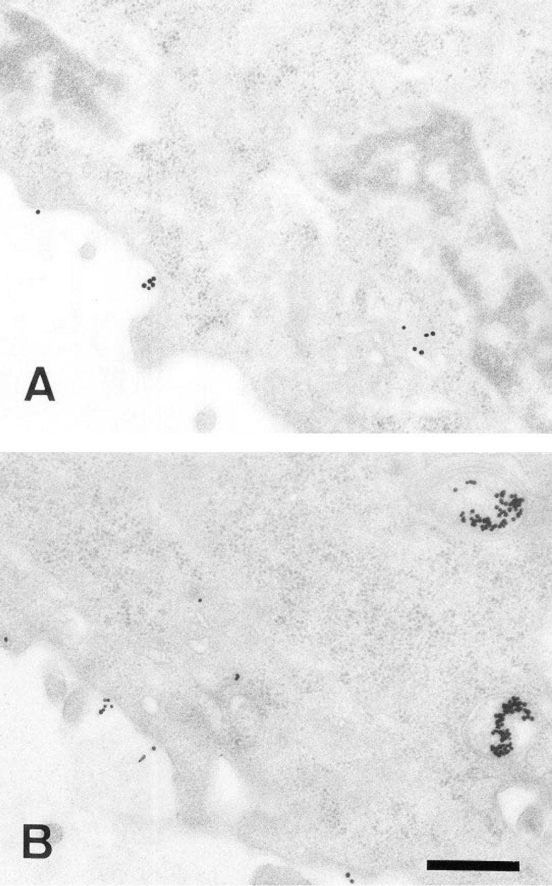
Binding and endocytosis of colloidal gold-LDL conjugates by cultured cells. CHO hLDLR cells were incubated with the conjugates for various times. Only 30-
Assessment of the amount of colloidal gold-LDL conjugates endocytosed at various times by CHO hLDLR cells using electron microscopy and ICP MS analysis a
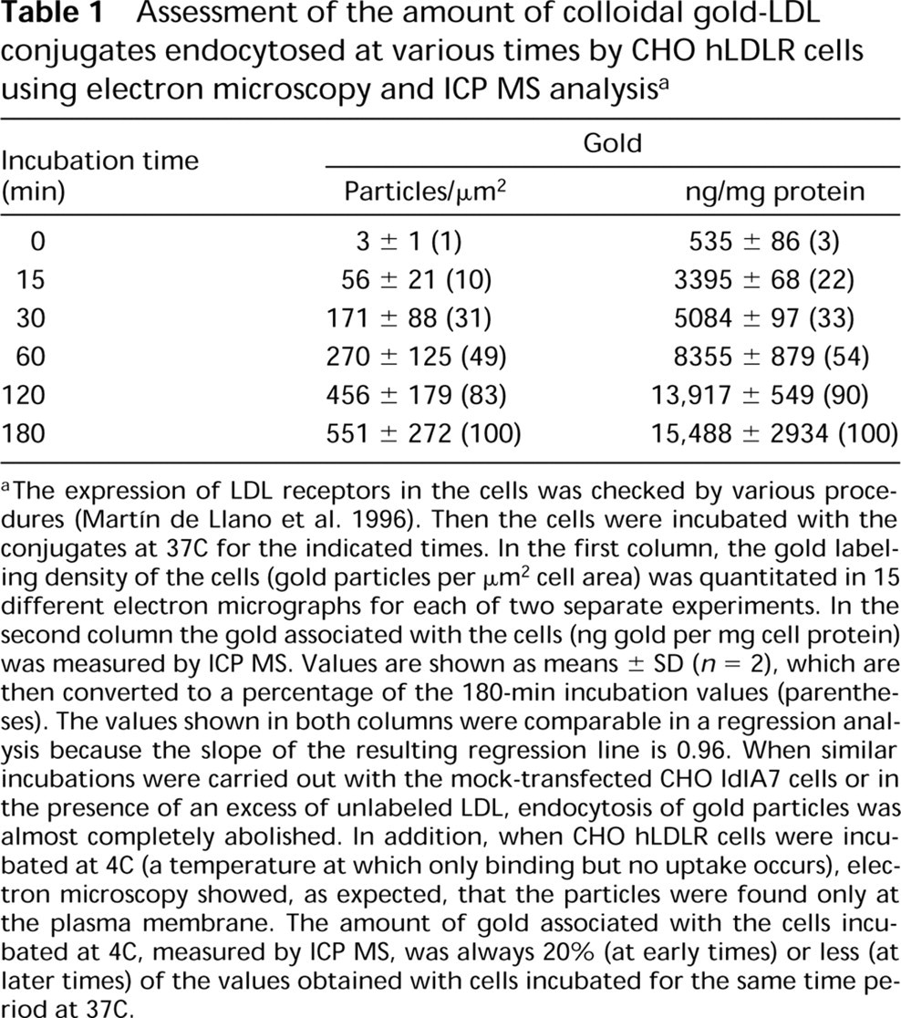
aThe expression of LDL receptors in the cells was checked by various procedures (Martín de Llano et al. 1996). Then the cells were incubated with the conjugates at 37C for the indicated times. In the first column, the gold labeling density of the cells (gold particles per μm2 cell area) was quantitated in 15 different electron micrographs for each of two separate experiments. In the second column the gold associated with the cells (ng gold per mg cell protein) was measured by ICP MS. Values are shown as means ± SD (n = 2), which are then converted to a percentage of the 180-min incubation values (parentheses). The values shown in both columns were comparable in a regression analysis because the slope of the resulting regression line is 0.96. When similar incubations were carried out with the mock-transfected CHO IdlA7 cells or in the presence of an excess of unlabeled LDL, endocytosis of gold particles was almost completely abolished. In addition, when CHO hLDLR cells were incubated at 4C (a temperature at which only binding but no uptake occurs), electron microscopy showed, as expected, that the particles were found only at the plasma membrane. The amount of gold associated with the cells incubated at 4C, measured by ICP MS, was always 20% (at early times) or less (at later times) of the values obtained with cells incubated for the same time period at 37C.
In summary, ICP MS combined with the use of various gold-labeled ligands complements electron microscopic studies, providing a rapid, inexpensive, and reliable method to estimate the expression of specific receptors and the endocytosis of various ligands. Such a method would allow easy investigation of the effects of various parameters (e.g., metabolic activity, cell proliferation), regulators, and drugs on the rate of endocytosis, and can also be of value in the clinical diagnosis of several endocrinologic disorders associated with reduced expression or activity of plasma membrane receptors.
Footnotes
Acknowledgements
Supported by FIS (grant 96/2063), GV (grant GV-97-VS-23-111) and DGICYT (grant PB94-1281).
We are grateful to Dr Miguel de la Guardia and José Soldevilla (Departamento de Química Analítica, Facultad de Ciencias Químicas, Universidad de Valencia) for their help with the ICP MS.