
Abstract
Recent advances in gold technology have led to probes with improved properties and performance for cell biologists: higher labeling density, better sensitivity, and greater penetration into tissues. Gold clusters, such as the 1.4-nm Nanogold, are gold compounds that can be covalently linked to Fab′ antibody fragments, making small and stable probes. Silver enhancement then makes these small gold particles easily visible by EM, LM, and directly by eye. Another advance is the combination of fluorescent and gold probes for correlative microscopy. Chemical crosslinking of gold particles to many biologically active molecules has made possible many novel probes, such as gold-lipids, gold-Ni-NTA, and gold-ATP.
Keywords
Colloidal Gold
Colloidal gold has been the label of choice for electron microscopy for some time, since it is made in convenient well-defined sizes and is rendered immunoreactive by adsorbing antibodies, as found by Faulk and Taylor (1971). Earlier records indicate that colloidal gold was made and used in the 1600s (Handley 1989), but their microscopes were very primitive.
Gold Clusters
A newer gold technology has emerged based on the use of gold clusters, which are gold compounds with a core of multiple gold atoms, with the gold atoms at the surface covalently attached to organic groups. Undecagold, Au11(P(C6H5)3)7, was first described by McPartlin and the structure solved by X-ray diffraction (McPartlin et al. 1969), in which they found the gold core to contain 11 gold atoms with a diameter of 0.82 nm. This cluster was made water soluble by altering the organic groups (Bartlett et al. 1978), then was derivatized to link to proteins (Reardon and Frey 1984; Safer et al. 1986). The structure of one such gold cluster is shown in Figure 1. Although usually prepared with only one linking group per gold particle, so that only one protein molecule is attached, it can be made with multiple linker arms to attach several other molecules, if desired.
A larger gold cluster was subsequently developed (Hainfeld and Furuya 1992) that has better visibility, the 1.4-nm Nanogold particle, which contains ∼67 gold atoms in its core and is also covalently linkable, like undecagold. Other chemists had previously described large gold and other metal clusters (Vargaftik et al. 1985; Schmid 1988).
In keeping with the objective of making the smallest immunoprobe for best penetration and highest density labeling, the Fab′ antibody fragment was chosen. Because the Fab′ fragment is one third the size of a full IgG molecule and because the gold cluster can be site-specifically attached to the end opposite the antigencombining region to preserve immunoreactivity, a probe that is significantly smaller than any IgG-colloidal gold probe can be made (Figure 2).
Discussion
Properties of Gold Clusters
What are the properties of these small gold cluster compounds and how do they differ from colloidal gold?
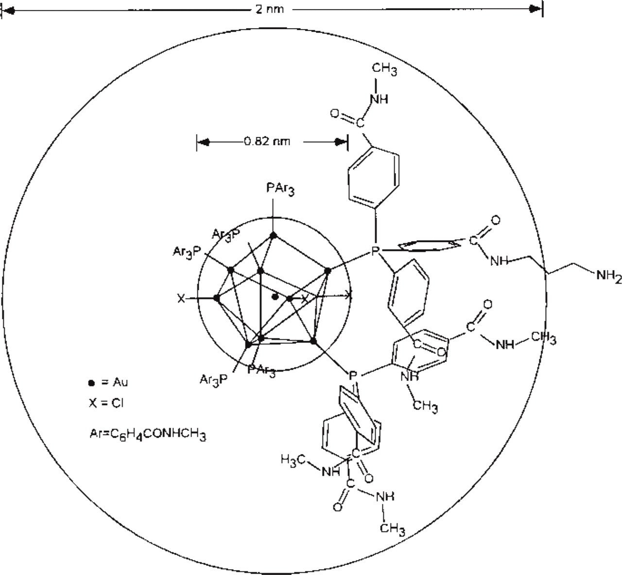
Structural diagram of monoamino-undecagold, with 11 gold atoms in the 0.82-nm core with 7 derivatized triphenylphosphines attached, one of which contains a terminal amino group for crosslinking.
Stability. The gold particle is covalently linked to the antibody or other molecule. They are therefore more stable than colloid gold immunoprobes where antibodies are simply adsorbed, and activity can be undiminished even after a year or more. In contrast, colloidal undergoes some antibody dissociation (Horisberger 1989; Kramarcy and Sealock 1991).
Penetration. The small gold cluster (1.4-nm Nanogold) coupled to an Fab′ fragment (5 nm) is smaller than an IgG (15 nm), giving excellent penetration into tissues (Takizawa and Robinson 1994); even 40 has been reported (Sun et al. 1995).
Better Labeling of Antigens. It has been recognized in many studies (Takizawa and Robinson 1994) that the smaller the gold particle, the more sites are labeled. Factors that explain this are as follows. A small particle does not obscure adjacent antigens and smaller probes diffuse better to sites. In addition, the gold cluster is attached at the hinge region cysteine, which is at the opposite end from the antigen binding site of the Fab′, so the gold does not interfere with antibody binding and native immunoreactivity is preserved (Hainfeld 1987). With colloidal gold, some antibody dissociates (Kramarcy and Sealock 1991), then competes for antigen binding sites, reducing those that then have gold. With gold clusters, the antibody is stably covalently attached, thus leading to more quantitative gold labeling.
Better Resolution. Gold clusters have even been used to mark specific sites of single biomolecules (Schnyder et al. 1995; Wenzel and Baumeister 1995; Zlotnick et al. 1997; Mosesson et al. 1998; Luo et al. 1999). There is no diffusion of reaction products as is seen with DAB.
Peptide and Small Molecule Conjugates Possible. Gold clusters can be covalently attached to almost any small molecule, including peptides (Segond von Banchet and Heppelmann 1995), ATP, nucleic acids (Skripkin et al. 1993), lipids, and carbohydrates (Lipka et al. 1983). Most of these conjugates are not possible with colloidal gold because they do not stably adsorb.
Small and Uniform Size. Because the clusters are actual compounds, they have a specific size (Hainfeld and Furuya 1992). In contrast, colloidal gold <3 nm or “ultrasmall gold” (a colloidal gold ∼1 nm) is very irregular (Hainfeld 1990).
No Aggregation. The small gold clusters do not aggregate even over time, so conjugates that contain one gold cluster attached to one Fab′ can be prepared (Hainfeld and Furuya 1992). In contrast, colloidal gold tends to be “sticky,” adsorbs multiple molecules, and particularly with small (1–3-nm) colloidal gold tends to aggregate (Hainfeld 1990), leading to large oligomeric gold-antibody structures. These lack many of the desirable properties of a small gold probe, such as improved penetration.
Chromatographically Purified. Using gel filtration, it is possible to isolate monomeric Fab′-single Nanogold conjugates and to eliminate excess gold and other species.
Use on Gels. Conjugates are small enough to run on gels. The gold retards proteins by the approximate weight of the gold (∼ kD for undecagold and ∼15 kD for Nanogold) (Weinstein et al. 1989; Hainfeld and Furuya 1995). One can then stain with Coomasssie and, in parallel with silver, track the labeling on a molecular basis (Gregori et al. 1997). In contrast, colloidal gold does not generally work in gels.
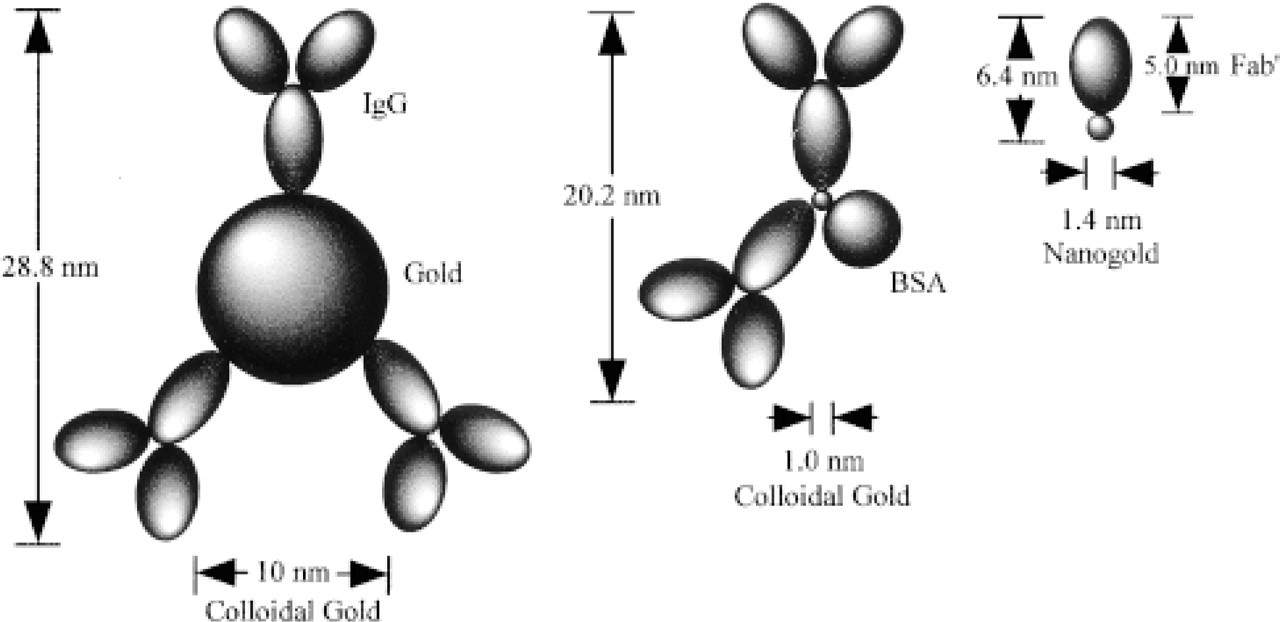
Size comparison of IgG-colloidal gold and Fab′-gold cluster probes. Left is schematic of maximal dimensions of IgG adsorbed to 10-nm colloidal gold. Center diagram is an “ultrasmall” 1.0-nm colloidal gold probe. Right diagram is of Fab′-Nanogold. Note that even using a small colloidal gold particle does not reduce dimensions significantly.
There are, however, some disadvantages of the gold cluster approach. First, silver enhancement is needed for many applications in order to “grow” the small gold size to more visible sizes (10–20 nm or larger). This is typically a simple procedure, however, just floating the grid on the developer for a few minutes. In addition, newer microscopes are able to visualize Nanogold without enhancement, such as on cell surfaces in the SEM. However, silver enhancement does add another variable, and achieving the desired mean product size may take several tries. In addition, if overdeveloped, some background appears. Second, the size distribution of metal particles after silver enhancement is larger than colloidal gold preparations with the same mean size. Finally, gold cluster conjugates are much more difficult to prepare than colloidal gold. Organic phosphines and gold clusters must be chemically synthesized and the gold and conjugates purified by several ultrafiltration and liquid chromatography runs.
Undecagold vs Nanogold vs Ultrasmall Gold
The larger Nanogold cluster (1.4-nm gold core) has several advantages over Au11 (0.8 nm): it is directly visible by TEM (and better SEMs) at high magnification, is stable in the beam, and it develops better with silver (Hainfeld and Furuya 1992). For most applications, therefore, Nanogold is the preferable choice.
It is possible to make colloidal gold in very small sizes, 1.0 nm or less, and proteins are adsorbed to these particles in the same way as with larger colloidal golds. Unfortunately these “ultrasmall gold” preparations have several disadvantages (Hainfeld 1990) compared to Nanogold: (a) the size distribution of gold particles is very large, usually ranging from 0.8 to 3.0 nm; (b) aggregation with antibodies tends to occur, so the conjugate, which contains multiple antibodies and gold particles is actually quite large, severely limiting penetration (Hainfeld 1990); and (c) the attachment is not covalent and is therefore less stable.
Labeling Reactions
The linking arm on the gold can be made to specifically react with thiols (e.g., cysteine residues on proteins), using a maleimide group, or with amino groups using an N-hydroxysuccinimide (Figure 3). Other specific reactions can be used to link to sugar groups in glycoproteins. These form stable covalent bonds and permit useful conjugates to be made, even with molecules that do not adsorb to colloidal gold.
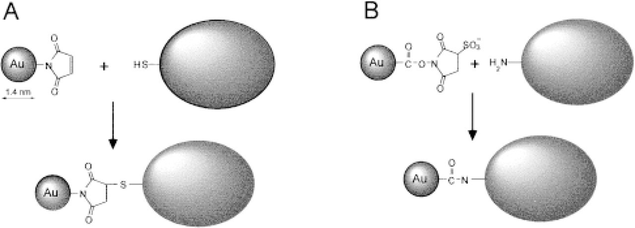
Diagram of chemical reactions that covalently link gold clusters to specific groups. (
Nanogold is available as reagents that react with free thiols or amines. These can be used to make conjugates with primary antibodies or other molecules. The procedure for labeling is quite simple, and consists of mixing the reactive gold reagent with the molecule to be labeled, then, after several hours, purifying the conjugate by gel filtration chromatography, ultrafiltration, or dialysis (to remove unreacted gold or un-labeled molecules). More specifically, the protocol for labeling is as follows:
Dissolve lyophilized Nanogold reagent by adding 1 ml water [contains dried buffer; final solution is PBS, pH 6.5 (maleimido) or 7.5 (Sulfo-NHS)].
Mix with ∼0.2 mg of protein to be labeled. If reacting with amines, no protein pretreatment is necessary. If making Fab′ or IgG conjugates with maleimido-Nanogold, these must be prepared. For IgG, reduce with mercaptoethanolamine, to expose hinge sulfhydryls, and column purify; for Fab′, digest with pepsin or ficin to get F(ab′)2, then reduce and column-purify (to remove reducing agent).
After 1–16 hr, gel-filter product (on, e.g., Pharmacia Superose 12 in PBS), to separate labeled protein from unreacted gold.
An example is the labeling of an Fab′ fragment, shown diagrammatically in Figure 4. An electron micrograph of Fab′ fragments labeled with Nanogold (after column chromatography to remove excess Nanogold reagent) is shown in Figure 5.
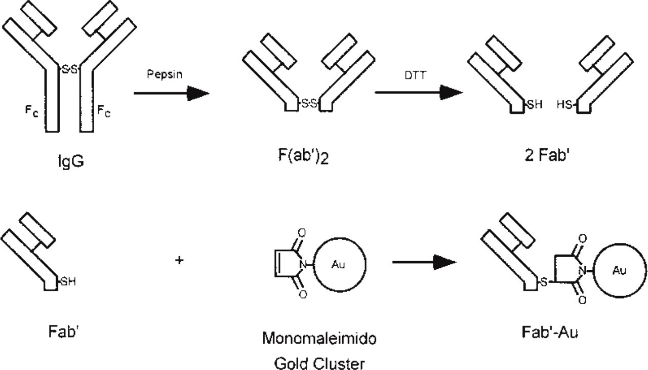
Schematic flow diagram for labeling the hinge thiol of an Fab′ antibody fragment with a gold cluster. IgG is first digested to F(ab′)2, then reduced to Fab′; gold cluster is then reacted with the free thiol at the hinge region.
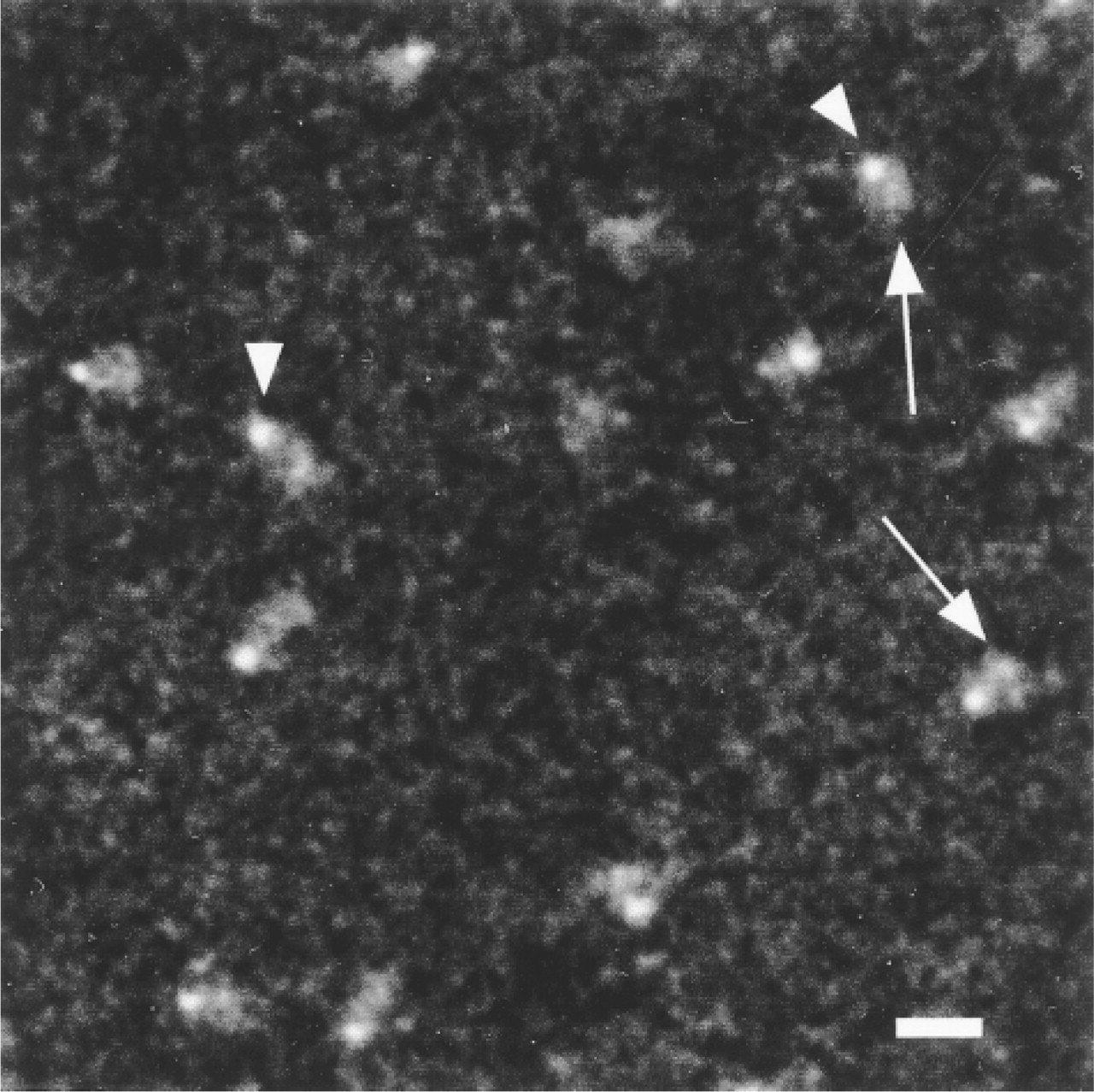
Darkfield scanning transmission electron micrograph of Fab′-Nanogold. Arrows point to Fab′ fragments and arrowheads to Nanogold particles. Bar = 10 nm.
Stoichiometry of Labeling
With colloidal gold, it is difficult to quantitate the amount of antibody bound per gold particle by UV-Vis spectroscopy because the gold dominates the spectral absorption. However, gold clusters are smaller and one Nanogold at 280 nm absorbs 1.4-fold as much as a single IgG molecule. In addition, the gold absorbs in the visible spectrum, whereas most proteins do not. Therefore, a simple calculation based on a UV-Visible spectrum (or measurements at two points, such as 280 and 420 nm) will yield the ratio of gold to antibody. For successful antibody labeling with Nanogold, this is close to 1:1. A more detailed discussion of the equations to use is found in Hainfeld (1989). For Nanogold, the extinction coefficients are 2.25 × 10⁵ M−1cm−1 at 280 nm and 1.12 × 10⁵ M−1cm−1 at 420 nm.
Silver Enhancement
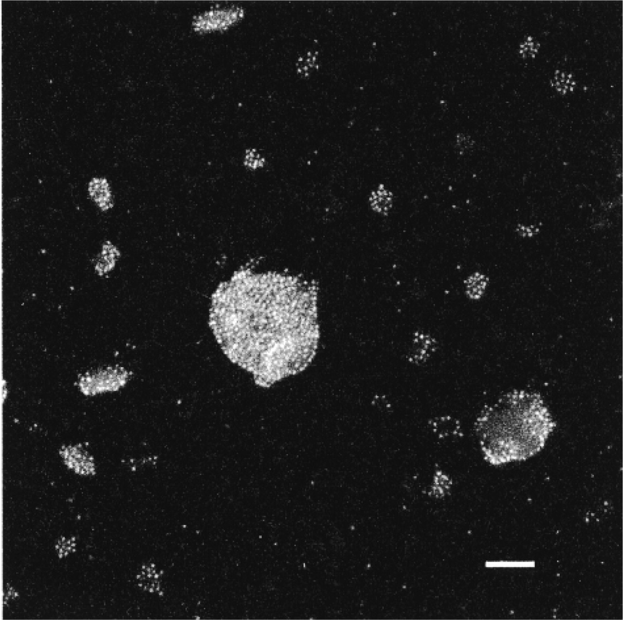
Silver enhancement, also called autometallography, is a process by which silver is deposited on the gold surface (Danscher et al. 1995). Because the metal particle acts as a catalyst, the plating is specific (Figure 6). Various silver salts, reducing agents, and moderators that can give better control (such as gum arabic), have been used; some formulations are better for EM or LM. The small gold particle can easily be grown to a useful size for EM (10–20 nm). Sometimes a larger size can be used for double labeling, e.g., to distinguish it from a smaller 10-nm colloidal gold (Takizawa and Robinson 1994), or so that labeling can be seen at very low EM magnification for easy screening of samples. Although very uniform particles have been reported after development (Krenács and Krenács, 1995), in general the distribution of particle sizes after silver enhancement is worse than that from a good colloidal gold preparation, say of 10-nm size. However, the typical size distribution is completely acceptable for most applications (Figure 7).
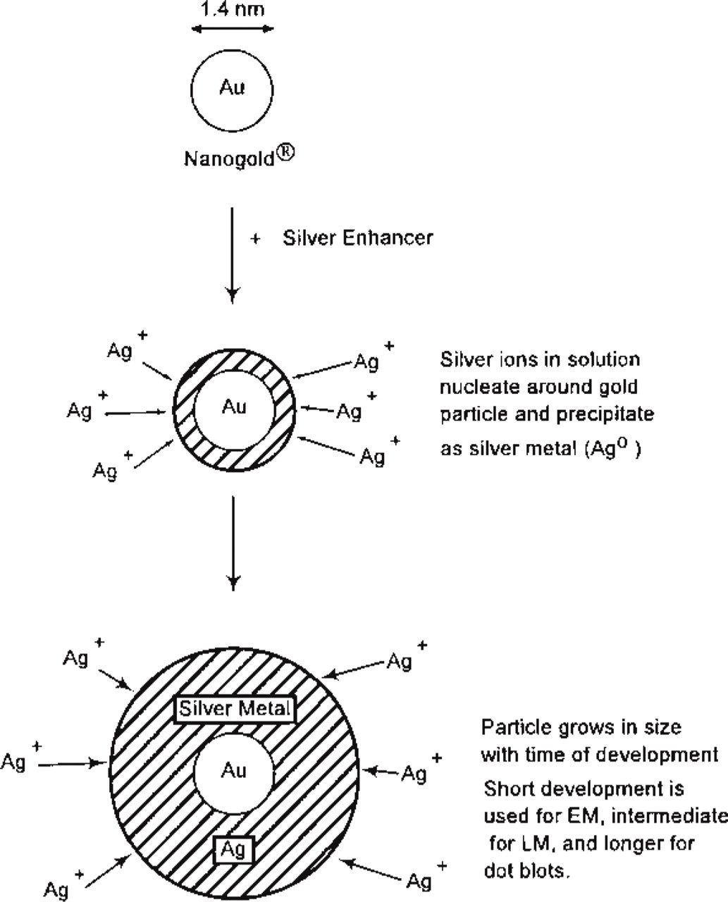
Diagram of silver enhancement process.
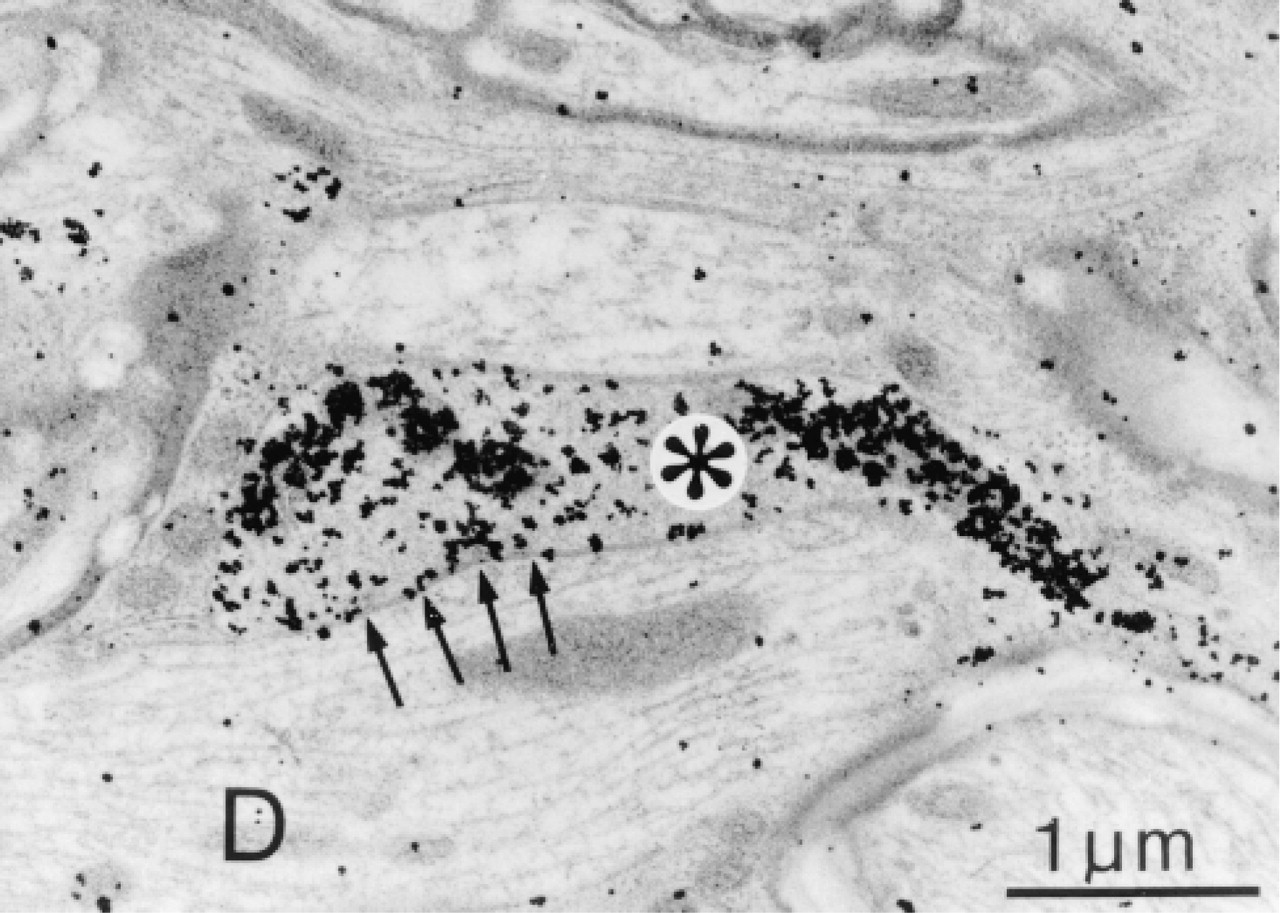
Electron micrograph after silver enhancement of Nanogold-Fab′ labeling, leading to ∼10-nm particles. Specimen shows GABA-containing terminals (asterisks) forming symmetrical synaptic specializations (arrows) with dendrites (D) in the thoracic spinal cord of the rat. Postembedding staining was done using GABA antiserum (Incstar 1:2000, 4C, 18 hr), Nanogold-goat anti-rabbit Fab′ (1:40 room temperature, 90 min), and intensified with HQ silver (Nanoprobes) for 6 min. Tissue was counterstained with lead citrate, and embedding resin was Durcupan (Fluka). Courtesy of S. Bacon, Oxford University, Dept. of Pharmacology, Oxford, UK.
Because osmium poststaining for EM can oxidize and reverse the silver staining, it was found that lower concentrations of OsO4 (0.1% for 30 min rather than 1%) does not affect the silver signal and still gives excellent staining (Burry et al. 1992). Another method has been to use gold toning, which coats the silver with gold, making it impervious to osmium (Sawada and Esaki 1994; Arai and Nagatsu 1995).
Electron Microscopy
Scanning Transmission Electron Microscopy (STEM)
The darkfield high-resolution STEM is one of the best instruments for visualizing small gold clusters clearly. It has been used in projects where single proteins or protein complexes have been labeled (Wilkinson et al. 1994; Yang et al. 1994; Mosesson et al. 1998; Luo et al. 1999). For example, an undecagold-Fab′ antibody to the carboxy terminus of the γ-chain of fibrinogen mapped the locus of this site on the fibrinogen molecule (Figure 8).
Labeling Peptides
Small peptides do not adsorb well to colloidal gold but can be covalently attached to gold clusters by reacting a cysteine -SH group, or an amino (-NH2) group (either the α-amino or one from a lysine residue) (Figure 9). In one example, the 40-amino-acid amyloid β-peptide, a peptide important in the development of Alzheimer's disease, was labeled with Nanogold (Gregori et al. 1997). The gold-labeled peptide was used to determine the peptide binding site in the proteasome (Figure 10).
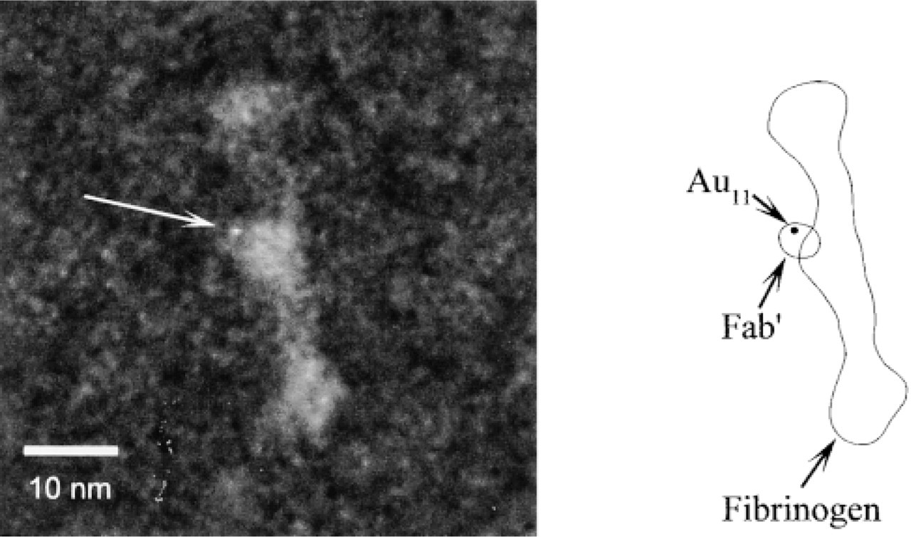
Darkfield scanning transmission electron micrograph of a single fibrinogen molecule with an undecagold (arrow)-labeled Fab′ targeted to the carboxy terminus of the γ-chain. Diagram at right identifies structures.
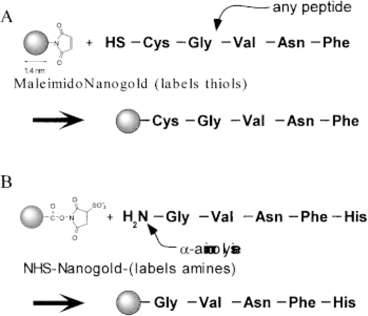
Scheme for labeling peptides with gold clusters. (
In another example, the 11-amino-acid peptide substance P (SP) was labeled with Sulfo-NHS-Nanogold (Segond von Banchet and Heppelmann 1995). This was then used to visualize binding in the rat spinal cord and on gels. Comparison with the previously used technique of labeling the peptide with 125I gave identical results and the controls behaved similarly, such as competing off the labeled SP binding with excess unlabeled SP. This work is notable for two reasons. First, it shows that even a small peptide retained activity after gold labeling. Second, the lengthy (months to develop an autoradiograph), hazardous (radioactive), and low-resolution 125I labeling could be replaced with gold cluster labeling, which gave results the same day, was non-hazardous, and gave high-resolution information.
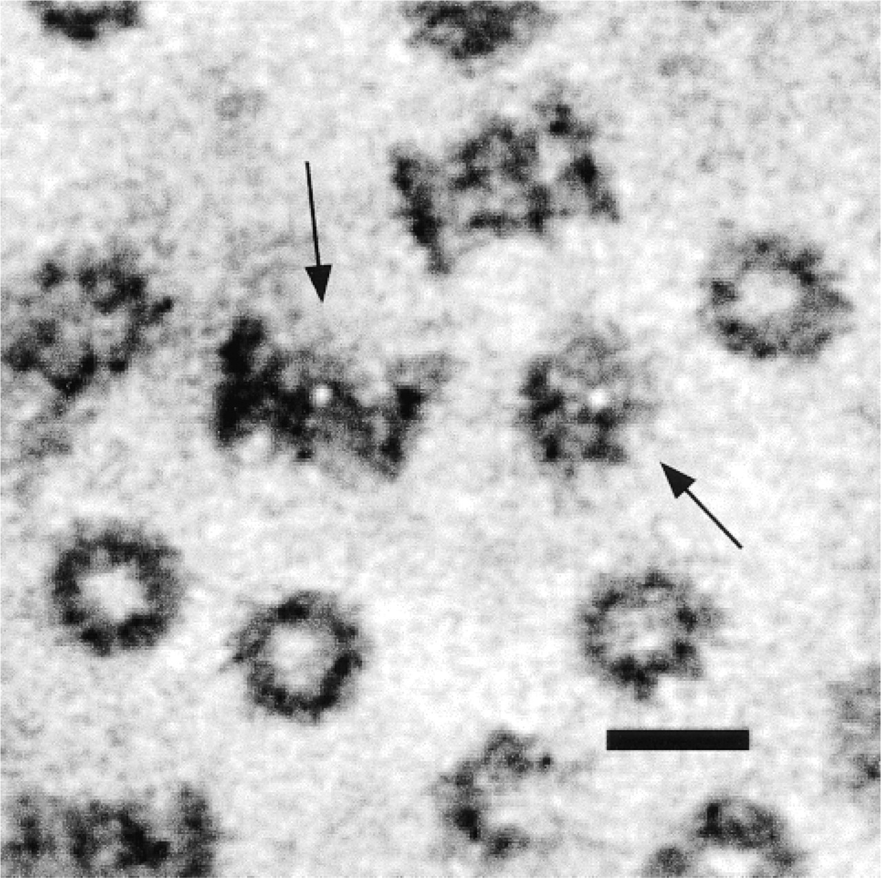
Negatively stained scanning transmission electron microscope image of proteasomes labeled with Nanogold-amyloid β protein after column purification. Sample stained with methylamine vanadate (NanoVan; Nanoprobes). Arrows point to proteasomes with gold clusters: bar = 10 nm. Sample prepared by L. Gregori, Dept. of Psychiatry and Behavioral Science, School of Medicine, State University of New York, Stony Brook, NY.
TEM
Many examples have now been reported in the literature of Nanogold labeling in tissue sections in the TEM, using both pre-embedding (Vandre and Burry 1992; Burry 1995; Nusser et al. 1995) and postembedding (Krenács and Krenács 1995). In general, the density of labeling is greater than with colloidal gold and the penetration many times better. One is best referred to this body of literature for specific details and protocols, which can be found in a reference list by topics on the Nanoprobes web site (http://www.nanoprobes.com). A useful comparative review of methods for the various techniques was recently published (Hainfeld and Powell 1997).
Light Microscopy
Use of silver-enhanced gold gives punctate, high-resolution detail that stands out against H&E and other cell stains. Because the small gold cluster immunoprobes or Nanogold-streptavidin penetrate into tissues up to 40 μm, this has led to many new applications for gold labeling for the LM. Although brightfield optics are usually adequate for viewing, reflected light (using crosspolarized epi-ilumination) can specifically identify the metal deposits because they repolarize the light upon reflection.
FluoroNanogold
For correlative light and electron microscopy, a dual label containing both a fluorophore and gold would be advantageous. The use of two separate immunolabels in parallel experiments (fluorescent in one and the gold in the other) has been unsatisfactory in many cases because different localizations are frequently seen. It also demands two preparations. Fluorescent and gold probes would also be useful for screening to see that labeling is successful at the LM level before proceeding (with the same sample) to the more extensive EM preparation. Another application of a dual probe is to follow a process in living cells, and when the fluorescent image indicates the time of interest, cells can be fixed and examined at the ultrastructural EM level. Combined fluorescent and colloidal gold probes have had limited success because gold particles strongly quench the fluorescence, and large gold particle sizes are worse. The quenching depends also on the distance between fluorophore and gold, so that they must be spaced apart at an appropriate distance.
Combined fluorescent and gold probes have been prepared by attaching both Nanogold and fluorescein (Powell et al. 1997; Robinson and Vandre 1997) or Cy3 (Keohane et al. 1999; Powell et al. 1999b) to Fab′ and streptavidin. It was found that attaching the fluorophore directly to Nanogold led to excessive quenching, so instead the Nanogold is attached to the hinge region thiol of an Fab′ antibody fragment, and the fluorophore to a reactive amino group (Figure 11) (Powell et al. 1997). By using an Fab′ fragment, the size of the whole conjugate is kept smaller than an IgG, so penetration into tissues and sections is excellent (Robinson and Vandre 1997). It has been possible with FluoroNanogold to visualize antigens in the same cell by light and electron microscopy (Powell et al. 1997, 1998; Takizawa et al. 1998). FluoroNanogold has also been made with streptavidin, so that biotinylated DNA or biotinylated primaries can be targeted. The original FluoroNanogold is Nanogold combined with fluorescein, which fluoresces green. Cy3-Nanogold is now available, which has a red color. Cy3 also has the advantages of better water solubility, greater brightness, and reduced bleaching (Powell et al. 1998, 1999b).
Two other reports in this issue detail applications of FluoroNanogold, and the reader is referred to them for further information and experimental details: T. Takizawa and J.M. Robinson, “FluoroNanogold is a bifunctional immunoprobe for correlative fluorescence and electron microscopy,” and J.M. Robinson, T. Takizawa, and D.D. Vandré, “Enhanced labeling efficiency using ultrasmall immunogold probes: immunocytochemistry.”
Metallosomes
Gold clusters, because they can be covalently linked with almost any other molecule, can be attached to phospholipids or fatty acids (Hainfeld 1996). This gives a long hydrophobic tail with the water-soluble gold on the head group (Figure 12). These molecules behave similarly to native lipids, and on sonication, for example, form micelles and vesicles (Figure 13). If they are mixed with unmodified lipids to “spike” liposomes, they can be used to follow the liposomes (Thurston et al. 1998), and have been so used to demonstrate targeted drug delivery (Adler-Moore 1994). The golden lipids also form monolayers on an air-water interface and can be compressed to form regular gold arrays (Hainfeld et al. 1999a).
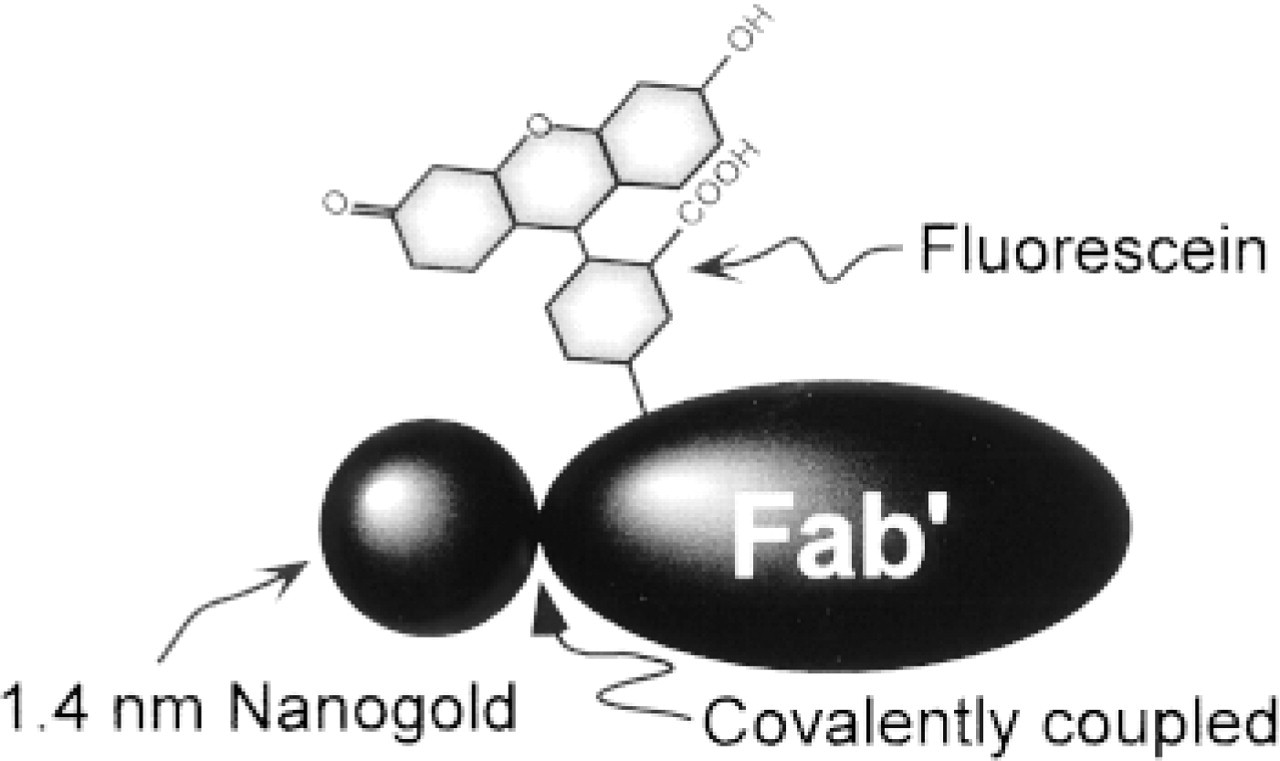
Diagram of construction of FluoroNanogold-Fab′ probe.
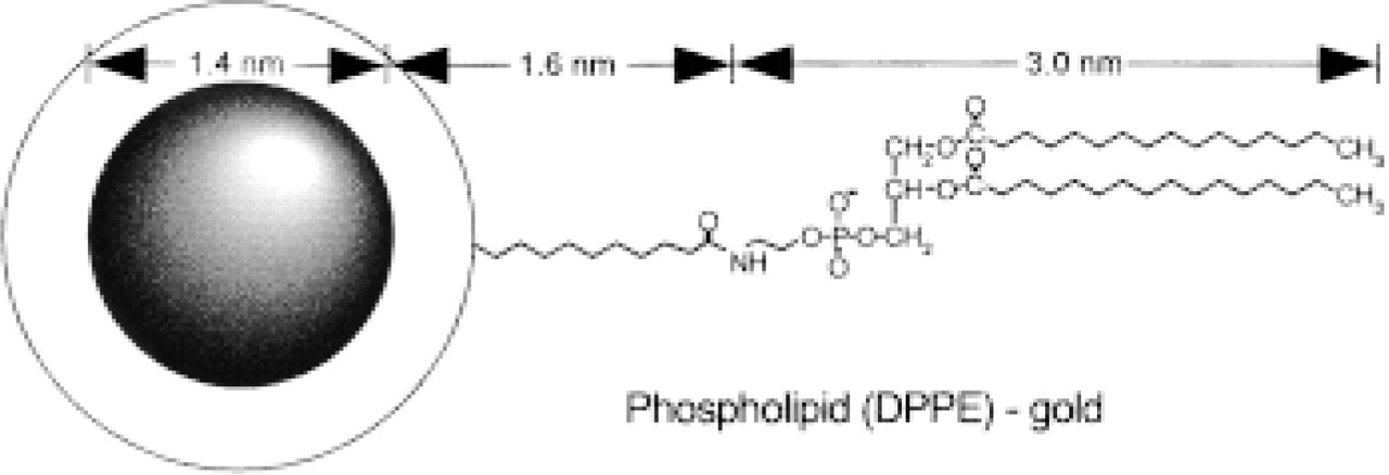
Scale diagram of Nanogold-phospholipid (dipalmatoyl-phosphatidylethanolamine, DPPE).
In Situ Hybridization
Certain genes or sequences can be detected in cells, and this is important for both basic studies and pathology. For example, HPV-16 (human papilloma virus) is highly correlated with cervical cancer (Cheung et al. 1999), and gene amplification of Her/2-neu is important in prognosis and treatment of breast cancer. Although fluorescent and colorometric ISH tests are used, gold detection gives an excellent black staining compatible with the use of other full-strength cell stains for LM. Compared with fluorescence, there is no autofluorescence to worry about, no expensive fluorescent optics, and the slide is permanent and does not bleach or fade with observation or storage. Using the CARD (catalyzed reporter deposition) method and streptavidin-Nanogold, single copy gene detection has been routinely achieved (Zehbe et al. 1997).
Gels and Blots
With silver enhancement, signals from immunogold targeting, for example, may be seen by eye on blots, as shown in Figure 14. Nanogold has shown to give particularly sensitive detection, rivaling or exceeding limits of fluorescent, radioactive, enzyme-colorometric, and chemiluminescent methods (Hainfeld and Furuya 1992). Another use, suggested by Burry (1995), is to spot dilutions of the Nanogold-antibody conjugate on nitrocellulose paper, then silver-develop. The graded response is used to determine the optimal silver enhancement time for EM or LM samples.
Colloidal gold is generally too large to enter into polyacrylamide gels. However, gold clusters (undecagold ∼5 kD and Nanogold ∼15 kD) attached to proteins shift the weight by approximately that amount. This means that Coomassie blue staining can be used to visualize proteins, and silver staining will reveal the gold-labeled bands (using a parallel gel, or a lane cut lengthwise, half for Coomassie, half for silver) (Hainfeld and Furuya, 1995; Gregori et al. 1997).
New Developments
Metal probes that can be covalently linked to biomolecules open up many possibilities for making novel probes for light and electron microscopy or other applications. Different molecules can be attached or different sized metal particles used, as well as a variety of metals in place of gold. Some of these are briefly described below.
Ni-NTA-Nanogold
Cloned proteins frequently have a 6x-His tag (6 histidine tail) engineered in, which binds to a nickel column for one-step purifications from cell lysates. The nickel is held to the column material via the chelator NTA (nitrilotriacetic acid). This chelator has been attached to Nanogold, then charged with Ni2+ ions, so that the Nanogold then binds to 6x-His tagged proteins (Figure 15) (Hainfeld et al. 1999c).
ATP-Gold
Darkfield scanning transmission electron micrograph of Nanogold-phospholipid after sonication. Bar = 20 nm.
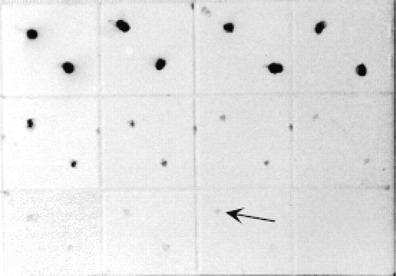
Sensitive dot blot using Nanogold-Fab′ and silver enhancement. Nanogold-antimouse Fab′ was incubated with dilutions of mouse IgG spotted on nitrocellulose, washed, and developed with LI Silver (Nanoprobes). Arrow points to spot detecting 0.1 pg of IgG. Lower right panel is control with just buffer spots.
ATP or its analogues linked to gold clusters can be used in several ways. One is to label nucleotide binding proteins. Another is to serve as a substrate for a polymerase or DNA synthesizer to incorporate gold into nucleic acid probes. An ATP-gold cluster conjugate has recently been described (Hainfeld et al. 1999b).
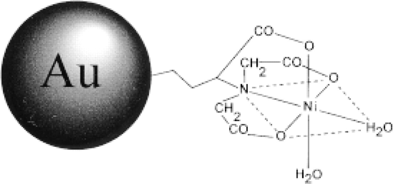
Schematic diagram of gold cluster with Ni-NTA group attached for binding 6xHis-tagged proteins.
Larger Platinum and Palladium Clusters
These clusters are based on 1,10-phenanthroline ligands instead of the phosphines used in gold clusters. Clusters ∼2 nm contain ∼200 heavy atoms, and clusters 2.4 nm (Pd561) and 3.6 nm (Pd2057) have been described. The advantages of these structures include the use of a different metal for spectroscopic detection and larger sizes for better visibility (Powell et al. 1999a).
Thiol-Gold
In addition to phosphine-stabilized gold particles, ligands bound to the gold core via a sulfur donor are also stable and may prove useful (Brust et al. 1994; Buining et al. 1997; Chen and Kimura 1999). An Ni-NTA cluster that binds 6x-His tags was prepared this way using a thiol-containing dipeptide (Hainfeld et al. 1999c).
Tetra Iridium
Clusters with four iridium atoms have been synthesized and linked to antibodies, viruses (Cheng et al. 1999), and other proteins (Figure 16). This small cluster may be useful for higher-resolution labeling and as an isomorphous replacement in the X-ray phasing of large unit cell protein crystals (Weinstein et al. 1989; Weinstein et al. 1999).
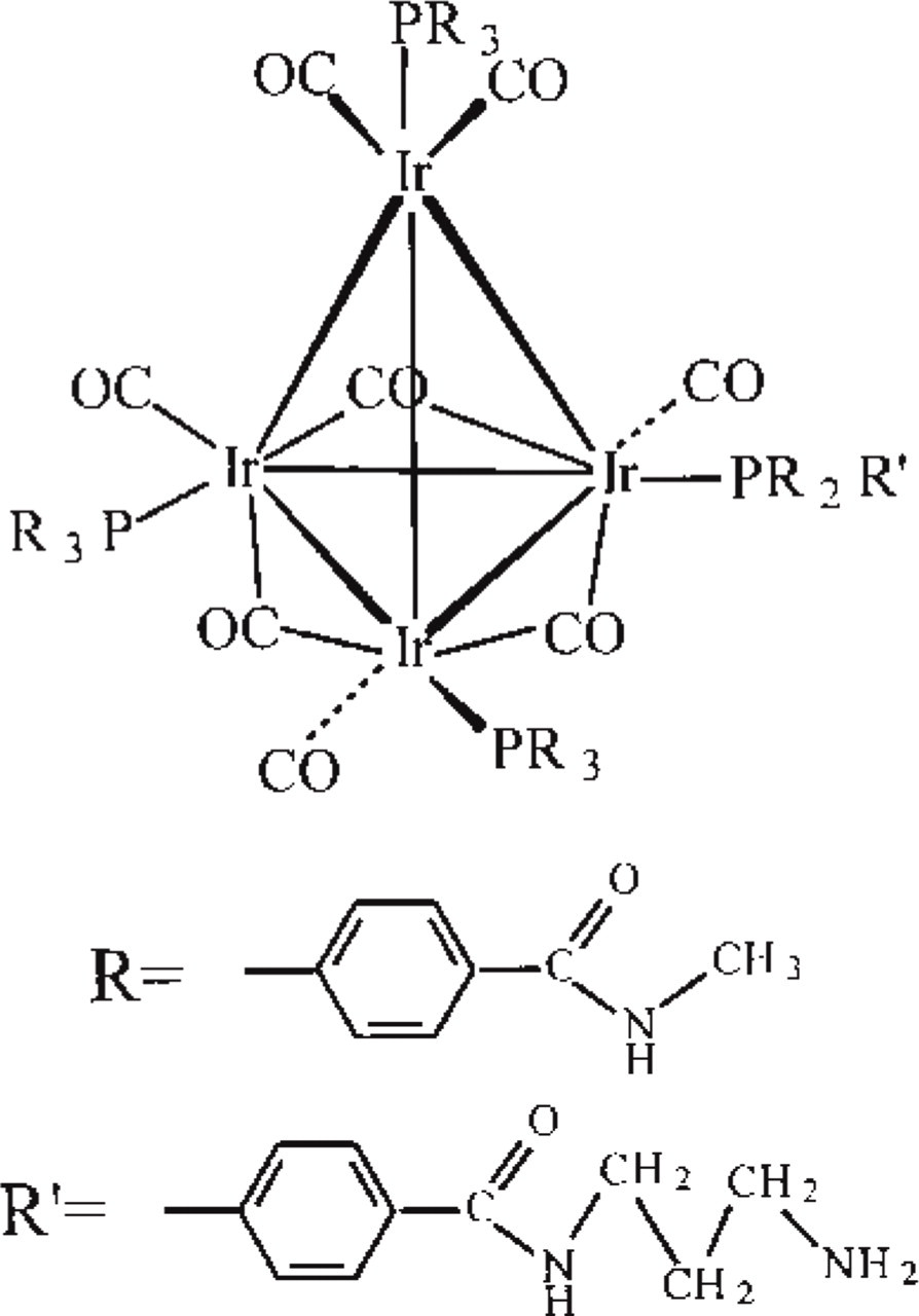
Tetra iridium cluster diagram, Ir4(CO)8(PR3)3(PR2R′). Dense metal Ir4 core is ∼0.5 nm, whereas the overall diameter, including the phosphines (PR3), is 1.7 nm.
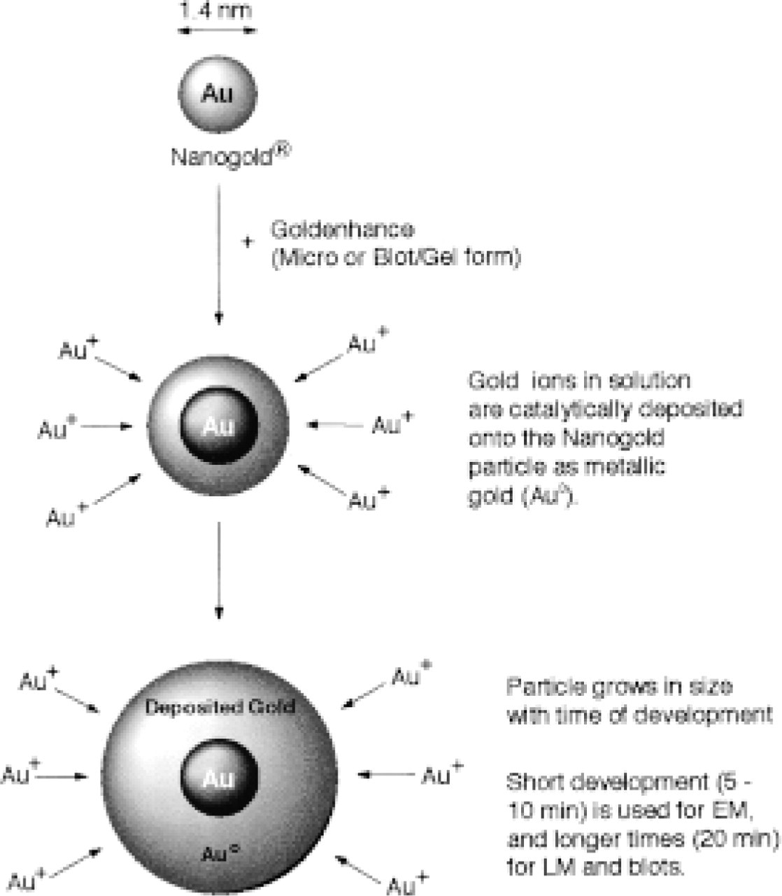
Gold development diagram. Small gold particle specifically nucleates additional gold deposition from solution, enlarging the particle for better detection.
Gold Developer
Silver enhancement has proved to be quite useful to amplify the signal from small gold particles. In a similar fashion, gold can be catalytically deposited on the targeted gold particles (Figure 17). Gold enhancement has a number of potential advantages compared to silver. It can safely be used before osmium tetroxide staining (it is inert to osmium), lower backgrounds are observed in many cases and, for SEM, gold gives a significantly higher backscatter signal (Hainfeld et al. 1999d).
Summary
Metal cluster labeling offers significant performance improvements over labeling with colloidal gold in the conventional manner, including more quantitative labeling and improved labeling of macromolecules and antigens in cells and tissues. The covalent crosslinking rationale and the ability to selectively modify the reactivity of metal cluster labels have enabled the preparation of new types of probes that are not feasible with colloidal gold. The development of combined fluorescent- and metal cluster-labeled probes has enabled more precise correlative microscopy, and the variety of different metals and cluster functionalities now available has significantly widened the applications of gold labeling.
Footnotes
Acknowledgements
Supported by the Office of Biological and Environmental Research of the US Department of Energy under Prime Contract No. DE-AC02-98CH10886 with Brookhaven National Laboratory, by National Institutes of Health Grant 2 P41 RR01777, and by NIH Small Business Innovation Research grants GM 48328, GM 49564, and GM 56090.
We wish to thank Dr Martha Simon, Ms Beth Lin, and Mr Frank Kito for STEM operation, and Dr Joseph Wall for helpful discussions. We are grateful to Carol M. R. Halsey, Vishwas Joshi, and Frederic Furuya for helpful experimental discussions, and to Gerhard W. Hacker for experimental data on gold enhancement and combined Cy3 and Nanogold probes.