
Abstract
We demonstrate a fluorescent ultrasmall immunogold probe, FluoroNanogold (FNG), to be a versatile reporter system for immunocytochemical labeling of ultrathin cryosections. FNG-labeled molecules in the same ultrathin cryosections can be resolved by two imaging techniques (i.e., fluorescence and electron microscopy). Lactoferrin, a marker protein for the specific granules in human neutrophils, was employed as the target for FNG immunolabeling. The spatial resolution of the fluorescence signal from FNG-labeled specific granules was compatible with that of silver-enhanced gold signal from the same granules in electron microscopy. Our results confirm that FNG can be used as a probe for highresolution correlation between immunofluorescence and electron microscopy.
Keywords
Recently, a unique ultrasmall immunogold probe, FluoroNanogold (FNG), has been developed for use as a secondary antibody in immunocytochemical applications (Powell et al. 1997). It consists of a 1.4-nm gold cluster compound to which antibodies and fluorochromes are covalently conjugated. FNG permits correlative microscopic observation of a sample stained in a single labeling procedure by multiple optical imaging techniques [e.g., fluorescence and differential interference contrast (DIC)] (Robinson and Vandré 1997). However, critical studies correlating the fluorescence signal from FNG in fluorescence microscopy and the silver-enhanced gold signal from the same FNG in electron microscopy have not been conducted.
The aim of the present study was to determine whether the utilization of FNG in immunocytochemistry on cryosections, especially ultrathin cryosections, will be useful for the correlation of immuno-LM and -EM signals. We have examined the utility of FNG as a secondary antibody for immunolabeling of lactoferrin (a marker protein for the so-called specific granules) in ultrathin, cryosectioned human neutrophils. Neutrophils contain abundant intracellular granules, which show variety in type and size. Therefore, detection of lactoferrin-containing specific granules requires high spatial resolution. The fluorescence signal from FNGlabeled specific granules in fluorescence microscopy corresponded precisely to silver-enhanced gold signal from the same granules in electron microscopy. Our results confirm that high-resolution correlation between fluorescence and electron microscopy can be achieved with ultrathin cryosections using FNG in immunocytochemistry.
Materials and Methods
Reagents
Except where noted, reagents were similar to those we have described previously (Takizawa and Robinson 1993,1994a,b). Rabbit anti-human lactoferrin (IgG fraction) was obtained from Cappel-Organon Teknika (Durham, NC). Goat antirabbit FluoroNanogold (affinity-purified goat anti-rabbit Fab' fragment conjugated to a molecular label containing both fluorescein and a 1.4-nm particle) (FNG) was purchased from NanoProbes (Stony Brook, NY). All immunological reagents were handled in accordance with the manufacturer's recommendations and used within the expiration date for each product. SlowFade-Light Antifade kit was from Molecular Probes (Eugene, OR).
Cell Isolation
Whole human blood was collected from healthy adult men after obtaining informed consent. Neutrophils were purified from whole blood in the unstimulated state, as described previously (Takizawa and Robinson 1993).
Preparation of Ultrathin Cryosections
Purified cells were fixed in suspension with 4% paraformaldehyde in 100 mM sodium cacodylate buffer, pH 7.4, containing 5% sucrose, washed in cacodylate buffer, embedded in gelatin, and then infiltrated with 2.3 M sucrose, as previously described (Takizawa and Robinson 1993,1994a,b). The gelatin blocks were then cut as ultrathin cryosections and collected on formvar film-coated EM grids. We used copper or nickel EM grids (Maxtaform “finder” grids; Graticules, Tonbridge, Kent, UK) which have markings so that the location of a given cell can be recorded by optical microscopy and readily found on reexamination by electron microscopy.
Immunocytochemistry on Ultrathin Cryosections with FNG
Cryosections mounted on EM grids were incubated for 1 hr at 22C in a solution containing 1% BSA and 10% normal goat serum in PBS to block nonspecific protein binding sites. Grids were incubated with anti-lactoferrin IgG (27 μg/ml) for 1.5 hr at 22C. The grids were then washed in five changes of PBS and incubated with goat anti-rabbit FNG (0.8 μg/ml Fab') for 1.5 hr at 22C. The grids were subsequently washed in at least five changes of PBS. All antibody solutions were diluted with PBS containing 1% BSA and 10% normal goat serum. Controls received the same treatment except for omission of the primary antibody.
Fluorescence Microscopy
The grids were examined by epifluorescence and DIC microscopy with an Olympus Provis AX80 equipped with a U-MCB photographic attachment and a PM-C35DX camera. The light source for fluorescence microscopy was a 100-W mercury lamp which was intensified with an AX-UCV conversion lens. The Olympus objective lenses employed were a X10 UPlanApo, NA 0.40, X20 UPlanApo, NA 0.70, and X100 UPlanApo, NA 1.35. The filter cube was an Olympus U-MWIBA with a 460–490-nm excitation filter and a 515–550-nm barrier filter. Photomicrographs were recorded on Fuji Provia 400 and Kodak T-Max 400 films which were exposed and developed at ASA 400 and 1600, respectively. The images recorded on these films were then captured on computer and magnified using Adobe Photoshop 4.0J without additional manipulation of the images.
For optical microscopic observation, the grids containing ultrathin cryosections were washed in SlowFade-Light Antifade solution containing 50% glycerol to retard photobleaching, immersed in 10 μl of the same solution on glass slides, overlaid with a glass coverslip (18 mm in diameter; number 1 thickness), and then immediately examined by optical microscopy. Observation of a given slide was carried out as rapidly as possible. Sections were located and their position on the grid was recorded with the aid of the X10 and X20 objective lenses; this was necessary for relocation of the same sections during EM observations.
Silver Enhancement of FNG and Electron Microscopy
After examination of FNG-labeled lactoferrin by optical microscopy, the grid was removed from the temporary slide and silver enhancement of FNG was carried out to visualize the gold particles at the electron microscopic level. Silver enhancement was achieved using the procedure developed by Burry and co-workers (for review see Burry 1995) as reported previously (Takizawa and Robinson 1993,1994a,b). After the silver enhancement process, the ultrathin cryosections were washed in distilled water, negatively stained with aqueous 2% phosphotungstic acid, pH 7.0, simultaneously covered with a small square of thin formvar film by the method of Sakai et al. (1995), and observed with a Hitachi H-7000 electron microscope (Hitachinaka, Japan) operated at 75–100 kV.
Results
The distribution of lactoferrin in ultrathin cryosectioned human neutrophils was determined with rabbit anti-lactoferrin followed by goat anti-rabbit FNG and was visualized by fluorescence microscopy (Figures 1A and 1D). Abundant granule-like fluorescent spots indicating the distribution of lactoferrin were present in the cells. The general morphology of cells in the sections was determined with DIC optics (Figure 1B).
Lactoferrin distribution in the same ultrathin cryosectioned cells was subsequently detected by electron microscopy after silver enhancement of FNG (Figures 1C and 1E). The negative staining-formvar technique reveals the fine structure of the ultrathin cryosections with high resolution and contrast. The subcellular distribution of lactoferrin was readily evident in these ultrastructural preparations (Figures 1E and 2C). Silver-enhanced FNG immunoprobes showed that lactoferrin was present in an intracellular granule compartment of high abundance in the cytoplasm of the cells (i.e., the specific granules), whereas other granules lacking lactoferrin were unlabeled (i.e., azurophillic granules) (Figure 2C).
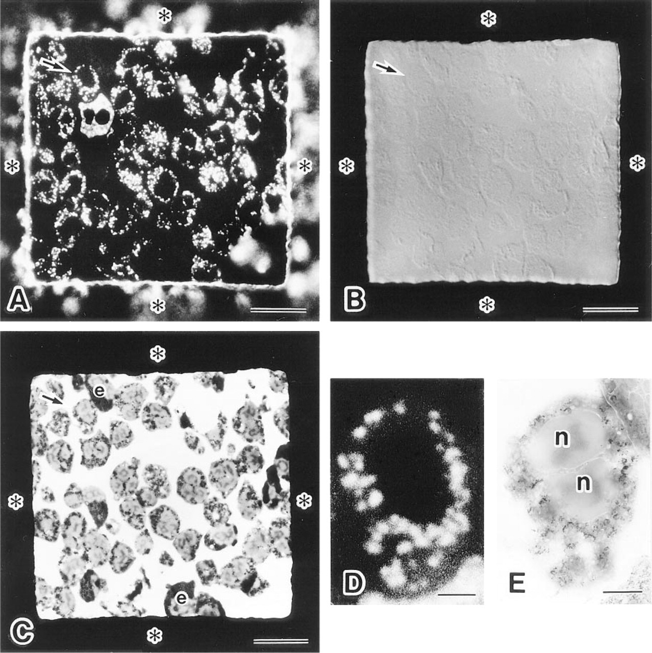
Localization of lactoferrin in a single ultrathin cryosection of human neutrophils by optical and electron microscopy using Fluoro-Nanogold (FNG) as the reporter system. (
There was a remarkable one-to-one relationship between fluorescent spots and specific granule profiles labeled with silver-enhanced FNG in ultrathin cryosections (Figure 2). Both larger and smaller specific granule profiles were detected by the fluorescence signal from FNG as well as the silver-enhanced FNG signal from the same granules. The fluorescence signal from FNG had a high spatial resolution. Fluorescent structures indicating two FNG-labeled specific granule profiles, which were near each other, were distinguishable as two fluorescent spots (Figures 2B and 2C). Therefore, the spatial resolution of fluorescence signal from FNG-labeled specific granules in fluorescence microscopy approached that of the silver-enhanced gold signal from the same granules in electron microscopy. However, irregular large, bright fluorescent spots were observed in some cells by fluorescence microscopy (data not shown). In these cases, the EM results revealed that those large spots were due to fusion of the fluorescence signals from several closely packed specific granules. Control samples lacking primary antibody displayed neither fluorescence nor silverenhanced gold labeling (data not shown).
Discussion
There has been wide use of immuno-LM in cell and molecular biological studies to determine the distribution of selected molecules in cells and tissues. Immunofluorescence probes have the advantage of high spatial resolution over other immunoprobes routinely employed for immuno-LM. In many cases, immunofluorescence provides sufficient resolution and sensitivity to answer the question being addressed. However, there are other cases in which additional resolution may be required to define more precisely the localization of specific molecules.
Immunoprobes for correlating immunofluorescence with immuno-EM have been developed. One is the conversion of the fluorescence signal to an electrondense form through the photo-oxidation of diaminobenzidine (Maranto 1982). Improvements to this methodology have been introduced (e.g., Deerinck et al. 1994). Recently a fluorescence-conjugated avidin-ABC complex method for detecting neurons injected with biotinylated molecules has also been introduced for neurobiological applications (Sun et al. 1998).
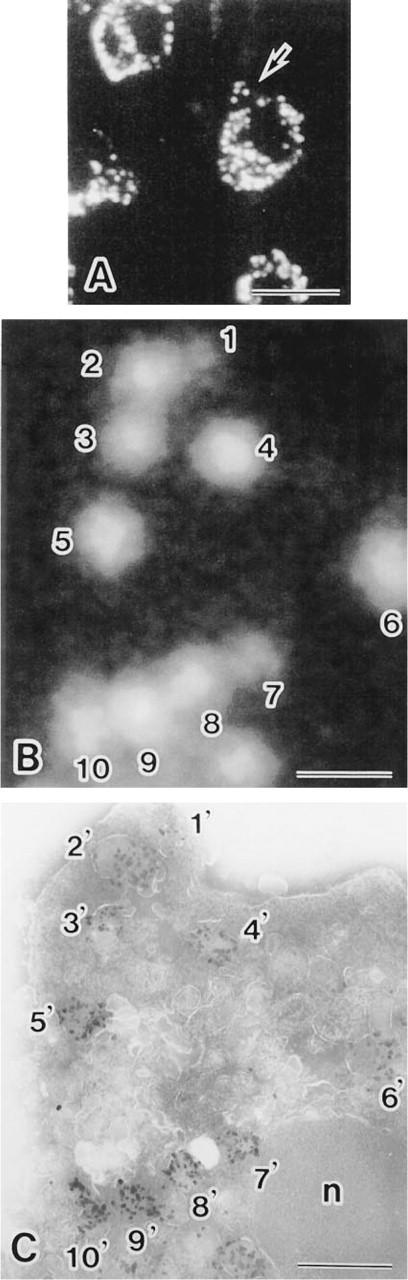
High-resolution comparison of fluorescence and silverenhanced gold signals from FNG in the same ultrathin cryosection of neutrophils. (
Another approach to correlative microscopy has been the development of fluorescently-labeled colloidal gold probes (Roth et al. 1980). This methodology has not been widely used because the colloidal gold tends to quench the fluorescence signal (e.g., Goodman et al. 1991). FNG was developed as an alternative immunoprobe (Powell et al. 1997). This fluorescent immunoprobe is distinct from the colloidal gold-fluorescent probes. FNG consists of a 1.4-nm gold particle to which antibodies (e.g., whole IgG, Fab' fragments) are covalently linked. Fluorochromes can also be conjugated to this immunoprobe. FNG has several advantageous properties as an immunoprobe for correlative microscopy: (a) the fluorescence signal does not appear to be reduced because of proximity to the gold cluster; (b) the fluorochrome and antibody do not readily dissociate from a 1.4-nm gold particles because they are covalently linked [dissociation of antibodies from colloidal gold immunoprobes has been reported (Kramarcy and Sealock 1991)]; and (c) FNG penetrates into cells as readily as conventional immunofluorescence probes (Robinson and Vandré 1997). Previous studies have shown FNG to be a valuable immunoprobe for correlative microscopy (Powell et al. 1997; Robinson and Vandré 1997). Although these approaches for correlative microscopy have been helpful for morphological analysis, they did not fully explore the capabilities of the methodology. In this study we have used human neutrophils as a model system and have shown the cellular distribution of lactoferrin in a single ultrathin cryosection examined by fluorescence microscopy and subsequently by electron microscopy.
In immunocytochemical studies employing semithin and ultrathin cryosections, fluorescence and colloidal gold immunoprobes are normally used. In a previous study, we utilized nonfluorescent ultrasmall immunogold particles (i.e., Nanogold) for the localization of lactoferrin-containing granules in ultrathin cryosections of human neutrophils. We found that Nanogold penetrates into cryosections to a greater extent than colloidal gold particles (i.e., 5-nm and 10-nm particles), which results in efficient immunolabeling (Takizawa and Robinson 1994a). We now use FNG as the secondary antibody along with the silver enhancement technique. The level of detection of lactoferrin with FNG was equivalent to that obtained with Nanogold. Moreover, the fluorescence signal from FNG was comparable to that of conventional fluorochrome-labeled antibodies (see Figures 1 and 2 in this study; Takizawa and Robinson 1994a). The relationship between fluorescence intensity and the number of silver-enhanced gold particles remains to be determined. However, the ability to predict the density of silver-enhanced gold particles on the basis of fluorescence intensities would also be a useful development.
In summary, we show FNG to be an ideal tool for immunocytolabeling of ultrathin cryosections. FNG-labeled molecules can be resolved by two imaging techniques (i.e., fluorescence microscopy and subsequent electron microscopy). The precise one-to-one relationship between the fluorescence signal and the silver-enhanced gold signal provides proof of principle for the use of FNG for high-resolution correlative microscopy.
Footnotes
Acknowledgements
Supported by grants from the Kazato Research Foundation (TT), the Naito Foundation (TT), the Nippon Foundation (TT), and by grants-in-aid for Scientific Research from the Ministry of Education, Science, Sports, and Culture of Japan (TT, KS). Partial support was also provided by NIH grant HD35121 (JMR).
We are deeply indebted to Ms Kiyomi Inose, Ms Chiaki Ishijima, and Ms Megumi Yatabe for excellent technical assistance.