
Abstract
We applied a fluorescent ultrasmall immunogold probe, FluoroNanogold (FNG), to immunocytochemistry on ultrathin cryosections. FNG has the properties of both a fluorescent dye-conjugated antibody for fluorescence microscopy and a gold particle-conjugated antibody for electron microscopy. Therefore, this bifunctional immunoprobe permits correlative microscopic observation of the same cell profiles labeled in a single labeling procedure by these two imaging methods. We demonstrate the utility of FNG as a secondary antibody for immunocytochemical labeling of myeloperoxidase (a marker protein for azurophilic granules) in ultrathin cryosectioned human neutrophils. Its detection requires high spatial resolution because neutrophils contain many cytoplasmic granules. There was a one-to-one relationship between fluorescent structures labeled with FNG and organelle profiles labeled with the same silver-enhanced FNG in ultrathin cryosections. Use of FNG immunocytochemistry on ultrathin cryosections is an ideal methodology for highresolution correlative fluorescence and electron microscopy and can provide unique information that may be difficult to obtain with a single imaging regimen.
Keywords
Immunofluorescence microscopy (immuno-FM) is a powerful technique often employed in studies related to cell structure and function. Immuno-FM permits determination of the distribution of molecules in cells and tissues by detecting the signal from fluorescent dye-conjugated immunoprobes (e.g., antibodies, protein A). Immuno-FM can also reveal the morphological relationship between multiple structures or molecules in the same cells by utilizing two or more fluorescent dyes with different spectral properties. Immuno-FM using semithin (≍0.5-μm) cryosections has been a reliable technique for light microscopic immunocytochemistry. Semithin cryosections are thin enough to permit detailed observation. In addition, immunoprobes penetrate into the sections without the need for permeabilizing agents. Immuno-FM using semithin cryosections enables us to make a survey of the distribution of specific molecules in cells and tissues by light microscopy. Moreover, in many cases it provides sufficient resolution and sensitivity to answer the experimental question being addressed. However, immunoFM using semithin cryosections does not provide ultrastructural detail on the localization of specific molecules. Immunoelectron microscopy (immuno-EM) using ultra-thin cryosections (≍90-nm) and immunogold probes provides a mechanism for obtaining information at the ultrastructural level. Therefore, immuno-FM of semithin cryosections and subsequent immuno-EM of ultrathin cryosections from the same sample blocks has been a regular approach for immunocytochemical labeling of cryosections, as initially introduced by Tokuyasu and associates (1984, 1985). Although this method has been valuable, correlative microscopy that enables the examination of the same cell profiles, not just the same sample blocks, by two imaging techniques (i.e., fluorescence and electron microscopy) would be highly desirable.
A unique fluorescent ultrasmall immunogold probe, FluoroNanogold (FNG), has been developed for use as a secondary antibody in immunocytochemical applications (Powell et al. 1997). It consists of an antibody (i.e., whole IgG, F(ab′)2, or Fab) to which a 1.4-nm gold cluster compound and fluorochromes are conjugated. Application of FNG to immunocytochemistry on ultrathin cryosections permits the correlative microscopic observation of the same cell profiles labeled in a single procedure by the two imaging techniques (Figure 1). Recently, we have shown FNG immunocytochemistry on ultrathin cryosections to be valuable for high-resolution correlation of fluorescence and electron microscopy. We have demonstrated the utility of FNG as a secondary antibody for immunocytochemical labeling of lactoferrin in ultrathin cryosectioned human neutrophils (Takizawa et al. 1998).
This article describes FNG immunocytochemistry on ultrathin cryosections, with special emphasis on the visualization of myeloperoxidase (MPO)-containing compartments in human neutrophils. The precise one-to-one relationship between the fluorescence signal and the silver-enhanced gold signal from the same FNG provides proof of principle for the use of FNG for high-resolution correlative microscopy.
Materials and Methods
Reagents
Except where noted, reagents were similar to those described previously (Takizawa et al. 1998). Bovine IgG, 1,4-diazabicyclo-[2.2.2]octane (DABCO), and p-phenylenediamine were obtained from Sigma Chemical (St Louis, MO). Monoclonal mouse anti-human MPO (clone MPO-7) was from Dako (Glostrup, Denmark). Goat anti-mouse FluoroNanogold (affinity-purified goat anti-mouse Fab′ fragment to which both fluorescein and a 1.4-nm gold particle are conjugated) (FNG) was purchased from NanoProbes (Stony Brook, NY). N-propyl gallate was supplied by Wako (Osaka, Japan). All immunological reagents were handled in accordance with the manufacturer's recommendations and used within the expiration date for each product.
Cell Isolation
Whole human blood was collected from healthy adult men after obtaining informed consent. Neutrophils were purified from whole blood in the unstimulated state as described previously (Takizawa and Robinson 1993).
Preparation of Ultrathin Cryosections
Purified cells were fixed in suspension with 4% paraformaldehyde in 100 mM sodium cacodylate buffer, pH 7.4, containing 5% sucrose for 120 min at 22C. Samples were then washed in the same buffer, embedded in gelatin, and then infiltrated with 2.3 M sucrose and frozen as previously described (Takizawa and Robinson 1993, 1994a). The gelatin blocks were then cut as ultrathin cryosections and collected on formvar film-coated nickel EM grids (Maxtaform “finder” grids; Graticules, Tonbridge, Kent, UK) (Takizawa and Robinson 1994b; Takizawa et al. 1998).
Immunocytochemistry on Ultrathin Cryosections with FNG
Cryosections mounted on EM grids were incubated for 1 hr at 22C in a solution containing 1% non-fat dry milk and 1 mg/ml bovine IgG in PBS to block nonspecific protein binding sites; 0.02% sodium azide was also present. Grids were incubated with anti-MPO IgG (1.3–2.6 μg/ml) for 3–4 hr at 22C. The grids were then washed in five changes of PBS and incubated with FNG (4–16 μg/ml Fab′) for 3–4 hr at 22C. The grids were then washed in at least five changes of PBS. All antibody solutions were diluted with PBS containing 1% non-fat dry milk, 1 mg/ml bovine IgG, and 0.02% sodium azide. Controls received the same treatment except for omission of the primary antibody.
Fluorescence Microscopy
The labeled grids were temporarily mounted in an anti-photobleaching solution between glass microscope slides and coverslips and then examined by epifluorescence and differential interference contrast (DIC) microscopy with an Olympus Provis AX80TR (Takizawa et al. 1998).
Photomicrographic images were recorded on Fuji Provia 400 film and then captured on computer and magnified using Adobe Photoshop 5.0J without additional manipulation of the images (Takizawa et al. 1998).
Silver Enhancement of FNG and Electron Microscopy
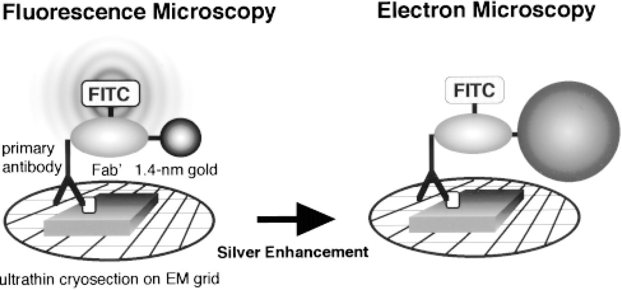
Schematic representation of correlative fluorescence and electron microscopy using ultrathin cryosections and FNG. Initially, the FNG-labeled ultrathin cryosection is mounted between a slide and a coverslip for viewing by fluorescence microscopy. Then the temporary slide is disassembled and the grid is subjected to the silver enhancement reaction. The sections are negatively stained before examination in an electron microscope.
After examination by optical microscopy, the slide preparations were disassembled so that the grids could be subjected to a silver enhancement procedure. The silver enhancement technique developed by Burry and associates (for review see Burry 1995) was employed to visualize the 1.4-nm gold particles at the electron microscopic level as reported previously (Takizawa and Robinson 1994a; Takizawa et al. 1998). After the silver enhancement process, the ultrathin cryosections were re-fixed with 2% glutaraldehyde-PBS, washed in distilled water, negatively stained by the method of Sakai et al. (1995), and observed with a Hitachi H-7000 electron microscope (Hitachinaka, Japan).
Results
The distribution of MPO in ultrathin cryosectioned human neutrophils was determined with anti-MPO followed by FNG as a secondary antibody and visualized by fluorescence microscopy (Figure 2A). Granule-like fluorescent spots indicated the distribution of MPO in the cells. The general morphology of cells in the sections was determined with DIC optics (not shown).
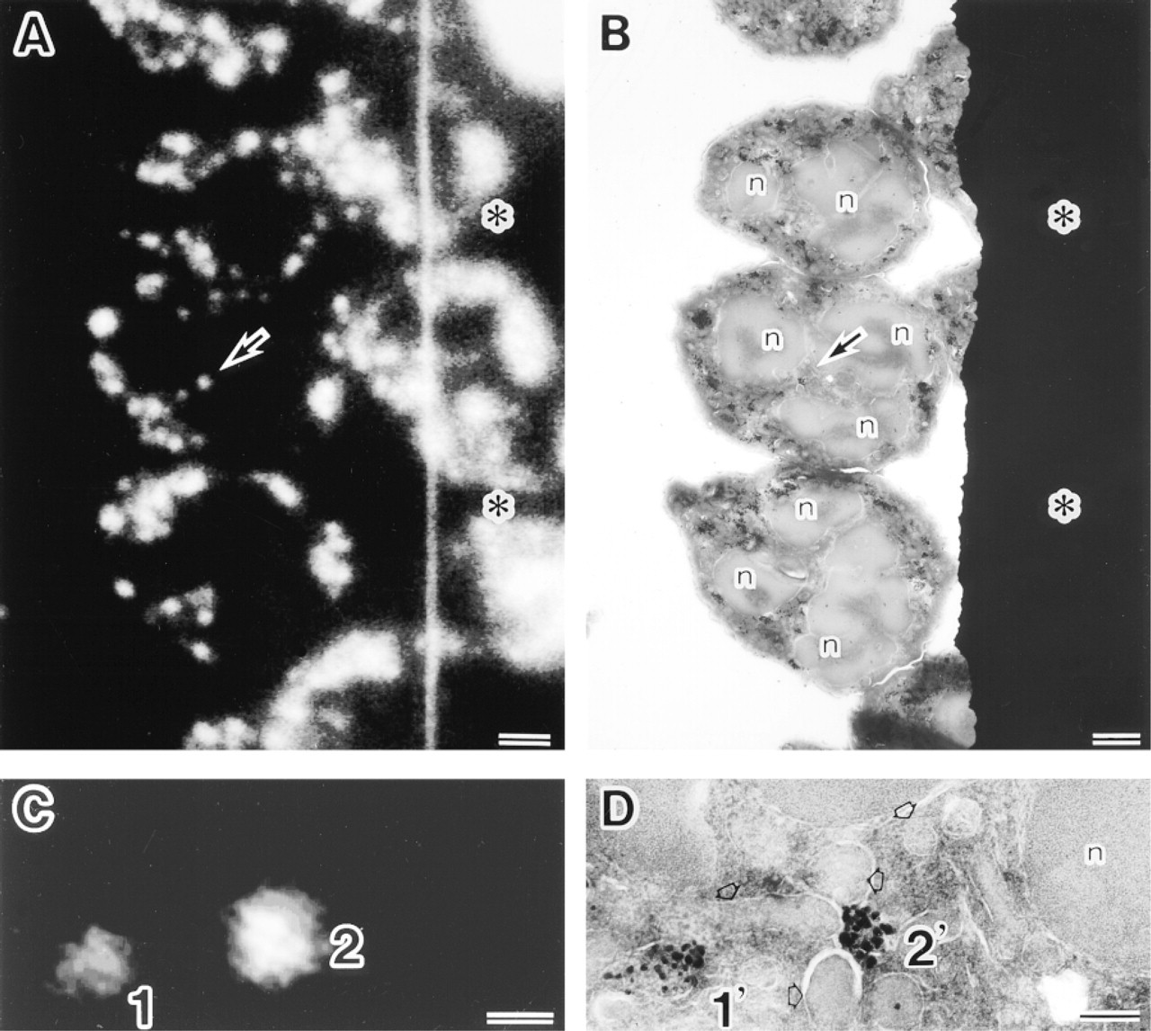
Localization of myeloperoxidase (MPO) in a single ultrathin cryosection of human neutrophils by optical and electron microscopy using anti-MPO and then FNG as the reporter system. (
The ultrastructural localization of MPO in the same ultrathin cryosectioned cells was subsequently detected by electron microscopy after silver enhancement of FNG and negative staining of cryosections (Figure 2B). The subcellular distribution of MPO was readily evident in these ultrastructural preparations. There is a remarkable one-to-one relationship between fluorescent spots and granule profiles labeled with silver-enhanced FNG in ultrathin cryosections (Figures 2C and 2D). Therefore, the spatial resolution of fluorescence signal from FNG-labeled compartments in fluorescence microscopy was compatible with that of silver-enhanced gold signal from the same compartments in electron microscopy. Control samples lacking primary antibody displayed neither fluorescence nor silver-enhanced gold labeling (not shown).
Reagents that retard photobleaching during fluorescence microscopic observation affected the quality of the immuno-EM results in ultrathin cryosections of neutrophils. When DABCO was used as an anti-photobleaching reagent, the silver-enhanced 1.4-nm gold particles were diminished even when a strong fluorescence signal was associated with the same structures (not shown). On the other hand, the anti-photobleaching reagents N-propyl gallate and p-phenylenediamine did not adversely affect the labeling intensity of silverenhanced FNG granules (not shown).
Discussion
Immunocytochemistry is a widely applied set of methods used in biomedical research. Detection of antibody binding to cell and tissue antigens requires a reporter system. The most widely applied reporter systems are fluorochromes, enzymes (e.g., horseradish peroxidase), and particulate probes (e.g., immunogold particles). Fluorescence labeling procedures are widely applied. However, in certain situations the precise localization of a fluorescence signal may be difficult to resolve owing to the contribution of fluorescence from above or below a given focal plane. This problem has been addressed by the examination of sectioned cells or tissues (e.g., cryosections at 0.5 μm thickness). Alternatively, the confocal microscope has been employed to minimize the out-of-focus fluorescence signal. Fluorescence microscopy is often sufficient to answer the question(s) being asked in a given experiment. Nevertheless, there are experimental situations in which the additional resolving power of the electron microscope is required.
A combination of immunofluorescence microscopy of semithin cryosections and subsequent immunoelectron microscopy of ultrathin cryosection from the same sample blocks has been beneficial to both immuno-FM and -EM (e.g., Takata et al. 1991). Recently, we have shown a more direct way of investigating the same molecules on ultrathin cryosectioned cells by fluorescence and electron microscopy using the bifunctional immunoprobe FNG (Takizawa et al. 1998). FNG combines the properties of a fluorescent dye-conjugated antibody for fluorescence microscopy and a gold particle-conjugated antibody for electron microscopy.
In the present study, we show FNG-labeled MPO-positive compartments in the same ultrathin cryosections by sequential fluorescence and electron microscopy. The spatial resolution of fluorescence signal from FNG-labeled granules was in agreement with that of silver-enhanced gold signal from the identical compartments in electron microscopy. We reconfirm that application of FNG to ultrathin cryosections can achieve high-resolution correlation between immunofluorescence and immunoelectron microscopy. It should also be noted that FNG facilitates multiple optical imaging techniques (Robinson and Vandré 1997).
Immunocytochemistry on ultrathin cryosections using FNG as the reporter system is time-intensive. Therefore, some precautions concerning the methodology are in order. First, it is crucial to prepare good ultrathin cryosections for immunocytochemistry (Takizawa and Robinson 1994b; Liou et al. 1996). Nickel “finder” grids are very useful so that the location of a given cell can be readily determined in both the fluorescence and electron microscopes. In addition, nickel grids are not adversely affected by the silver enhancement procedure. Anti-photobleaching reagents used as the mounting medium during fluorescence microscopy should be chosen carefully. For example, DABCO may cause dissociation of a 1.4-nm gold particle form FNG. We recommend that N-propyl gallate or p-phenylenediamine be employed as anti-photobleaching reagents. The time course for the silver enhancement reaction is also crucial to achieve adequate particle enhancement while minimizing the background signal (Robinson and Vandré 1997).
Ultrathin cryosections are an excellent substrate for immunofluorescence labeling (Ishiko et al. 1998; Takizawa et al. 1998; Pombo et al. 1999). Use of ultrathin cryosections has the added beneficial effect of increasing axial resolution in fluorescence microscopy (Ishiko et al. 1998; Pombo et al. 1999). We show FNG immunocytochemistry on ultrathin cryosections to be an ideal methodology for correlative microscopy and for bridging the resolution gap between fluorescence and electron microscopy. FNG is a single but bifunctional immunoprobe. FNG can serve as a fluorescence probe for immunofluorescence microscopy and simultaneously as a particulate probe for immunoelectron microscopy. Correlative microscopy should provide unique information that may be difficult to obtain with a single imaging regimen.
Footnotes
Acknowledgements
Supported by Grants-in-Aid for Scientific Research and project grants of Center for Molecular Medicine of Jichi Medical School from the Ministry of Education, Science, Sports, and Culture of Japan (TT) and by grants from NIH (HD35121) and the American Heart Association (8807785) (JMR).
We thank Dr James F. Hainfeld of Nanoprobes, Inc. and Dr Richard W. Burry of Ohio State University for their encouragement of this study. We also thank Dr Shigeo Ookawara, Dr Takashi Yashiro, Ms Kiyomi Inose, Ms Megumi Yatabe, and Ms Michiyo Soutome of Jichi Medical School for their help.