
Abstract
The biotinyl–tyramide protocol recently introduced for sensitive light microscopic immunocytochemistry was applied to electron microscopy and revealed various tissue antigens with high resolution. The protocol consists of an indirect method in which thin tissue sections are incubated successively within a specific primary antibody, followed by a biotinylated secondary antibody, streptavidin–HRP, and then finally with biotinyl–tyramide. The reaction product appears as a dense filamentous material that is deposited over particular cellular compartments. The labeling obtained for the antigens tested, amylase and heat-shock protein 70 in pancreatic acinar cells, insulin in pancreatic β-cells, and carbamoyl phosphate synthetase and catalase in liver tissue, was found to be highly specific, with the labeling for each antigen confined to its particular cellular compartment. Background levels and nonspecific deposition of the staining were negligible. The use of biotinyl–tyramide therefore appears to be an alternative sensitive technique for immunoelectron microscopy.
S
Recently, a novel approach for revealing antigen–antibody complexes, the tyramide signal amplification technique, has been reported. First introduced for immunoassays (Bobrow et al. 1989, 1991), it was then extended to immunocytochemistry at the light microscopic level (Adams 1992; Berghorn et al. 1994; Merz et al. 1995; Sanno et al. 1996; Shindler and Roth 1996; Plenat et al. 1997). This technique is based on the ability of HRP from an HRP–avidin complex (or any HRP–protein complex) to catalyze the deposition of biotinyl–tyramide onto a surface displaying biotinylated antigen–antibody complexes. This deposition is believed to take place at the site of the enzyme reaction, thus leading to good resolution (Bobrow et al. 1992). The development of fluorescent tyramide reagents has made possible the application of this technique to fluorescent microscopy, either in immunostainings (Berghorn et al. 1994; De Haas et al. 1996; Hunyady et al. 1996; Plenat et al. 1997; van Gijlswijk et al. 1997; Van heusden et al. 1997) or in in situ hybridization (Kerstens et al. 1995; Raap et al. 1995; De Haas et al. 1996; Macechko et al. 1997; Schmidt et al. 1997; van Gijlswijk et al. 1996,1997; Zehbe et al. 1997).
In the present study, we report the use of the tyramide signal amplification approach (TSA) for immunoelectron microscopy. Application of the technique on thin sections of tissues and examination at the electron microscopic level have revealed the presence of a dense reaction product at the antigen-antibody reaction sites. This novel technique appears to be simple, reliable, and of high specificity and good resolution. It provides an alternative to existing techniques and could be used in combination with others for multiple staining experiments.
Materials and Methods
Tissue Processing
Small fragments of normal rat pancreatic and liver tissues were fixed by immersion in 0.1 M phosphate-buffered 1% glutaraldehyde for 2 hr at 4C. After fixation, the tissue fragments were washed in the phosphate buffer and processed for low-temperature embedding in Lowicryl as previously described (Bendayan 1995). Ultrathin sections were cut and mounted on Parlodion–carbon-coated nickel grids and processed for immunocytochemistry using the TSA (Tyramide Signal Amplification) as the electron-dense marker.
Antisera and Reagents
Five different primary antibodies were used to assess the TSA technique: a rabbit anti-human amylase and a mouse monoclonal anti-heat shock protein 70 (HSP-70) from Sigma Chemicals (St Louis, MO), a guinea pig anti-porcine insulin from ICN (Montreal, Quebec, Canada), a rabbit anti-carbamoyl phosphate synthetase (CPS) (Bendayan and Shore 1982), and a rabbit anti-catalase (Bendayan and Reddy 1982). The specificity of these antibodies was previously demonstrated by immunochemical and immunocytochemical techniques (Bendayan and Reddy 1982; Bendayan and Shore 1982; Bendayan 1984,1989; Velez–Granell et al. 1994). The secondary antibodies were a biotin-conjugated goat anti-rabbit IgG (GAR-biotin), a biotin-conjugated goat anti-mouse IgG (GAM–biotin) (Sigma), and biotin-conjugated goat anti-guinea pig IgG (GAG–biotin) (Amersham Life Science; Oakville, Ontario, Canada). Streptavidin–HRP (SA-HRP), biotinyl–tyramide (BT), and the amplification diluent were kindly provided by NEN Life Science Products (Renaissance TSA-Indirect kit; NEN Life Science Products, Boston, MA).
Immunocytochemistry
Labeling was carried out by floating the grids with tissue sections face down on drops of 1% ovalbumin in 10 mM PBS, pH 7.2, for 30 min at room temperature (RT) and then transferring them directly onto a drop of one of the specific primary antibodies for 2 hr at RT or overnight at 4C. The antibodies were used at the following dilutions: anti-amylase 1:10; anti-HSP-70 1:10; anti-insulin 1:200; anti-CPS 1:50; and anti-catalase 1:200. The grids were then rinsed with PBS for 15 min, transferred to the 1% ovalbumin solution for 30 min, and then incubated on a drop of one of the corresponding specific biotinylated secondary antibodies for 60 min at RT: GAR–biotin 1:800 for anti-amylase, anti-CPS, and anti-catalase; GAG–biotin 1:500 for the anti-insulin; and GAM–biotin 1:500 for the anti-HSP-70. Tissue sections were then rinsed with PBS for 15 min, transferred to the ovalbumin solution for 15 min, and then incubated on a drop of streptavidin–HRP diluted 1:500 with PBS for 30 min at RT. The grids were then washed for 15 min with PBS and incubated for 10 min at RT with biotinyl–tyramide diluted 1:50 in 1 x amplification diluent. After washing with PBS and distilled water, the tissue sections were stained with uranyl acetate and examined with a Philips 410SL electron microscope.
The specificity of the labeling with tyramide was assessed by several control experiments: omission of either the primary or secondary antibody and omission of the streptavidin–HRP or the biotinyl–tyramide steps, in four separate experiments.
Results
Application of the TSA technique in combination with specific primary antibodies on thin tissue sections prepared for electron microscopy led to the deposition of a dense filamentous material on structures known to contain the different antigens. The specificity of the labelings appeared to be excellent and is supported by the results obtained using various antigens located in different cells and/or different cell compartments. Incubation of pancreatic tissue with the anti-amylase antibody (Figure 1) led to staining of different compartments of the acinar cells, such as the rough endoplasmic reticulum, the Golgi cisternae, the condensing vacuoles, and the zymogen granules. The dense filamentous reaction product was present on the cell compartments with different intensities, the highest being over the zymogen granules known to contain high amounts of amylase. Other compartments, such as mitochondria or nuclei, were devoid of any staining (Figure 1). The use of the anti-insulin antibody resulted in staining of the pancreatic B-cells, particularly in those compartments involved in secretion, the rough endoplasmic reticulum, the Golgi apparatus, and the immature and mature secretory granules (Figure 2). Staining for HSP-70 was found in the Golgi apparatus and condensing vacuoles of the pancreatic acinar cells and was particularly concentrated in the trans-Golgi network (Figure 3). For the hepatic cells, labeling for carbamoyl phosphate synthetase was restricted to the mitochondria (Figure 4) and that for catalase to the peroxisomes (Figure 5). In all cases, the reaction product appeared as thin filamentous deposits that overlaid the labeled structures without masking their morphological features. Considering the different labeled compartments, the reaction product remained within the limits of each compartment, thus reflecting good resolution. The specificity of the results, as demonstrated by the very low levels of background, was excellent, the labeling being restricted to the corresponding compartments. It was also supported by the control experiments. Omission of the primary or secondary antibodies in the labeling protocol led to absence of staining except for the presence of some random dense spots (Figure 1C). This indicates that application of the TSA protocol without a specific antibody does not generate any staining (Figure 1C). Similar results were obtained when the streptavidin–HRP or the biotinyl–tyramide step was omitted. Nonspecific deposition of the different reagents therefore appears to be negligible. The technique appears reliable and sensitive. However, some precautions must be taken to generate intense and specific stainings. The different solutions should be as fresh as possible, particularly the diluted tyramide solution, which should not be more than 2 weeks old. The use of an old solution affects the chemical reactions and generates labeling of low intensity. Incubation with 1% ovalbumin appears to interfere with the HRP–tyramide reaction and therefore should not be carried out before incubation with the biotinyl–tyramide.
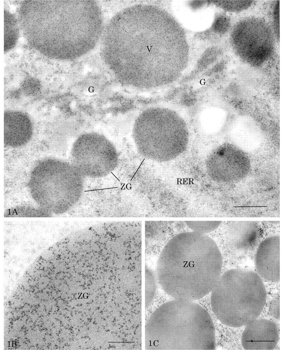
Thin section of rat pancreatic acinar cell labeled for amylase by the TSA technique. Dense filamentous deposits, revealing amylase antigenic sites, are present over the rough endoplasmic reticulum (RER), the Golgi apparatus (G), the condensing vacuoles (V), and the zymogen granules (ZG). Bar = 0.5 μm. (
Discussion
The TSA approach was originally introduced for detection of antigens on solid-phase immunoassay (Bobrow et al. 1989, 1991) and was then adapted to histochemistry for sensitive detection of tissue antigens at the light microscopic level (Adams 1992; Berghorn et al. 1994). In the present work we have extended the TSA protocol for the ultrastructural localization of tissue antigens by electron microscopy. The technique is an indirect one in which a specific antigen–antibody complex is revealed by a biotinylated secondary antibody, which in turn is reacted with streptavidin–HRP. This HRP reacting with hydrogen peroxide catalyzes the oxidative condensation of tyramide by a free radical mechanism. This activated tyramide binds covalently to the tyrosine residues of the proteins surrounding the HRP. Moreover, since the half-life of the tyramide radicals is very short, the deposition occurs very close to the activating enzyme (Bobrow et al. 1992). Therefore, the HRP catalyzes the deposition of tyramide at the surface of the tissue section, giving rise to an electron-dense deposit at the site of the specific antigen–antibody complex. Details of the biochemical reactions taking place during oxidation of the tyramine have been reported to generate a brown pigment, which was found to contain dityramine and more extensively oxidized and polymerized derivatives (Gross and Sizer 1959). That such polymerized derivatives correspond to our dense filamentous reaction product found at the electron microscopic level remains to be confirmed. Application of this TSA protocol in combination with specific antibodies resulted in the staining of cell compartments with a dense filamentous deposit. This deposit, generated by the HRP–tyramide reaction, appeared to be confined to the labeled structures and thus yielded labeling of good resolution. Because of differences in molecular interactions and reactions, it is quite difficult to perform comparative evaluations between this TSA technique and other techniques. Because it is a reaction product rather than a particulate marker, there are difficulties in providing quantitative evaluations of labeling intensities, as is the case with the colloidal gold marker. Specificity of the results is always based on the characteristics of the primary and secondary antibodies. However, for the TSA reagents, levels of background as well as nonspecific deposition of the reagent product were negligible. Therefore, TSA appears to represent an alternative technique for immunoelectron microscopy that can be applied either for the simple detection of antigens or in combination with other techniques for multiple labeling experiments.
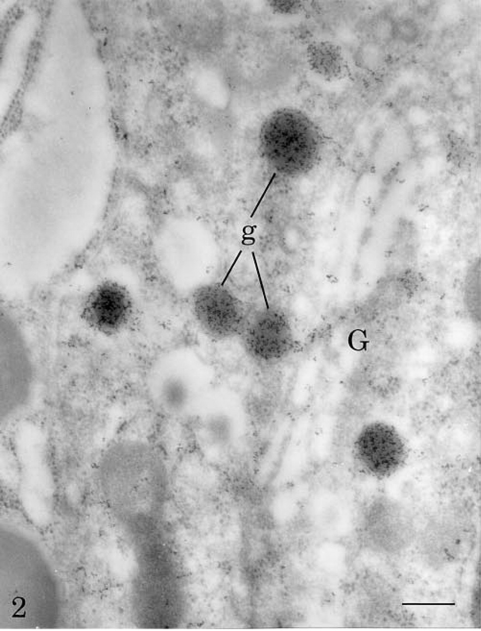
Thin section of rat pancreatic B-cell labeled for insulin by the TSA technique. Dense filamentous reaction product, revealing insulin antigenic sites, is present over the Golgi apparatus (G) and the secretory granules (g). Bar = 0.25 μm.
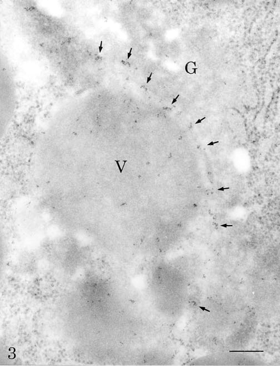
Localization of heat shock protein 70 in rat pancreatic acinar cell with the TSA technique. Labeling by the electron-dense deposits is present mostly in the trans-cisternae of the Golgi apparatus (G) and in the condensing vacuoles (V). Bar = 0.25 μm.
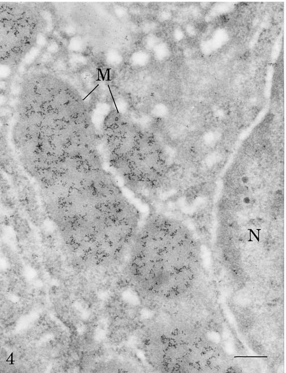
Thin section of rat liver tissue labeled for carbamoyl phosphate synthetase by the TSA technique. Labeling by dense filamentous deposits is present over mitochondria (M). Little background is seen over the nucleus (N) and the cytoplasm. Bar = 0.25 μm.
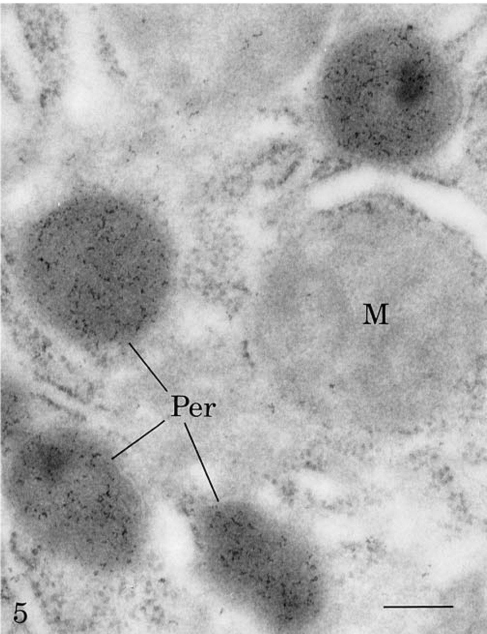
Thin section of rat liver cell labeled for catalase by the TSA technique. Labeling by the dense reaction product is present over the peroxisomes (Per). Background levels over mitochondria (M) and cytoplasm are very low. Bar = 0.25 μm.
Footnotes
Acknowledgements
Supported by grants from the Medical Research Council of Canada.
We would like to express our gratitude to Patricia Mayer from NEN Life Science Products (Boston, MA) for her kindness in providing us with the different reagents and her interest in this study.