
Abstract
Methylmethacrylate (MMA) embedding is routinely used for histomorphometry of undecalcified bone preserved by prolonged immersion in ethanol, a procedure that yields poor ultrastructural detail. Because microwave irradiation (MWI) facilitates penetration of fixatives, we have investigated whether it can improve preservation by ethanol. Rat tibiae, some labeled with tetracycline, and a human iliac crest biopsy were immersed in 70% ethanol and dehydrated, both under MWI, for a total processing time of ~7 hr. Controls were not irradiated, and all specimens were embedded in MMA at 4C. They were then processed for histomorphometry, histochemistry, structural analysis, and immunolabeling. The results showed that histological preservation was improved with MWI. Static bone formation and resorption parameters and rate of mineral apposition were similar to those of conventionally processed specimens. Mineral distribution, as visualized by von Kossa staining and backscattered electron imaging, was not affected. Alkaline phosphatase and tartrate-resistant acid phosphatase activity, as well as immunolocalization of bone sialoprotein and osteopontin, were readily visualized. Ultrastructurally, osteopontin exhibited a typical distribution in mineralization foci, between calcified collagen fibrils, and at cement lines. These data show that MWI improves preservation and permits application of a broad spectrum of analytical methodologies on the same bone sample while considerably reducing processing time.
Keywords
A
Electron microscopy has led to a more-detailed understanding of cell and tissue organization and of structure-function relationships. Furthermore, it is an important tool for the diagnosis of diseases difficult to clarify with the light microscope (Djaldetti et al. 1987). Ultrastructural analysis of bone biopsies allows a better definition of pathological elements and better understanding of their histogenesis, and has played a determining role in the characterization of a variety of bone-related neoplasms (Walaas and Kindblom 1990; Goh et al. 2001; Marucci et al. 2002; Suh et al. 2002).
MMA embedding of calcified or soft tissues has mostly been used for analyses at the light microscope level; its application in electron microscopy is not well documented (Baskin et al. 1992). In a recent report, we have shown that MMA is suitable for the ultra-structural immunolabeling of non-collagenous bone matrix proteins but that conventional ethanol fixation, widely used for histological procedures in the laboratory and hospital settings, yields inconsistent and, at best, suboptimal cellular preservation (Laboux et al. 2003). Microwave irradiation (MWI) facilitates the penetration of fixatives and improves antigen detection (discussed in Arana-Chavez and Nanci 2001). Therefore, we applied this approach to optimize preservation by ethanol and verified whether irradiation affects histological reactions for which MMA is conventionally used. The results demonstrate that MWI significantly reduces the processing time of calcified bone samples, improves preservation, and permits fluorochrome labeling for dynamic histomorphometry as well as a variety of light and electron microscope analyses.
Materials and Methods
Tissue Processing
Four male Wistar rats weighing ~250 g (Charles River Canada; St-Constant, QC, Canada) were injected through the jugular vein twice at 3-day intervals with tetracycline hydrochloride (25 mg/kg of body weight; Sigma-Aldrich Canada, Oakville, ON, Canada). Before each injection, the animals were anesthetized by IP administration of 0.6 ml of a 1:1:2 mixture of Hypnorm (fentanyl citrate and fluanisone; Janssen Pharmaceutica, Beerse, Belgium), Versed (midazolam; Hoffmann-LaRoche, Mississauga, ON, Canada), and distilled water. Four days after the last injection the rats were anesthetized with Halotane (MTC Pharmaceuticals; Cambridge, ON, Canada) and decapitated, and tibiae were quickly dissected and placed in a beaker containing 40 ml 70% ethanol. Another three rats weighing ~100 g and two weighing ~200 g, having not received tetracycline, were similarly sacrificed, and tibiae were also immersed in ethanol. For MWI (Wagenaar et al. 1993; Massa and Arana-Chavez 2000; Arana-Chavez and Nanci 2001), the beaker was then positioned in a larger container filled with crushed ice. The container was placed in a PELCO 3440 MAX laboratory microwave oven set at 100% (Ted Pella; Redding, CA) and the tissues were irradiated for four periods of 6 min in fresh changes of ethanol. The temperature probe of the oven was submerged in the ethanol and the temperature programmed to a maximum of 37C. The samples were kept in 70% ethanol at 4C for 5 hr and then dehydrated in 80%, 90%, 95%, and three 100% ethanol changes for periods of 15 min, with a 120-sec MWI on ice at each step. Tissues were infiltrated at 4C with a mixture of 80% MMA (J-T Baker; Phillipsburg, NJ) from which hydroxyquinone was removed, 20% N-dibutylphtalate, and 0.4% (weight) benzoyl peroxide (Theuns et al. 1993; Erben 1997). The above resin mixture was polymerized, also at 4C. by addition of 0.1% N,N-dimethylalanine. As controls, controlateral tibiae were dissected and immersed quickly in 70% ethanol for 1 week, then classically dehydrated and similarly embedded in MMA.
Procedures for animal handling were approved by the “Comité de Déontologie de l'Université de Montréal.”
Human Bone Biopsy
Two contiguous transiliac crest biopsies were obtained from a 75-year-old osteoporotic male patient who had received tetracycline for dynamic histomorphometry. The biopsies were placed in 70% ethanol within minutes after their removal. About 1 hr later, one of them was subjected to three 5-min MWI cycles on ice, at 100% setting of the oven and the temperature programmed to a maximum of 37C. The specimen was immediately dehydrated in 80%, 90%, and three 100% changes of ethanol for periods of 30 min, with 5 min MWI on ice at each change. The other biopsy was kept in 70% ethanol for 1 week and then conventionally dehydrated and embedded in MMA as above. Procedure and patient consent forms for the biopsies were approved by the Ethics Committee of Hôpital St. Luc.
Bone Histomorphometry
Undecalcified MMA-embedded rat tibiae from conventionally fixed (n = 4) and MWI-treated (n = 4) samples and the two human transiliac biopsies were sectioned longitudinally with a Leica Polycut-E horizontal microtome. Five- and 8-μm-thick sections were cut at 60-μm intervals and mounted on gelatin-coated glass slides. The 5-μm-thick sections were deplasticized with toluene, rehydrated with decreasing concentrations of ethanol, and stained according to the Goldner's trichrome protocol. The 8-μm-thick sections were not deplasticized and were visualized unstained by epifluorescence for localization of tetracycline labeling.
Structural and static parameters of cancellous bone remodeling were quantified at the secondary spongiosa of the proximal metaphysis. Dynamic bone formation measurements were carried out at the extremity of the epiphysieal region. This region was selected to avoid autofluorescence associated with cartilage and weakly mineralized bone that is frequently observed in metaphyseal trabecular bone in young growing rats. Histomorphometry was also carried out on the cancellous bone of the human biopsies. Measurements were performed with a semiautomatic image analyzing system consisting of a Leica Polyvar light microscope equipped with a camera lucida and digitizing tablet linked to a computer. The data were acquired and analyzed using the OsteoMeasure software (Osteometrics; Decatur, GA). Nomenclature and abbreviations of histomorphometric parameters follow the recommendations of the American Society for Bone and Mineral Research (Parfitt et al. 1987).
Histochemistry
Detection of alkaline phosphatase (ALP) and tartrate-resistant acid phosphatase (TRACP) activity was carried out on 5-μm-thick sections according to the method of Liu et al. (1987). Naphthol-AS-TR was used as substrate for both enzymes, and Fast Blue BB salt (Sigma-Aldrich) and pararosaniline were used as couplers for ALP and TRACP, respectively.
Immunohistochemistry
Five-μm-thick sections were deplasticized with toluene and rehydrated through decreasing concentrations of ethanol to distilled water. Sections were first treated with peroxidase blocking reagent from the DAKO EnVision + System, horseradish peroxidase (HRP) (DAB) Kit (DAKO; Carpinteria, CA), blocked for 20 min with 5% powdered skimmed milk in 0.01 M phosphate buffered saline (PBS), and then incubated for 1 hr with polyclonal rabbit anti-osteopontin (OPN) antibody (LF-123; courtesy of L.W. Fisher, NIDCR, Bethesda, MD) or with anti-bone sialoprotein (BSP) antibody (LF-100; courtesy of L.W. Fisher). Sections were then washed in 0.01 M PBS containing 0.01% Tween-20, and incubated for 30 min with labeled polymer, HRP anti-rabbit antibody (DAKO En-Vision). The sections were then washed with PBS-Tween followed by distilled water, treated with DAB chromogen (DAKO EnVision) for 6 min, and then counterstained with methyl green. Finally, the sections were dehydrated in ethanol, passed in xylene, and covered with a glass coverslip mounted with Permount (Fisher Scientific; Fair Lawn, NJ). Controls consisted of omitting the primary antibody and incubation with a variety of unrelated primary antibodies. All incubations were carried out in a moist environment at room temperature.
Processing for Light and Transmission Electron Microscopy
Semithin sections (1 μm) for light microscopy were cut with a diamond histoknife on a Reichert Ultracut E microtome and stained with toluidine blue. Some sections were processed for mineral detection by the von Kossa method and lightly counterstained with toluidine blue. For ultrastructural observations, thin sections (~100 nm) were cut with a diamond knife and mounted on Formvar–carbon-coated nickel grids. All thin sections used for morphology or immunolabeling were stained with uranyl acetate and lead citrate before examination with a JEOL JEM 1200EX-II operated at 60 kV.
Postembedding Colloidal Gold Immunolabeling
Grid-mounted sections were processed for postembedding protein A-gold immunolabeling (reviewed in Bendayan 1995). Briefly, sections were floated for 15 min on a drop of 0.01 M PBS, pH 7.2, containing 1% ovalbumin (Oval) (Sigma-Aldrich) to saturate nonspecific binding sites, and then transferred to and incubated for 1 hr on a drop of a chicken egg yolk anti-rat OPN antibody (diluted 1:50 in PBS; Nanci et al. 1996). Sections were then rinsed with PBS, blocked with PBS–Oval for 15 min, and incubated with a rabbit anti-chicken gold-conjugated secondary antibody (Cappel Research Products; Scarborough, ON, Canada) diluted 1:5 with PBS, pH 8. As controls, sections were incubated with the secondary antibody only. All procedures were performed at room temperature. After the secondary antibody, grids were washed thoroughly with PBS, rinsed with distilled water, and air dried.
Backscattered Electron Imaging
Tissue blocks faced for cutting sections were directly examined using a backscattered electron detector in a JEOL JSM-LV 6460 variable pressure scanning electron microscope operated at 15 kV and a pressure of 25–50 Pa.
Results
Histology on Semithin Sections
Conventional ethanol preservation and dehydration of tibiae for MMA embedding yielded poor tissue preservation that was more apparent with the larger bones from older rats. Although there were regions that showed acceptable morphology, in general, osteoblasts appeared contracted and exhibited poorly definable outlines (Figure 1A). On the other hand, in MWI bone, osteoblasts on bone-forming surfaces generally appeared as plump cells with a well-defined cuboidal outline (Figure 1B). They were closely apposed to the surface of spicules and trabecules (Figure 1B). Visually, there was no significant difference in von Kossa staining between tibiae processed by the two methods and, as expected, mineral was identified in the mineralized bone and calcified cartilage (cf. Figures 1A and 1B). Backscattered electron imaging showed a similar distribution of mineral but also revealed regions of variable mineral content as well as cement lines between layers of mineralized matrix (Figures 1C and 1D).
Ultrastructural Appearance
At the ultrastructural level, there were dramatic regional variations in the quality of preservation in conventionally processed tibiae. Many cells exhibited an extracted cytoplasm and disrupted cell membrane, and appeared as cellular ghosts. Other cells appeared to have better retained their integrity, but the level of preservation was still unacceptable (Figure 2A). They were contracted, and the cytoplasm was vacuolated or its content appeared to have coagulated into unidentifiable masses. Organelles were difficult to recognize and, when identifiable, mitochondria were burst. The chromatin was generally compacted.
The morphological preservation of bone-associated cells was improved by the use of MWI. Although poorly preserved cells with extracted cytoplasm were also encountered, there were now osteoblasts with a recognizable intracellular organization (Figure 2B). There was less evidence of shrinkage, and membranes were not well preserved, but mitochondria and organelles implicated in protein synthesis and secretion could be defined. Typical patterns of heterochromatin and euchromatin were present. Calcified bone matrix showed similar ultrastructural features irrespective of whether the tibiae were irradiated with microwaves. However, in MWI samples, mineralization foci in osteoid seemed more evident and cross-banding along unmineralized collagen fibrils was crisper (see Figure 6B).
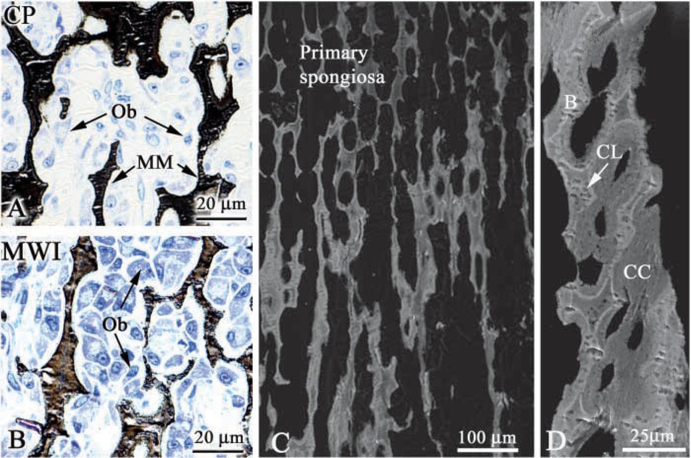
Light micrographs of the primary spongiosa in the rat tibia after (
Histomorphometric Analysis
There was no readily apparent histological difference between rat tibiae and human bone biopsies processed conventionally or by MWI (Figure 3). Tetracycline labels were not affected by MWI and were clearly distinguishable (Figures 1E, 1F, and 1H). As shown in Table 1, there were no significant quantitative differences in structural static parameters of bone formation and resorption as well as dynamic bone formation parameters for tibiae processed with and without MWI.
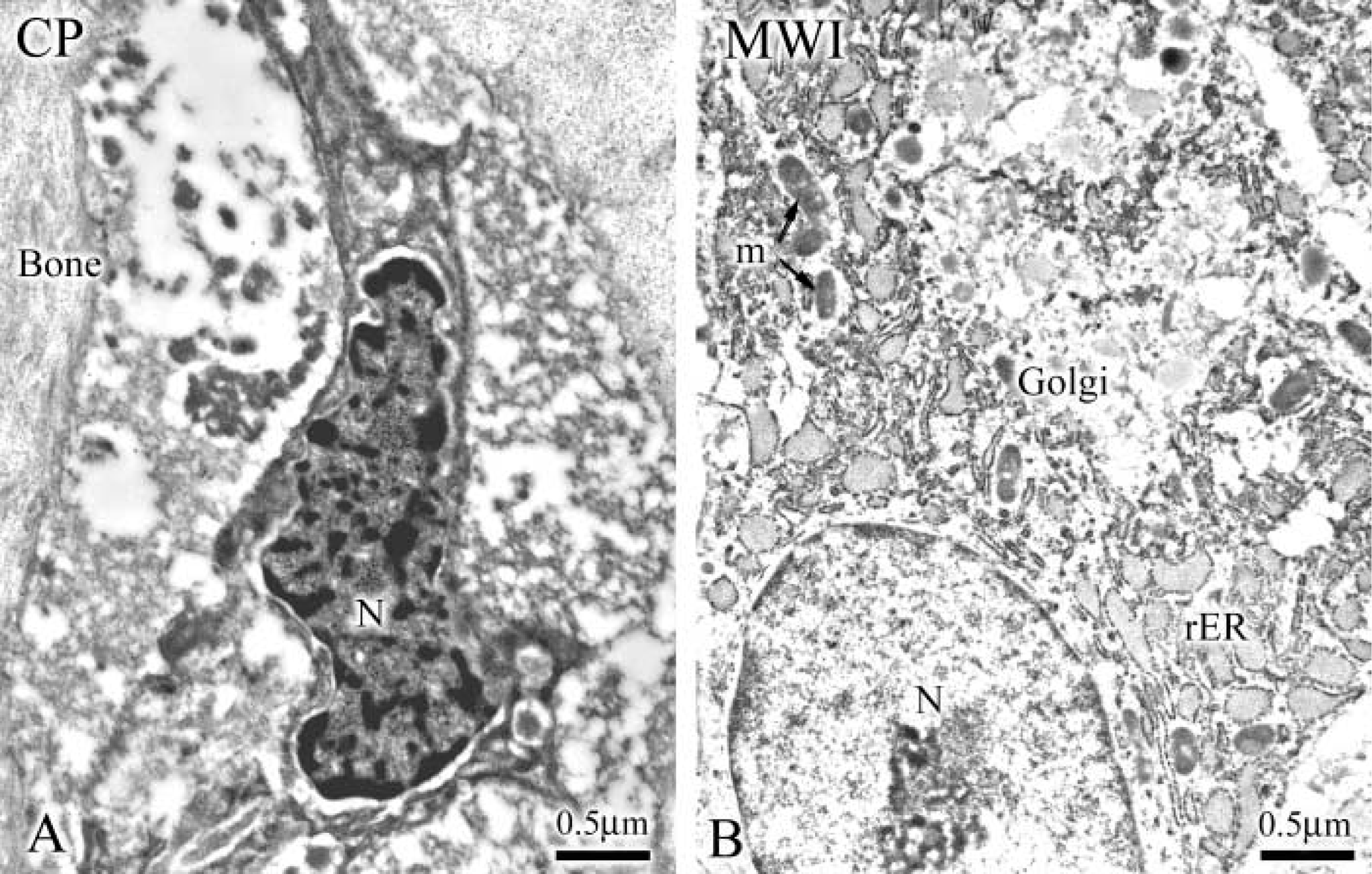
Electron micrographs illustrating the ultrastructural preservation obtained with (
The human biopsies were from a 75-year-old male suffering from osteoporosis treated with bisphosphonate for 1 year. Because the patient continued to sustain new fractures, bone biopsy was performed to ensure the absence of additional bone disease. Compared with normal values, results from histomorphometrical analysis of both biopsies (with or without MWI) led to the diagnosis of cortical osteoporosis with low bone remodeling but without mineralization defect (Table 2).
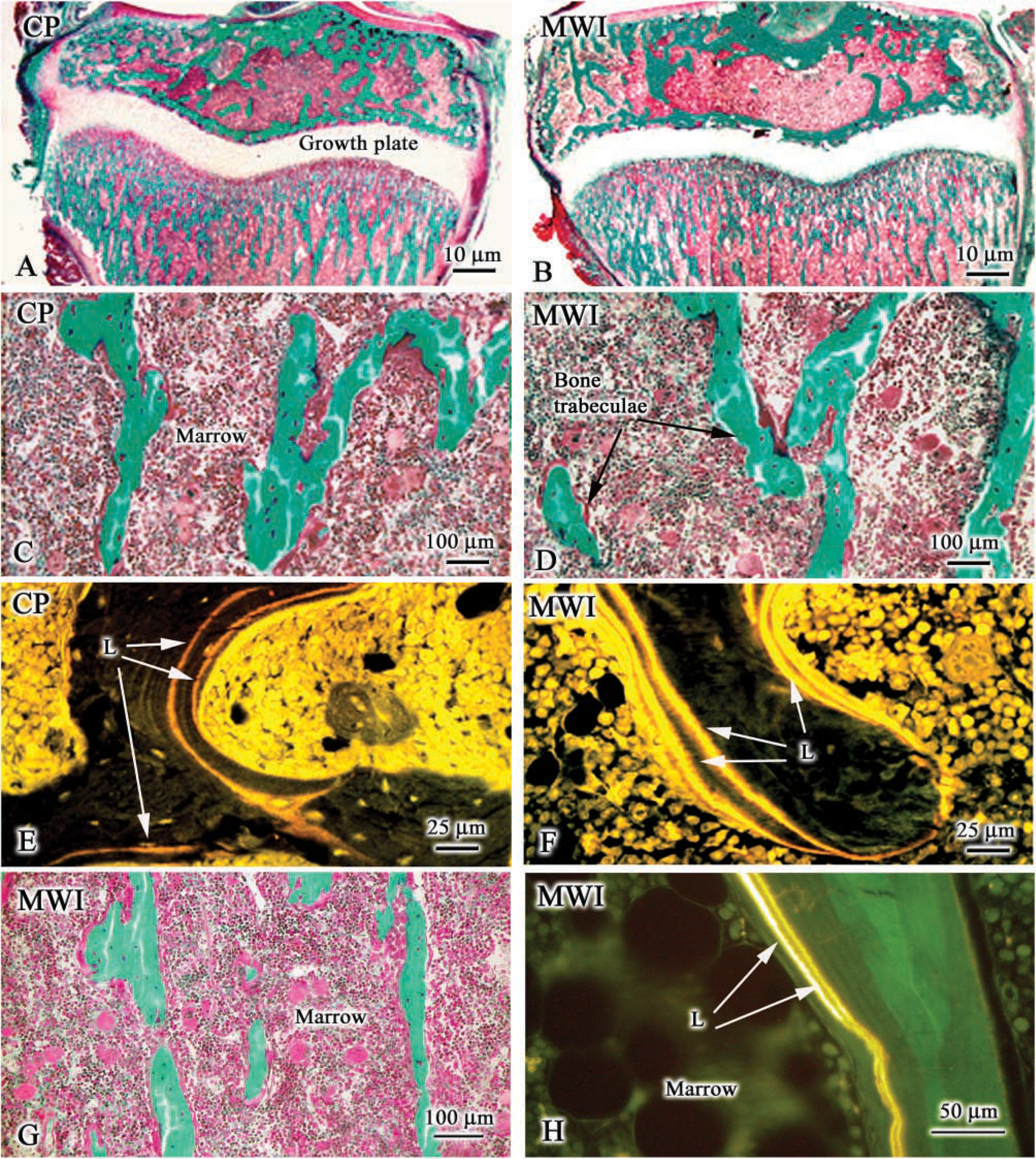
Light micrographs of bone samples used for static and dynamic histomorphometry. (
Histomorphometric analysis of ethanol-fixed rat tibiae processed with (MWI) or without microwave irradiation (conventional) a
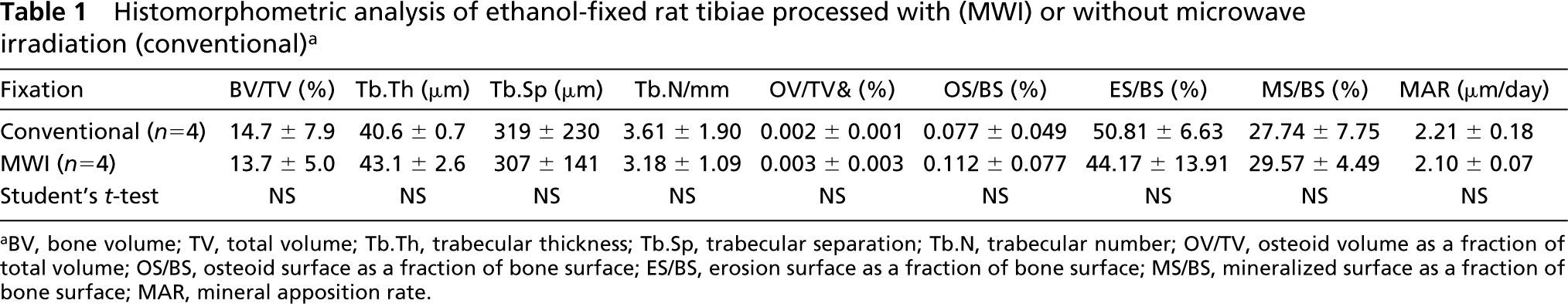
aBV, bone volume; TV, total volume; Tb.Th, trabecular thickness; Tb.Sp, trabecular separation; Tb.N, trabecular number; OV/TV, osteoid volume as a fraction of total volume; OS/BS, osteoid surface as a fraction of bone surface; ES/BS, erosion surface as a fraction of bone surface; MS/BS, mineralized surface as a fraction of bone surface; MAR, mineral apposition rate.
Histochemistry
ALP and TRACP activities, as well as immunolabeling for BSP and OPN, were all clearly detectable on sections of human and rat MWI bone samples (Figures 4 and 5). The observed immunostaining was abolished under control incubation conditions.
Immunogold Labeling in MWI Bone
The distribution of OPN in calcified bone matrix (Figure 6) was similar to that described previously using aldehyde-fixed bone embedded in an acrylic resin (reviewed in Nanci 1999). Gold particles accumulated as clusters among the mineralized collagen fibrils and between layers of calcified tissues. These areas probably correspond to the interfibrillar patches of non-collagenous matrix proteins and cement lines that are readily apparent in decalcified specimens but not in calcified ones. Gold particles were also observed over mineralization foci in the osteoid tissue. Only a few randomly distributed gold particles were observed under control conditions.
Discussion
MWI was used to facilitate the processing of undecalcified bone samples and to allow application of both light and electron microscope analytical procedures to the same sample. The results obtained from young and older rats and from a human bone biopsy led to four major conclusions. First, MWI does not affect any of the conventional procedures and analyses carried out on MMA-embedded bone. Results of histomorphometric measurements on tibiae preserved with ethanol, with or without MWI, and embedded in MMA were not significantly different. In addition, tetracycline double labeling was qualitatively and quantitatively comparable in both rat and human samples. Second, histochemical detections of ALP, TRACP, BSP, and OPN were not altered by MWI. Third, MWI sufficiently improves tissue preservation to permit ultrastructural analysis and immunolabeling. Fourth, the dehydration time is significantly shortened from 1 week to ~7 hr. This last aspect is of particular importance because the outlined MWI procedure allows biopsies to be processed more rapidly, and thus diagnostic results can be obtained earlier. This may not be of critical importance in diagnosing chronic diseases such as osteoporosis but may represent a determinant factor for patients with neoplasias. The latter patients will benefit from earlier detection and treatment. Electron microscopy also provides additional detail that will permit a better characterization and staging of a disease, again essential elements in establishing the therapeutic protocol.
Histomorphometric analysis of ethanol-fixed human bone biopsies processed with (MWI) or without microwave irradiation (conventional) a
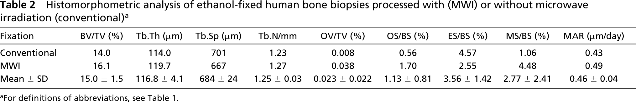
aFor definitions of abbreviations, see Table 1.
One major problem with fixation by immersion is that the quality of preservation depends on the speed at which the fixative penetrates the sample. The centers of samples, particularly larger ones such as biopsy specimens, are usually less well preserved. MWI facilitates and accelerates the penetration of fixatives, in our case ethanol, into samples, reducing the diffusion rate effect on preservation (Wagenaar et al. 1993; Massa and Arana-Chavez 2000). In addition, it has been proposed that MWI stabilizes tissue proteins, but as yet it is not clear whether any crosslinking is induced in the presence of ethanol or whether the stabilizing effect observed simply results from temperature-induced protein coagulation (Moran et al. 1988; discussed in Visser et al. 1992). In any case, our results demonstrate that tissue preservation is improved by MWI of ethanol-immersed bone samples. The resulting structural details are comparable to those that are usually obtained when tissues are simply immersed in mild fixatives and then embedded in acrylic resins.
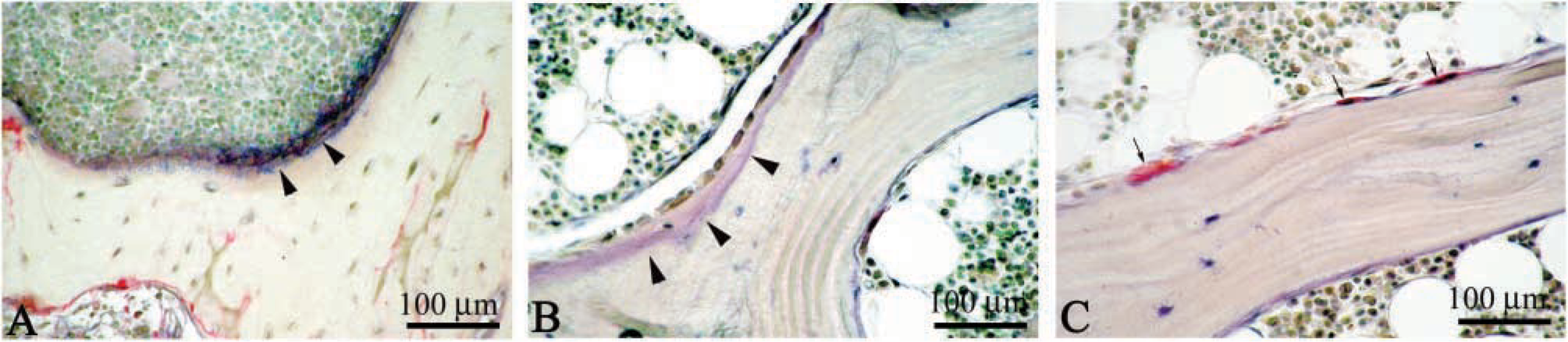
Histochemical detection of alkaline phosphatase activity (purplish staining indicated by arrowheads) in (
Clearly, for both experimental and clinical analyses, it would be very useful to cross-match information obtained by various techniques from the same sample. Transgenic/knockout animals are proving to be very powerful models for understanding the function of proteins and how alteration in their production may, directly or indirectly, lead to disease. Such animal models are complex to produce and maintain, and hence are limited in availability. Any methodology that allows extraction of maximal information from precious tissue samples will certainly help improve our understanding of the function of gene products and phenomena that lead to pathological alterations.
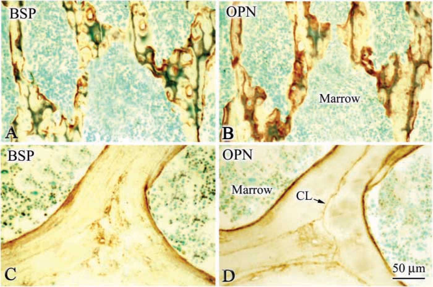
Immunohistochemical preparations for bone sialoprotein (BSP) and osteopontin (OPN) on rat (
Characterization of the biochemical profile of bones and variations induced by pathological alterations is complicated by the fact that this profile varies according to the anatomic location and also at the level of the microenvironment (discussed in Nanci 1999). The organic matrix of bone consists mainly of collagen type I and small amounts (~10%) of several non-collagenous matrix proteins, which play major roles in cellular and extracellular events (reviewed in Boskey 1996; Gehron Robey 1996). Disease-induced variations in the minor non-collagenous components would be difficult to determine biochemically. Immunocytochemistry is a form of “biochemistry on section” that allows detection of small quantities of a constituent and its localization with respect to others at the site where they accumulate and likely express their action, in normal and diseased bone. Furthermore, the localization of matrix proteins at the ultra-structural level is important for determining their relationship with osteoblasts and osteoclasts and with sites of mineralization events. The fact that sections of MMA-embedded tissues can be deacrylated is likely to enhance the sensitivity of immunolabeling and the detection of low-abundance epitopes, such as membrane proteins or low-level expression gene products. It also permits investigation of mRNA expression by in situ hybridization (Saito et al. 1999). Because the ethanol-MMA combination preserves mRNAs, deacrylated MMA sections of ethanol-fixed tissues could, in theory, also be used for in situ RT-PCR and mRNA extraction from laser-dissected samples. Normally, laser dissection is carried out on paraffin-embedded tissues. In the case of calcified tissues, this requires an additional decalcification step, which may result in the loss and/or deterioration of the mRNA. On the other hand, MMA-embedded calcified tissues can readily be cut at various thicknesses and, after deacrylation, would be amenable to mRNA detection and extraction methodologies. The ability to carry out all the above evaluations on the same specimen offers the advantage of correlating macroscopic data, bone formation and resorption parameters, molecular markers of cell activity, and mineral status and composition of the organic matrix.
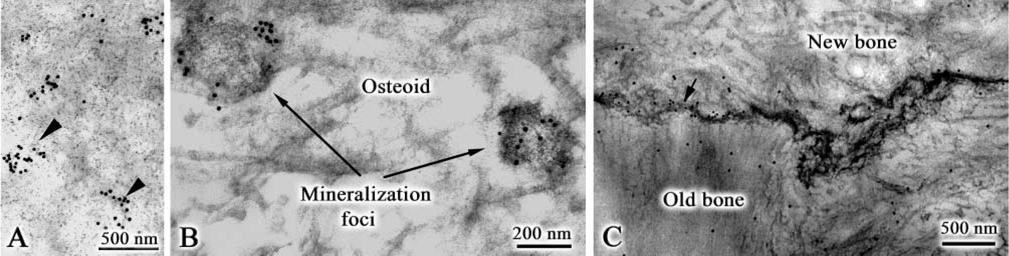
Immunocytochemical preparations from microwave-processed rat bone labeled for osteopontin. (
In summary, in this study, we suggest a protocol for processing calcified bone samples, which essentially consists of treating with MWI samples conventionally immersed in 70% ethanol. This simple protocol requires only minor adaptations to current laboratory procedures and significantly shortens tissue-processing time. It broadens the spectrum of analytical methodologies that can be applied to the same sample and allows more structural and compositional information to be obtained. This enhanced approach makes it possible to fully achieve the concept of “molecular histomorphometry” expounded by Parfitt (1994), which is the integration of conventional histomorphometry with molecular information to provide new insights into the physiology and pathology of bone.
Footnotes
Acknowledgements
Supported by an operating grant to A.N. from the Canadian Institutes of Health Research.
We are grateful to Sylvia Francis Zalzal (Université de Montréal) for her help with backscattered electron imaging, to Micheline Fortin (Université de Montréal) for assistance with tissue sectioning and immunolabeling, and to Roxane Carrier and Claire Deschěsnes (Hôpital St Luc) for technical participation in histomorphometric preparations. O.L. was the recipient of an INSERM/Canadian Institutes of Health Research award (International Scientific Exchanges IRSC/INSERM; XIF-40990).